One of the most familiar features of echinoderm functional morphology and physiology is their remarkable facility for regeneration (Emson and Wilkie, 1980). Regeneration in echinoderms is found throughout the phylum and forms an integral part of their adaptive repertoire. For example, in asteroids it fulfils not only a repair facility but also, in some species, allows for asexual reproduction (Emson and Wilkie, 1980; Thorndyke et al., 1999). In both ophiuroids and holothuroids it underlies an important ‘sacrificial defence’ purpose. When holothuroids are threatened by predators they will eject much of their gastrointestinal tract and replace it by proliferative activity arising largely from the mesenteric epithelium (Garcia-Arraras et al., 1998; Garcia-Arraras et al., 1999). The phenomenon of regeneration has perhaps reached it greatest adaptive development in some ophiuroid families. The amphiurid group of brittlestars includes benthic species such as Amphiura filiformis, which form a significant and important part of the biomass (Sköld and Rosenberg, 1996). Their feeding mechanism makes them especially vulnerable to predation by flatfish and crustaceans, and they are a crucial part of the benthic food chain being a primary source of food for bottom-dwelling fish. Estimates predict that in certain areas of the Skaggerak their lost arms contribute as much as 300 metric tonnes of biomass per year (Sköld and Rosenberg, 1996). Following arm loss, the missing part is replaced rapidly by regeneration, and it is common to find natural populations in which almost every individual present shows evidence of one, two or more current arm regeneration events (Thorndyke et al.,
1999). While it is clear that regeneration is an entirely natural phenomenon, several questions deserve attention. For example, to what extent are the events of arm autotomy and the subsequent regeneration process stressful, as with other environmental challenges such as temperature variation that impact upon an animal’s physiology (Feder, 1999a; Patruno et al., 2000a)?
The intracellular protein turnover experienced by echinoderms during the arm regeneration process has been recently studied (Patruno et al., 2000a). Molecules such ubiquitin and heat-shock proteins (for example Hsp70) were used to determine whether any changes in their expression are correlated with a particular phase of regeneration. We know from other studies that Hsps, which are encoded by highly conserved families of genes, play key roles not only in the correct folding and degradation of proteins but also during development (Becker and Craig, 1994). A specific example is Drosophila melanogaster, where small increases in Hsp70 levels during development enhance thermotolerance (Feder, 1999b); however, if overexpression of the Hsp70 gene is induced, larval mortality increases and development slows down (Krebs and Feder, 1997). Therefore, it might be expected that Hsps is involved in regeneration ‘stress’ (Patruno et al., 2000a; Feder, 1999b). Furthermore, regeneration can involve rapid and considerable growth phases where, in some individuals of A. filiformis, rates of arm replacement can approach 0.04 mm per day (J. Mallefet and M. C. Thorndyke, unpublished observations). Here, growth factors and other JEB3070
The study of regeneration in armed echinoderm species, including crinoids, ophiuroids and asteroids, is attracting increasing attention. Recent interest has focused on the presence and potential role of growth factors, including members of the nerve growth factor (NGF) and transforming growth factor-beta (TGF-β) families, in the regenerative process and their possible relationship to the normal developmental (ontogenetic) regulatory cascade. In addition, the expression patterns of the heat-shock family of stress proteins (Hsps) during regeneration are also
important. Their role forms part of a normal stress response to the trauma of autotomy in combination with a putative function in tissue remodelling and associated protein turnover during regeneration. The temporal dynamics of the stress response may also be strongly indicative of environmentally adaptive pressures operating on these systems.
Key words: regeneration, echinoderm, stress protein, growth factor, bone morphogenic protein (BMP).
Summary
Introduction
GROWTH FACTORS, HEAT-SHOCK PROTEINS AND REGENERATION IN
ECHINODERMS
M. PATRUNO1, M. C. THORNDYKE1,*, M. D. CANDIA CARNEVALI2, F. BONASORO2 ANDP. W. BEESLEY1 1School of Biological Science, Royal Holloway, University of London, Egham TW20 OEX, UK and
2Department of Biology, University of Milan, Milano Via G. Celoria 26, 20133 Italy
*Author for correspondence (e-mail: m.thorndyke@rhul.ac.uk)
regulators of cell proliferation and differentiation, such as members of the transforming growth factor-beta family of proteins (TGF-β), probably play an important part in the process. TGF-β family members are a diverse group of secreted proteins widely involved in many aspects of growth regulation (Kingsley, 1994; Hogan, 1996). They may act as humoral, paracrine or autocrine agents in all life cycle stages from embryogenesis to adult wound healing.
The purpose of the current study was to explore the putative role(s) of growth factors and Hsps in echinoderm regeneration and assess the extent to which regeneration is a stress response that might be related to a natural environmental niche. That is, could there be a difference in the regeneration stress response between those animals living in a normally more stressful (changing) environment and those from more stable environments?
Materials and methods
Samples of crinoids and ophiuroids were collected from both Bangor (North Wales) and Kristineberg Marine Research Station (Sweden). Samples of Asterias rubens collected at Bangor from the intertidal zone during a low tide (intertidal group) were processed in situ or transported and maintained in a circulating artificial seawater system as described (Moss et al., 1998). Other samples of A. rubens collected by scuba divers in Wales and Sweden were processed immediately or kept in a circulating seawater system (benthic group).
Thermal stress
To determine the effect of thermal stress on expression of heat-shock proteins (Hsps), some normal (non-regenerating) animals were kept at a higher temperature compared to natural conditions (water temperature in the Gulf of Taranto was 15 °C at 20 m depth and at Kristineberg, 12 °C at the same depth). In order to induce a temperature-related stress response these animals were subjected to a gradual increase in temperature. They were transferred to pre-heated tanks of seawater (from 1 to 5 °C higher than their natural habitat) and left for at least 30 min followed by subsequent recovery for 1 h at normal temperature. Samples of crinoids and ophiuroids were prepared from whole arms (normal and regenerating) and, at later stages, from regenerating blastema only. Samples of asteroids were prepared from whole normal arms and whole regenerating arms. The radial nerve cord (rnc) was also dissected and analysed for ubiquitin and hsp70 immunoreactivity. ‘Whole arm preparation’ refers to the complete arm with the rnc removed.
Gel electrophoresis and western blotting
Samples were homogenised in 10 mmol ml−1 sodium
phosphate buffer (pH 7.0) containing the following protease inhibitors: leupeptin, pepstatin, chymostatin and antipain (all at 1 mg ml−1 final concentration) and
phenylmethylsulphonylfluoride (0.2 mmol l−1 final
concentration). The calcareous deposits present in the homogenates were sedimented by low-speed centrifugation at 1000 gav for 1 min. Prior to electrophoresis, protein
determinations were made using a Bio-Rad protein assay (Bio-Rad Labs). Equal amounts of protein extract (from 20 to 60µg per lane, depending on the antibody used) were then separated by sodium dodecyl sulphate (SDS)-polyacrylamide gel electrophoresis (Laemmli, 1970). Gels were stained with Coomassie Blue for protein visualization. Western blotting was essentially as described (Towbin et al., 1979).
Immunodevelopment of western blots with RHUb1 anti-ubiquitin monoclonal antibody (mAb, neat hybridoma supernatant) was carried out as described (Flann et al., 1997; Mimnaugh et al., 1999). The anti-Hsp72 antibody (Stressgen SPA-810) was diluted 1:500 (v/v) in blocking solution and blots incubated overnight in the primary antibody. The anti-Hsp70 antibody (Sigma, clone BRM-22) was provided by Dr V. Matranga (C.N.R. Palermo, Italy) and used as suggested by the manufacturer. Following incubation in primary antibody, blots were washed in phosphate-buffered saline (PBS) and incubated in secondary antibody, anti-mouse horseradish peroxidase-conjugate (Dako) diluted 1:2000 (v/v) in blocking solution for 1 hour. Blots were washed in PBS (1×15 min followed by 4×5 min) and immunoreactivity visualized using the enhanced chemiluminescence method (Amersham or Pierce). The nitrocellulose membrane was silver stained following immunodevelopment with antibodies (Kovarik et al., 1987). Densitometric analysis of blots (at least three replicate samples for each stage) measured the area under the densitometrically scanned peak utilising a UVP-gel documentation and analysis system. For protein extracts all values quoted are expressed relative to normal arms (P0; 100 %).
Immunocytochemistry
The samples were fixed in 4 % paraformaldehyde, then processed for embedding in resin and subsequent sectioning according to established techniques (Candia Carnevali et al., 1998). After mounting semithin sections on gelatinized slides, they were treated with a resin-removing mixture (1 h in sodium ethanolate). Subsequently sections were rinsed with absolute ethanol, washed in distilled water and then immersed for 7 min in sodium periodate (fresh 1 % solution). Next, sections were treated for 30 min with 0.3 % (v/v) H2O2in phosphate-buffered
Labs). Visualization was by incubation in 0.05 % (w/v) 3,3′-diaminobenzidine (DAB, Sigma). For fluorescence visualisation, the ABC reagent was replaced with fluorescein-or Texas Red-conjugated avidin D (Vectfluorescein-or Labs). DAB preparations were dehydrated and mounted in DPX. Fluorescence preparations were washed and mounted in Vectashield (Vector Labs).
Observation was by a Zeiss Axioplan with fluorescence attachments and by a Leica TCS-4D confocal laser-scanning microscope (CLSM). Controls were conducted by replacing primary antiserum with non-immune sera from the animal in which the primary antiserum was raised or omitting the primary antibody and incubating in PBS and 1 % (v/v) normal NGS. Nuclear labelling was confirmed by using DAPI (Molecular Probes) staining. The specificity of the anti-ubiquitin antibody is well established (Flann et al., 1997).
Some immunocytochemistry was also carried out on paraffin sections, but with the following modifications. Slides were rinsed with xylene and dehydrated in an ascending ethanol series. The step with sodium periodate was omitted and in the blocking step, NGS was used at 5 % (v/v).
Results and Discussion
Is regeneration a stressful event for all echinoderms? A complete and functional regrowth of the arm following amputation depends on many factors. Probably, one of the most important is the site of amputation along the proximal–distal axis of the arm. A traumatic amputation that does not follow the natural autotomy plane may involve more complex reparative/regenerative mechanisms and would therefore be slower than a non-traumatic autotomy (Candia Carnevali and Bonasoro, 1995). Whichever is the chosen regenerative ‘pathway’, substantial tissue rearrangement and
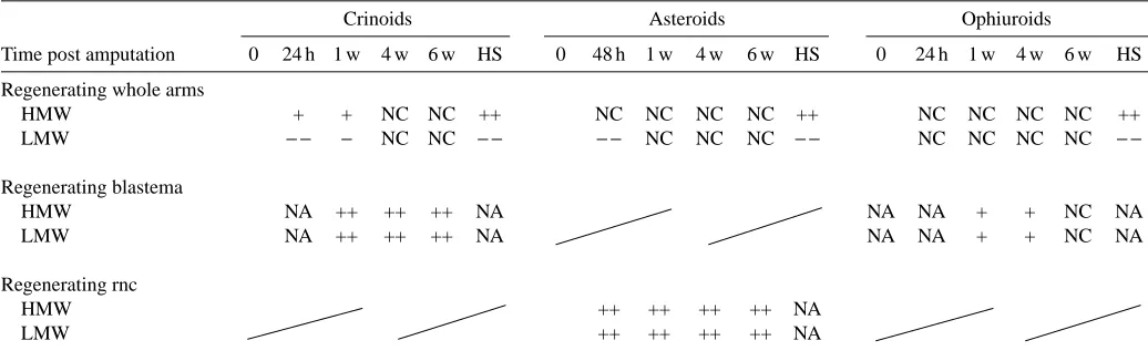
extensive cellular proliferation/differentiation is observed in all the echinoderm species so far investigated. During the arm regeneration process, these animals may experience initial stress followed by a massive intracellular protein turnover. Table 1 summarises Hsps expression detected in the echinoderm species investigated in this laboratory.
Using an immunochemical approach, two major points emerge from this study. One is the indication that regenerating tissues alone behave differently in terms of Hsp expression compared with the rest of the arm. Indeed, in rapidly regenerating tissues such as the crinoid blastema or the nerve cord in asteroids, the levels of ubiquitin conjugates increase noticeably and in general the pattern is similar to that seen in phylogenetically related systems such as the developing larvae of sea urchins (Pickart et al., 1991). In particular, there is an increase in different isoforms of ubiquinated histones at 10 and 32 h post-fertilization in the pluteus larva stage of Strongylocentrotus purpuratus (Jasinskiene et al., 1995). Other examples of ubiquinated histone upregulation are observed during spermatogenesis in the chicken (Agell and Mezquita, 1988) and in transformed human cells, in which immunoreactivity is localised to clusters within the nucleus (Vassilev et al., 1995).
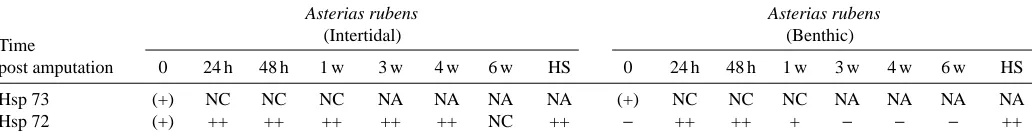
[image:3.612.49.567.517.680.2]The other major point is that the prolonged expression period of Hsp72 (Table 2) during regeneration in intertidal asteroids may reflect the more stressful amputation experienced by starfish compared to brittlestars or featherstars. In crinoids and ophiuroids, Hsp72 is present only briefly after amputation. This short expression period suggests that these animals are not dramatically affected by arm amputation, since this is a more natural phenomenon resulting from high rates of fish predation (Sköld and Rosenberg, 1996). The constitutive form of the Hsp70 family (Hsp73) remained constant throughout the regenerative process, confirming its basic role in cellular metabolism (Table 2).
Table 1. Summary of the changes in levels of ubiquitin conjugates from different regenerating tissues and in heat-treated animals
Crinoids Asteroids Ophiuroids
Time post amputation 0 24 h 1 w 4 w 6 w HS 0 48 h 1 w 4 w 6 w HS 0 24 h 1 w 4 w 6 w HS
Regenerating whole arms
HMW + + NC NC ++ NC NC NC NC ++ NC NC NC NC ++
LMW − − − NC NC − − − − NC NC NC − − NC NC NC NC − −
Regenerating blastema
HMW NA ++ ++ ++ NA NA NA + + NC NA
LMW NA ++ ++ ++ NA NA NA + + NC NA
Regenerating rnc
HMW ++ ++ ++ ++ NA
LMW ++ ++ ++ ++ NA
Ubiquitin conjugates were detected by western blotting.
Is the expression of Hsps in echinoderms dependent on an environmental gradient of stress?
In nature, organisms show a variation in the stress response that is dependent on an environmental gradient of stress. Indeed, one important matter is whether organisms from environments with little stress have a different stress response from that of animals living in a more changeable environment. Although some studies (reviewed by Feder and Hofmann, 1999) confirmed that increasing levels of expression of Hsps are positively correlated with stressful environments, the picture is not clear. For example, Hsp expression varies seasonally in some fish (Feder and Hofmann, 1999) and intertidal invertebrates (Hofmann and Somero, 1995). Furthermore, studies on ubiquitin conjugation and Hsp72 expression in Asterias rubens collected from a stable (benthic) or variable (intertidal) environment showed (1) no differences in the level and pattern of ubiquitin conjugates between the two populations, (2) a marked elevation of Hsp72 expression in the intertidal compared with the benthic animals and (3), following amputation, that Hsp72 levels were elevated for longer in the intertidal than in the benthic animals. In contrast, crinoid and ophiuroid samples, collected from different (North Sea and Mediterranean Sea) but stable environments, showed no difference in Hsp expression during normal growth and regeneration. These data confirm that the stress response may indeed be environment-dependent in echinoderms as proposed by Feder and Hoffman (Feder and Hoffman, 1999; Hoffman, 1999), but also show how such a conserved mechanism is intimately correlated with multiple patterns of Hsp expression in all phyla.
TGF-βhomologues are present in crinoid echinoderms Growth factors are a comprehensive group of polypeptides, known as multifunctional hormones, which are involved in fundamental cell activities such as proliferation, differentiation and maintenance (Cross and Dexter, 1991). A peculiar characteristic of growth factors is their ability to display multiple properties depending on what has been called the ‘cellular context’, e.g. according to the type of target cells involved and their synergistic/antagonistic interactions with other factors (Massagué and Wotton, 2000).
[image:4.612.44.560.99.167.2]Our recent investigation of ‘putative’ growth factors important for the regenerative process in crinoids has concentrated on TGF-β1 because of its importance in wound healing in both embryonic and adult vertebrates (Nodder and Martin, 1997; O’Kane and Ferguson, 1997). Other studies support the idea that TGF-β is an important molecule involved in epithelial–mesenchymal interactions, which are fundamental for pattern determination in regenerating systems (Ferretti and Gèraudie, 1998). One of the most familiar roles for TGF-βis in wound repair following injury. Here, it plays a part in a variety of associated processes including the regulation of extracellular matrix (ECM) structure and function, cell migration and adhesion. These factors are, therefore, prime candidates for a role in regeneration. The considerable amino acid identity exhibited by mature proteins of the TGF-β superfamily (Fig. 1) has allowed the characterization of echinoderm homologues. To summarise, it seems that TGF-β1 is a good candidate for one of the growth factors involved in the initialization of wound Table 2. Summary of Hsp levels during the arm regenerative process and in heat-shocked Asterias rubens from benthic and
intertidal zones
Asterias rubens Asterias rubens
Time (Intertidal) (Benthic)
post amputation 0 24 h 48 h 1 w 3 w 4 w 6 w HS 0 24 h 48 h 1 w 3 w 4 w 6 w HS
Hsp 73 (+) NC NC NC NA NA NA NA (+) NC NC NC NA NA NA NA
Hsp 72 (+) ++ ++ ++ ++ ++ NC ++ − ++ ++ + − − − ++
(+), presence of Hsp72-Hsp73; +, moderate increase compared with control; ++, large increase compared with control; NC, no change compared with control; −, not detected; NA, not assessed.
HS, heat-shocked animals.
Fig. 1. Alignment of the C-terminal regions of some TGF-βsuperfamily members. Alignment begins at the first conserved cysteine of each molecule and extends to the C terminus. GenBank accession numbers are: (1) Homo sapiens BMP-4, BAA06410.1; (2) Oryctolagus cuniculis, AAB96785.1; (3) Dama species BMP-2, CAA05033.1; (4) Branchiostoma floridae, AAC97488.1; (5) Halocynthia roretzi, BAA31132.1; (6)
healing and also for activating the proliferation of migratory elements.
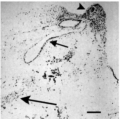
The presence and distribution in normal and regenerating arms of Antedon mediterranea and Antedon bifida of both a TGF-β-like molecule and its receptors have been detected by immunochemical studies (Patruno et al., 2000b). Increasing amounts of immunolabelling and western blot detection of these molecules at early stages of regeneration suggest that their involvement is important at early regenerative stages (Fig. 2; M. Patruno, M. Thorndyke and P. Beesley, unpublished observations). Less obvious candidates, though possibly more significant, are members of the bone morphogenetic protein (BMP) subfamily, since these are a group of secreted morphogens that play a crucial and central role early in the establishment of body axes (Hogan, 1996). Moreover, they later participate in the regulation of patterning and cell identity in the developing nervous system (Dale et al., 1999; Nguyen et al., 2000). It is not surprising then that BMPs have been identified in echinoderms. Here, emphasis has been placed on the very early stages of development, where BMPs have been shown to be involved in regulating the position of the ectoderm/endoderm boundary as well as epidermal/ nonepidermal differentiation (Angerer et al., 2000). To date, BMP homologues have been found only in four echinoderm species. Three BMP2/4 homologues have been identified in echinoids Tripneustes gratilla (TgBMP2/4) (Hwang et al., 1997), Strongylocentrotus purpuratus (SpBMP2/4) (Angerer et al., 2000) and Lytechinus vulgaris (LvBMP2/4) (C. Y. Logan and D. R. McClay, unpublished; Accession Number
AF119712); one BMP2/4 homologue from the asteroid Asterias rubens (C. Lelong, M. Mathieu and P. Favrel, unpublished; Accession Number CAB63584); and one BMP5/7 homologue from Strongylocentrotus purpuratus (Ponce et al., 1999) and univin (Stenzel et al., 1994), also from Strongylocentrotus purpuratus. In situ hybridisation and mRNA microinjection in S. purpuratus confirmed that BMP signalling might be considered as a developmental coordination system homologous with that of vertebrates. In particular, these experiments suggest that BMP2/4 might have a significant role in establishing morphogenetic gradients in echinoderms (Angerer et al., 2000; Angerer and Angerer, 2000). In contrast, the functional role of BMPs during growth in adult echinoderms is still unknown. In this respect, however, our work reported here implicates a role for TGF-β-like factors in adult regeneration. Clearly then, such factors are prime candidates for a role (or roles) in regenerative development. Indeed, new data suggest that a recently characterised crinoid BMP homologue (AnBMP2/4) (Patruno et al., 2000b) might be upregulated during regeneration and so play a part in the regulation of patterning and cell lineage specification that occurs at this time.
This work was supported by an Anglo-Italian/British Council–Murst exchange grant, University of London Central Research Fund, Royal Holloway Research Strategy Fund and a Thomas Holloway Studentship (M.P.). We are especially grateful to the Director and staff at Kristineberg Marine Research Station (Sweden) and a grant from the EU Large-Scale Facility Fund for funding part of this work.
References
Agell, N. and Mezquita, C. (1988). Cellular content of ubiquitin and
formation of ubiquitin conjugates during chicken spermatogenesis.
Biochem. J. 250, 883–889.
Angerer, L. M. and Angerer, R. C. (2000). Animal–vegetal axis
patterning mechanisms in the early sea urchin embryo. Dev. Biol.
218, 1–12.
Angerer, L. M., Oleksyn, D. W., Logan, C. Y., McClay, D. R., Dale, L. and Angerer, R. C. (2000). A BMP pathway regulates
cell fate allocation along the sea urchin animal–vegetal embryonic axis. Development 127, 1105–1114.
Becker, J. and Craig, E. A. (1994). Heat-shock proteins as molecular
chaperones. Eur. J. Biochem. 219, 11–23.
Candia Carnevali, M. D. and Bonasoro, F. (1995). Arm
regeneration and pattern formation in crinoids. In Echinoderm
Research 1995 (ed. R. Emson, A. B. Smith and A. Campbell), pp.
245–253. Rotterdam: Balkema.
Candia Carnevali, M. D., Bonasoro, F., Welsch, U. and Thorndyke, M. C. (1998). Arm regeneration and growth factors
in Crinoids. In Echinoderms: San Francisco (ed. R. Mooi and M. Telford), pp. 145–150. Rotterdam: Balkema.
Cross, M. and Dexter, T. M. (1991). Growth factors in
developmental transformation and tumorigenesis. Cell 64,
271–280.
[image:5.612.57.293.71.305.2]Dale, K., Sattar, N., Heemskerk, J., Clarke, J. D. W., Placzec, M. and Dodd, J. (1999). Differential patterning of ventral midline
cells by axial mesoderm is regulated by BMP7 and chordin.
Development 126, 397–408.
Emson, R. H. and Wilkie, I. C. (1980). Fission and autotomy in
echinoderms. Oceanogr. Mar. Biol. Annu. Rev. 18, 155–250.
Feder, M. E. and Hofmann, G. E. (1999). Heat-shock proteins,
molecular chaperons, and the stress response: evolutionary and ecological physiology. Annu. Rev. Physiol. 61, 243–282.
Feder, M. E. (1999a). Organismal, ecological, and evolutionary
aspects of heat-shock proteins and the stress response: established conclusions and unresolved issues. Am. Zool. 39, 857–864.
Feder, M. E. (1999b). Engineering candidate genes in studies of
adaptation: the heat-shock protein Hsp70 in Drosophila
melanogaster. Am. Nat. 154, 55–66.
Ferretti, P. and Gèraudie, J. (1998). Cellular and Molecular Basis of Regeneration: from Invertebrates to Humans. Chichester: John
Wiley & Sons Ltd.
Flann, S., Hawkes, R. B., Riederer, B. M., Rider, C. C. and Beesley, P. W. (1997). Changes in ubiquitin immunoreactivity in
developing rat brain: a putative role for ubiquitin and ubiquitin conjugates in dendrite outgrowth and differentiation. Neuroscience
81, 173–187.
Garcia-Arraras, J. E., Estrada-Rodgers, L., Santiago, R., Torres, I., Diaz-Miranda, L. and Torres-Avillan, I. (1998). Cellular
mechanisms of intestine regeneration in the sea-cucumber
Holothuria glaberrina Selenka (Holothuroidea: Echinodermata). J. Exp. Zool. 281, 288–304.
Garcia-Arraras, J. E., Diaz-Miranda, L.,Torres, I. I., File, S., Jimenez, L. B., Rivera-Bermudez, K., Arroyo, E. J. and Cruz, W. (1999). Regeneration of the enteric nervous system in the sea
cucumber Holothuria glaberrina. J. Comp. Neurol. 406, 461–475.
Hofmann, G. E. and Somero, G. N. (1995). Evidence for protein
damage at environmental temperatures: seasonal changes in levels of ubiquitin conjugates and hsp70 in the intertidal mussel Mytilus
trossulus. J. Exp. Biol. 198, 1509–1518.
Hofmann, G. E. (1999) Ecologically relevant variation in induction
and function of heat shock proteins in marine organisms. Am. Zool.
39, 889–900.
Hogan, B. L. M. (1996). Bone morphogenetic proteins:
multifunctional regulators of vertebrate development. Genes Dev.
10, 1580–1594.
Hwang, S. P. L., Chen, C. A. and Chen, C. P. (1999). Sea urchin
TgBMP2/4 gene encoding a bone morphogenetic protein closely related to vertebrate BMP2 and BMP4 with maximal expression at the later stages of embryonic development. Biochem. Biophys. Res.
Comm. 258, 457–463.
Jasinskiene, N., Jasinskas, A. and Langmore, J. P. (1995).
Embryonic regulation of histone ubiquitination in the sea urchin.
Dev. Genet. 16, 278–290.
Kingsley, D. M. (1994). The TGF-βsuperfamily: new members, new receptors, and new genetic tests of function in different organisms.
Genes Dev. 8, 133–146.
Kovarik, A., Hlubinova, K., Vrbenska, A. and Prachar, J. (1987).
An improved colloidal silver staining method of protein blots on nitrocellulose membranes. Folia Biol. 33, 254–257.
Krebs, R. A. and Feder, M. E. (1997). Deleterious consequences of
Hsp70 overexpression in Drosophila melanogaster larvae. Cell
Stress Chaperones 2, 60–71.
Laemmli, U. K. (1970). Cleavage of structural proteins during the
assembly of the head of bacteriophage T4. Nature 227, 680–685.
Massagué, J. and Wotton, D. (2000). Transcriptional control by the
TGF-/Smad signalling system. EMBO J. 19, 1745–1754.
Mimnaugh, E. G., Bonvini, P. and Neckers, L. (1999). The
measurement of ubiquitin and ubiquitinated proteins.
Electrophoresis 20, 418–428.
Moss, C., Hunter, A. J. and Thorndyke, M. C. (1998). Patterns of
bromodeoxyuridine incorporation and neuropeptide immunoreactivity in the regenerating arm of the starfish Asterias
rubens. Phil. Trans. R. Soc. Lond. B 353, 421–436.
Nguyen, Vu. H., Trout, J., Connors, S. A., Andermann, P., Weinberg, E. and Mullins, M. C. (2000). Dorsal and intermediate
neuronal cell types of the spinal cord are established by a BMP signalling pathway. Development 127, 1209–1220.
Nodder, S. and Martin, P. (1997). Wound healing in embryos: a
review. Anat. Embryol. 195, 215–228.
O’Kane, S. and Ferguson, M. W. J. (1997). Transforming growth
factor βs and wound healing. Int. J. Biochem. Cell. Biol. 29, 63–78.
Patruno, M., Beesley, P., Thorndyke, M. C., Bonasoro, F. and Candia Carnevali, M. D. (2000a). Changes in ubiquitin conjugates
and Hsp72 levels during arm regeneration in echinoderms. Mar.
Biotech. (in press).
Patruno, M., Graham, A., McGonnell, I., Candia Carnevali, M. D., Bonasoro, F., Beesley, P. W. and Thorndyke, M. C. (2000b).
Occurrence and expression of a novel transforming growth factor-beta homologue in Crinoids. In Proceedings of 10thInternational
Echinoderm Conference, Dunedin 2000 (ed. M. Barker). Rotterdam: Balkema (in press).
Pickart, C. M., Summers, R. G., Shim, H. and Kasperek, M.
(1991). Dynamics of ubiquitin pools in developing sea urchin embryos. Dev. Growth Diff. 33, 587–598.
Ponce, M. R., Micol, J. L., Peterson, K. J. and Davidson, E. H.
(1999) Molecular characterization and phylogenetic analysis of SPBMP5-7, a new member of the TGF-βsuperfamily expressed in sea urchin embryos. Mol. Biol. Evol. 16, 634–645.
Sköld, M. and Rosenberg, R. (1996). Arm regeneration frequency
in eight species of Ophiuroidea (Echinodermata) from European sea areas. J. Sea Res. 35, 353–362.
Stenzel, P., Angerer, L. M., Smith, B. J., Angerer, R. C. and Vale, W. W. (1994). The univin gene encodes a member of the
transforming growth factor-β superfamily with restricted expression in the sea urchin embryo. Dev. Biol. 166, 149–158.
Thorndyke, M. C., Chen, W-C., Moss, C., Candia Carnevali, M. D. and Bonasoro, F. (1999). Regeneration in echinoderms: cellular
and molecular aspects. In Echinoderm Research 1998 (ed. M. D. Candia Carnevali and F. Bonasoro), pp. 159–164. Rotterdam: Balkema.
Towbin, H., Staehelin, T. and Goedon, J. (1979). Electrophoretic
transfer of proteins from polyacrylamide gels to nitrocellulose sheets: procedure and some applications. Proc. Natl. Acad. Sci.
USA 76, 4350–4354.
Vassilev, A. P., Rasmussen, H. H., Christensen, E. I., Nielsen, S. and Celis, J. E. (1995). The levels of ubiquitinated histone H2A