IMAGING CALCIUM WAVES IN EGGS AND EMBRYOS
ISABELLE GILLOT ANDMICHAEL WHITAKER
Department of Physiology, University College London, Gower Street, London WC1E 6BT
Summary
Sea urchin eggs and those of most other deuterostomes are activated at fertilization by an increase in cytoplasmic free calcium concentration ([Ca2+]
i) that triggers the onset of
the embryonic cell division cycles. We can image the calcium wave using fluorescent calcium indicator dyes and confocal microscopy. There are two components to the [Ca2+]
i increase at fertilization. The first is due to a rapid calcium influx caused by a
calcium action potential; this leads to a small increase in [Ca2+]
ijust beneath the plasma
membrane with spherical symmetry. After a latent period of some 15s, there is a second large and rapid increase in [Ca2+]
ilocalized to the region of sperm–egg contact: during
the latent period [Ca2+]
idoes not change but within 1s of the end of the latent period
[Ca2+]
ireaches 2mmol l21. The calcium wave then spreads across the egg with a velocity
of 5mms21. Behind the advancing wavefront, the calcium concentration is uniformly
high, even within the egg nucleus, though there are no indications that intranuclear calcium concentration differs from [Ca2+]
i. [Ca2+]ifalls uniformly towards resting levels
over the next 500s. In cases where there is an apparent inhomogeneity in [Ca2+]
iin either
the cortex or the nucleus, we find that the calcium indicator dye is inhomogeneously distributed. This appears to be due to uptake of the indicator dye (Fluo-3), probably into mitochondria. The artefact can be avoided by using a dextran-conjugated dye.
Introduction
Fertilization in many species and in all but a very few vertebrates is marked by an activating wave of calcium that sweeps through the cytoplasm from the point of sperm entry (Whitaker and Swann, 1993). In sea urchin eggs, the wave is caused by the dual activation of both inositol trisphosphate (InsP3)- and cyclic ADP ribose
(cADPr)-ryanodine-gated calcium release channels in the endoplasmic reticulum of the egg (Galione et al. 1993). Activation results in calcium-induced calcium release (CICR) from both sets of channels.
We do not know how the explosive calcium release is triggered by the sperm. A simple mechanism in which interaction of egg and sperm stimulates phosphoinositide messenger production via a GTP binding protein (Jaffe et al. 1988) appears to be ruled out (Whitaker and Swann, 1993). The earliest detectable response at fertilization is the fusion of egg and sperm (McCulloh and Chambers, 1992); it is possible that the calcium wave is started by an activating messenger that diffuses from the sperm into the egg (Whitaker et al. 1989).
Signal transduction at fertilization is a problem in space as well as in time: the sperm is tiny compared with the egg and in the first few seconds only a very small region of the egg can be directly influenced by the sperm. The fertilization calcium wave also evolves in time and space, as do calcium signals in other cells (Berridge, 1993). In large cells like eggs (100mm in diameter), conventional fluorescence imaging techniques with calcium indicator dyes such as Fura-2 cannot provide adequate spatial resolution (Silver et al. 1991). Confocal microscopy does not suffer the same limitation; indeed, it was developed to overcome it. We can use confocal calcium-imaging microscopy to obtain images of [Ca2+]iin sea urchin eggs at a few micrometres resolution deep within the egg. We can
image calcium in the region of sperm–egg contact and show that the calcium wave sweeps through the middle of the egg. However, one disadvantage of the present visible-wavelength calcium indicator dyes used for confocal calcium imaging is that they are single-wavelength probes. They lack the built-in correction for dye distribution of a ratio dye like Fura-2. This disadvantage is more than outweighed by the advantage of being able to image calcium at high resolution inside cells, but is does mean that confocal calcium images need to be interpreted with caution.
Images of the calcium wave at fertilization in sea urchin eggs
The first sign of fertilization in images of cytoplasmic calcium is a sudden increase in [Ca2+]ijust beneath the plasma membrane (Fig. 1). Many seconds later, the calcium wave
begins at the point of sperm–egg interaction and then spreads across the egg. The early cortical increase in [Ca2+]iis very small (50nmol l21) compared with the calcium wave
itself (3mmol l21). It is due to calcium influx through voltage-gated calcium channels
(McDougall et al. 1993). Fusion of the sperm with the egg (McCulloh and Chambers, 1992) introduces ion channels in parallel with the egg membrane resistance (Dale et al. 1978) and depolarizes the egg, causing it to fire a calcium action potential (Chambers and De Armendi, 1979). The small elevation of [Ca2+]
iclose to the plasma membrane can be
detected using the confocal calcium-imaging technique, but is not evident using conventional calcium-imaging techniques.
After the calcium influx, very little seems to happen for 10–15s. Even in confocal sections where we can detect the exact point of sperm–egg interaction (Fig. 2), [Ca2+]
i
does not increase during this period. The apparently uneventful interval between sperm–egg fusion and the start of the calcium wave is known as the latent period. The only biochemical event that has been shown to occur during the latent period is a increase in protein tyrosine phosphorylation (Ciapa et al. 1991). Certainly, [Ca2+]
i does not
increase, so we cannot invoke calcium-triggered CICR as a mechanism for initiating the calcium wave. A model that might fit is a sperm-induced increase in InsP3 or cADPr
concentration that sensitizes the calcium release channels to the resting [Ca2+]i. A very
local increase in either would not be detectable using biochemical methods, as the region close to the sperm is less than 0.05% of the egg volume. In support of this idea, heparin (the InsP3 antagonist) has been shown to increase the duration of the latent period
The calcium wave passes through the centre of the egg in the equatorial section shown in Fig. 1. Behind the wavefront, [Ca2+]
i is apparently uniform. There is no obvious
difference in [Ca2+]iin the cortex or the nucleus, contrary to previous reports (Stricker et al. 1992; Shen and Buck, 1993). The uniformity suggests that the calcium store (probably
the endoplasmic reticulum: Terasaki and Sardet, 1991) is itself fairly uniformly distributed within the egg (Terasaki and Jaffe, 1991). However, the ryanodine receptor is localized to the egg cortex (McPherson et al. 1992). That the wave is uniform while the localization of one of the calcium release channels is non-uniform indicates that the ryanodine- and InsP3-receptor release channels are equally capable of propagating the
[image:3.595.106.383.227.422.2]calcium wave with similar kinetics (Galione et al. 1993). It may be that the cortical
Fig. 4. Uptake of Fluo-3 into cytoplasmic organelles. (A) 3min after microinjection of Fluo-3, the nucleus is bright relative to the cytoplasm. At 25min, the nucleus appears dark and the cytoplasm takes on a more granular appearance. At 26min, digitonin was added to the egg to allow diffusion of cytoplasmic dye out of the egg. At 28min and thereafter, most of the dye signal remained. The cytoplasm is very evidently granular in appearance and the nucleus is dark. This is consistent with uptake of dye into organelles. (B) The fertilization calcium wave in an egg allowed to sit for 20min after microinjection of Fluo-3. Sequestration of the dye has occurred and, as a result, the relative increase in the signal from the nucleus appears to be greater than that from the cytoplasm. Compare this with Fig. 3B, where the egg was fertilized soon after injection: the nucleus is far more prominent here and the ratio increase in the cytoplasm smaller, because although sequestered dye contributes significantly to the resting signal, it can no longer respond to the free cytoplasmic calcium concentration ([Ca2+]
i).
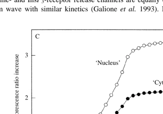
(C) An illustration of the effects of sequestered dye. The upper trace is a whole-cell record of fluorescence changes at fertilization (‘nuclear’ signal). The lower trace consists of the same data as the upper trace, but after addition of an unchanging signal equal to the resting fluorescence (‘cytoplasm’ signal). The unchanging signal reduces the apparent sensitivity of the dye to calcium. This explains why the nuclear signal appears greater than the cytoplasmic signal after dye sequestration, since the nucleus lacks sequestering organelles. It also explains why the relative change in the dye signal in response to [Ca2+]i decreases as dye is
sequestered. C
3
2
1
‘Nucleus’
‘Cytoplasm’
0 20 40 60 80 100
localization of the ryanodine receptor is also connected with the initiation of the calcium wave.
Ratio imaging in the confocal microscope
The distribution of calcium indicator dyes in the cytoplasm is not uniform. The signal from the egg cortex is more intense than the signal deeper within the egg and the signal from the nucleus is more intense than that from the cytoplasm (Fig. 3). The more intense cortical signal may result from an inhomogeneity in the optical system rather than in dye distribution. Laser light passing into the egg and emitted light emerging from it travel a greater distance through the egg cytoplasm than light travelling to and from the egg cortex. Scattering by the cytoplasm may lead to lower intensities inside the egg than at the periphery, even with uniform cytoplasmic dye concentrations. Conversely, the more intense signal in the nucleus seems to be due to the presence of more dye there.
Because dye concentrations and optical paths are not uniform, simple confocal images of fertilization give the misleading impression (Stricker et al. 1992; Berridge, 1993) that the cortical and nuclear calcium concentrations rise higher than that in the rest of the cytoplasm during the fertilization wave (Fig. 3). There is no visible wavelength ratio calcium indicator dye yet available to sidestep the dye distribution and pathlength problems; the alternative, confocal calcium imaging with ultraviolet light, is not yet readily available or convenient. It is possible to use two-dye methods (Berger and Brownlee, 1993; Lipp and Niggli, 1993). However, provided dye distribution is unchanged during the experiment, a straightforward way to overcome the problem is to divide each experimental image pixel-by-pixel by a control image taken just prior to the experiment (Fig. 3B). This technique has previously been used with true ratio dyes under conditions in which it was impractical to use rapid dual-wavelength excitation (Silver et
al. 1989). Using this correction method, [Ca2+]ican be seen to be uniform through the egg
(Fig. 3B), as it was in Fig. 1 which is also displayed as a ratio image.
Internalization of calcium indicator dyes
Calcium indicator dyes can be sequestered in intracellular compartments. The sequestered dye obviously no longer reports free cytoplasmic calcium concentration. If the sequestered dye were not fluorescent, this would pose no problems, but Fig. 4 shows that the signal from the sequestered dye is comparable to, or even larger than, the dye signal from the resting cytoplasm. In non-imaging applications, sequestration of this sort leads to an underestimate of [Ca2+]iincreases, but is otherwise not too serious. In imaging
very time-dependent (Fig. 4). Shortly after microinjection, as we showed above, there is a bigger dye signal in the nucleus than the cytoplasm. Sequestration leads to a loss of dye from the cytoplasm and nucleus and an increase in organelles. At a later time, the resting signal in the nucleus decreases relative to that in the cytoplasmic region, while the signal from the cytoplasmic region (cytoplasm proper + organelles) increases. Imaging the calcium wave soon after microinjection gives a nuclear calcium signal similar to that from the cytoplasm (Fig. 3B). Imaging the calcium wave after 20min of sequestration gives a very large apparent increase in ‘nuclear’ calcium that is due to sequestration, and so is spurious (Fig. 4B,C).
It is possible to stratify eggs to separate the mitochondria from the endoplasmic reticulum (Harvey, 1956; Eisen and Reynolds, 1985). Most of the sequestered dye seems to be in mitochondria (Fig. 5). The Fluo-3 indicator used in Figs 3–5 is particularly prone to this sequestration artefact. We think that this is because of its large dynamic range: it can show a 40-fold enhancement at saturating calcium concentrations (Minta et al. 1989). We also think that the dye saturates in the mitochondria because calcium concentration is high there: its signal will increase 30- to 40-fold. This means that uptake of only 2–4% of the total dye will give a signal more-or-less equivalent to the true resting cytoplasmic signal and thus halve the apparent calcium concentration increase. Calcium Green-1 has a smaller dynamic range (14-fold: Haughland, 1992) and may be less prone to sequestration: sequestration artefacts (and thus large increases in ‘nuclear calcium’ relative to the cytoplasm) are not prominent (compare Figs 1 and 4B).
Sequestration can be avoided by using indicator dyes conjugated to dextran. 10000 Mr
Calcium Green-1–dextran enters the nucleus. A fertilization calcium wave imaged using Calcium Green-1–dextran appears uniform (Fig. 6) and is comparable to the image obtained with Calcium Green-1 alone. With Calcium Green-1–dextran, there is no evidence of sequestration up to 20min after injection of the dye.
Binding and diffusion of indicator dyes
Another possible complicating factor is the binding of dyes to cytoplasmic constituents. One way of testing for this is to measure a diffusion constant for the dye within the egg. We can do this using confocal imaging. Fig. 7 shows the evolution of the dye signal in time and space after pressure-microinjection. Pressure microinjection introduces a spherical source of dye of between 5 and 10mm in radius in the centre of the egg. The dye within the sphere then diffuses into the cytoplasm. Fitting a diffusion equation (Crank, 1975) to these data give diffusion constants of 7.931028±2.7131028cm2s21 (mean and S.D., N=3) for dextran-conjugated Calcium
Green-1 and 5.031028±1.5131028cm2s21(mean and S.D., N=3) for Calcium Green-1
itself. The diffusion constant for the 10000 Mrdextran is similar to that for a similarly
Conclusion
Confocal imaging of the fertilization calcium wave has demonstrated that [Ca2+]idoes
not increase significantly in the region of sperm–egg fusion until 15s or more after cytoplasmic continuity has been established between sperm and egg. The interval is comparable to the time then taken for the calcium wave to cross the egg. Behind the advancing wavefront, [Ca2+]i is uniform: in particular, although calcium concentration
rises in the nucleus, it is to levels comparable with those in the cytoplasm, and [Ca2+] iin
the egg cortex is similar to [Ca2+]ideeper within the egg.
Isabelle Gillot has a Fellowship from the Ministère de la Recherche et de l’Espace. The work was supported by a grant from The Wellcome Trust.
References
BERGER, F. ANDBROWNLEE, C. (1993). Ratio confocal imaging of free cytoplasmic calcium gradients in
polarizing and polarized Fucus zygotes. Zygote 1, 9–15.
BERRIDGE, M. J.(1993). Inositol trisphosphate and calcium signalling. Nature 361, 315–325.
CHAMBERS, E. L. AND DE ARMENDI, J. (1979). Membrane potential, action potential and activation
potential of the eggs of the sea urchin Lytechinus variegatus. Expl Cell Res. 122, 203–218.
CIAPA, B., BORG, B. ANDEPEL, D. (1991). Polyphosphoinositides, tyrosine kinase and sea urchin egg
activation. In Proceedings of the Seventh International Echinoderm Conference, Sept. 9–14, Atami, Japan. Biology of the Echinodermata (ed. Y. Yanagisawa, I. Yasumasu, C. Oguro, N. Suzuki and T. Motokawa), pp. 41–50. Rotterdam: A. A. Balkema.
CRANK, J.(1975). The Mathematics of Diffusion. Oxford: Clarendon Press.
CROSSLEY, I., WHALLEY, T. ANDWHITAKER, M. J. (1991). Guanosine 5-thiotriphosphate may stimulate
phosphoinositide messenger production in sea urchin eggs by a different route than the fertilizing sperm. Cell Regulation 2, 121–133.
DALE, B., DEFELICE, L. J. ANDTAGLIETTI, V. (1978). Membrane noise and conductance increase during
single spermatozoon–egg interactions. Nature 275, 217–219.
EISEN, A. ANDREYNOLDS, G. T.(1985). Source and sinks for the calcium released during fertilization of
single sea urchin eggs. J. Cell Biol. 100, 1522–1527.
GALIONE, A., MCDOUGALL, A., BUSA, W. B., WILLMOTT, N., GILLOT, I. ANDWHITAKER, M. J. (1993).
Redundant mechanisms of calcium-induced calcium release underlying calcium waves during fertilization of sea urchin eggs. Science (in press).
HARVEY, E. B. (1956). The American Arbacia and Other Sea Urchins. Princeton, NJ: Princeton
University Press, pp. 132–134.
HAUGHLAND, R. P. (1992). Handbook of Fluorescent Probes and Research Chemicals. Eugene, OR:
Molecular Probes Inc., pp. 113–122.
JAFFE, L. A., TURNER, P. R., KLINE, D., KADO, R. T. ANDSHILLING, F. (1988). G-proteins and egg
activation. Cell Differ. Dev. 25 (Suppl.) 15–18.
LIPP, P. AND NIGGLI, E. (1993). Ratiometric confocal Ca2+measurements with visible wavelength
indicators in isolated cardiac myocytes. Cell Calcium 14, 359–372.
MCCULLOH, D. H. ANDCHAMBERS, E. L. (1992). Fusion of membranes during fertilization: increases of
sea urchin egg’s membrane capacitance and membrane conductance at the site of contact with the sperm. J. gen. Physiol. 99, 137–175.
MCDOUGALL, A., GILLOT, I. ANDWHITAKER, M. J. (1993). Thimerosal reveals calcium-induced calcium
release in unfertilized sea urchin eggs. Zygote 1, 35–42.
MCPHERSON, S. M., MCPHERSON, P. S., MATTHEWS, L., CAMPBELL, K. P. ANDLONGO, F. J. (1992).
Cortical localization of calcium release channel in sea urchin eggs. J. Cell Biol. 116, 1111–1121. MINTA, A., KAO, J. P. Y. ANDTSIEN, R. Y.(1989). Fluorescent indicators for cytosolic calcium based on
rhodamine and fluorescein chromophores. J. biol. Chem. 264, 8171–8178.
SHEN, S. S. ANDBUCK, W. R. (1993). Sources of calcium in sea urchin eggs during the fertilization
SILVER, R. A., LAMB, A. G. ANDBOLSOVER, S. R. (1989). Calcium hotspots caused by L-channel
clustering promote morphological changes in neuronal growth cones. Nature 343, 751–754.
SILVER, R. A., WHITAKER, M. J. ANDBOLSOVER, S. R.(1991). Intracellular ion imaging using fluorescent
dyes: artefacts and limits to resolution. Pflügers Arch. 420, 595–602.
STRICKER, S. A., CENTONZE, V. E., PADDOCK, S. W. ANDSCHATTEN, G.(1992). Confocal microscopy of
fertilization-induced calcium dynamics in sea urchin eggs. Devl Biol. 149, 370–380.
TERASAKI, M. ANDJAFFE, L. A.(1991). Organization of the sea urchin egg endoplasmic reticulum and its
reorganization at fertilization. J. Cell Biol. 116, 929–940.
TERASAKI, M. ANDSARDET, C. (1991). Demonstration of calcium uptake and release in sea urchin eggs
by cortical endoplasmic reticulum. J. Cell Biol. 115, 1031–1037.
WHITAKER, M. J. ANDSWANN, K.(1993). Lighting the fuse at fertilization. Development 117, 1–12.
WHITAKER, M. J., SWANN, K. ANDCROSSLEY, I. B. (1989). What happens during the latent period at