James M. Provenzale and Nancy B. Allen
PURPOSE: To demonstrate the spectrum of CT and MR imaging findings in patients with Wegener granulomatosis and to determine how often these findings could be attributed to either direct extension from paranasal or orbital disease sites, remote granulomas, or central nervous system (CNS) vasculitis. METHODS: We retrospectively reviewed the CT or MR studies of 15 patients with Wegener granulomatosis. RESULTS: Abnormal findings were seen in 7 patients (5 examined with MR imaging, 2 with CT). Findings included dural thickening and contrast enhancement (3 pa-tients), infarcts (2 papa-tients), regions of hyperintense signal on T2-weighted MR images (2 papa-tients), and abnormal MR signal in the brain stem (2 patients). Three patients with imaging findings of dural enhancement and thickening were thought to have remote granulomatous lesions involving the dura. No patients had extension from sites external to the CNS or clinical findings suggestive of CNS vasculitis. CONCLUSION: The spectrum of CT and MR findings in Wegener granulomatosis includes dural thickening and enhancement, cerebral infarction, and MR signal abnormalities in the brain stem and white matter. Presumed remote granulomatous lesions were the most common causes of CNS findings in this study. Complications related to non-CNS disease (eg, hypertension, endocarditis) also appear to have played a role in some patients.
Index terms: Granuloma; Vasculitis
AJNR Am J Neuroradiol17:785–792, April 1996
Wegener granulomatosis is a rare systemic granulomatous vasculitis that commonly (22% to 54% of cases) involves the nervous system, usually in the form of peripheral or cranial neu-ropathy (1– 4). Involvement of the brain and meninges is reported in only 2% to 8% of pa-tients (1, 4, 5). The most frequent clinical man-ifestations of brain involvement are cerebral in-farction (approximately 4% of patients) and seizures (3, 4). Three major mechanisms have been proposed for nervous system involvement: granulomatous invasion by contiguous exten-sion from sites external to the central nervous system (CNS) (eg, extension of nasal, parana-sal, or orbital granulomata into the meninges or brain); CNS granulomatous vasculitis; and
re-mote granulomatous lesions (eg, granulomata within brain parenchyma or dura) (3). Because Wegener granulomatosis is rare and brain in-volvement is present in only a small percentage of patients, series reporting neuroradiologic findings in this disease are few (6). We reviewed the computed tomography (CT) and magnetic resonance (MR) findings in a series of patients with Wegener granulomatosis to document the spectrum of imaging findings and to determine how often these findings could be caused by local extension from sites external to the CNS or by CNS vasculitis or remote granulomatous lesions.
Materials and Methods
Review of a database of 100 patients with Wegener granulomatosis seen at our institution revealed 15 patients who had had neuroradiologic examinations. Of these, we identified 7 patients whose neuroradiologic examinations produced abnormal findings. In addition, we reviewed the CT and MR examinations of 12 patients with orbital, para-nasal, or nasal Wegener granulomatosis involvement for possible intracranial extension. No intracranial disease was found in any of these patients. All patients with intra-cranial disease met clinical and American College of Received May 13, 1995; accepted after revision October 24.
Presented at the 33rd Annual Meeting of the American Society of Neuroradiology, Chicago, April 1995.
From the Departments of Radiology (J.M.P.) and Medicine (N.B.A.), Duke University Medical Center, Durham, NC.
Address reprint requests to James M. Provenzale, MD, Department of Radiology, Box 3808, Duke University Medical Center, Durham, NC 27710.
AJNR 17:785–792, Apr 1996 0195-6108/96/1704 –0785
qAmerican Society of Neuroradiology
Rheumatology criteria for diagnosis of Wegener granulo-matosis (7), which includes the presence of vasculitis on skin, lung, or orbital tissue on biopsy specimens (5 pa-tients); necrotizing granulomata in the nasal cavity, para-nasal sinuses, orbits, kidneys, or lungs (6 patients); glo-merulonephritis or renal failure (5 patients); airway or pulmonary disease (6 patients); and the presence of circulating antineutrophil cytoplasmic autoantibodies (c-ANCA) (3 patients). The systemic and neurologic fea-tures are summarized in Table 1. Neurologic feafea-tures in-cluded stroke or transient ischemic attacks (4 patients), headaches (2 patients), optic neuropathy (2 patients), tinnitus or hearing loss (2 patients), diplopia (1 patient), seizures (1 patient), and encephalopathy (1 patient). Two patients died of causes related to Wegener granulomatosis (patients 5 and 7).
In five patients, MR imaging was performed with a 1.5-T system. T1-weighted images were obtained before and after administration of contrast material in all five patients with imaging parameters of 500/10 –14/1 (repetition time/ echo time/excitations). T2-weighted images were ob-tained by using parameters of 2000 –2500/40, 80/0.75–2. Two patients were examined with CT only (1 after admin-istration of contrast material).
Results
In six patients, the diagnosis of Wegener granulomatosis was established or thought probable before their imaging examinations. In one case (patient 4), who had a history of chronic renal failure and hypertension, a pon-tine stroke occurred 1 year before onset of typical non-CNS manifestations of Wegener granulomatosis.
The neuroradiologic findings are reported in Table 1. Three patients were found to have dif-fuse dural thickening and abnormal contrast en-hancement (Figs 1–3). Following the definition used by other authors (8, 9), we defined dural contrast enhancement as curvilinear sheetlike or plaquelike enhancement over the cerebral convexities, along the falx cerebri or the tento-rium cerebelli and not following the convolu-tions of the gyri, as in leptomeningeal enhance-ment. Abnormal hyperintense signal of the brain stem on T2-weighted MR images was seen in two patients. One patient with abnormal MR signal of the brain stem (patient 6) had a history of third and sixth nerve pareses, which were most likely due to multiple cranial neuropathies. The region of abnormal signal seen at MR im-aging involved most of the pons and was more widespread than would be expected from the clinical symptoms alone (Fig 4).
One patient had sudden onset of
unrespon-siveness due to bilateral acute middle cerebral artery infarctions as seen on CT scans. Because of the severe neurologic disability resulting from the infarcts, cerebral angiography was not per-formed. This patient died 3 days later and at autopsy was found to have marantic endocar-ditis. Histologic examination of the brain dem-onstrated the bilateral infarctions and patent ce-rebral arteries. The infarctions were presumed to be caused by a cardiogenic embolism with subsequent fragmentation of emboli accounting for the patency of the arteries.
In one patient with seizures (patient 5), mul-tiple bilateral regions of hyperintense signal consistent with infarctions were seen within the cerebral cortex and subjacent white matter. This patient did not have clinical evidence of active Wegener granulomatosis at other sites. There-fore, CNS vasculitis was thought to be unlikely as the cause of the MR imaging abnormalities, and cerebral angiography was not performed. Immunosuppressive therapy, previously used in this patient, was not restarted and the seizures responded to anticonvulsants alone.
One patient (patient 4) had multiple conflu-ent regions of hyperintense white matter signal on T2-weighted images and a small pontine infarct, which were consistent with the patient’s long-standing history of poorly controlled hy-pertension. Two other patients (patients 5 and 6) also suffered from hypertension, which was well controlled.
Discussion
The triad of necrotizing granulomatous le-sions of the upper or lower respiratory tracts, a generalized necrotizing vasculitis of both arter-ies and veins, and glomerulonephritis, now re-ferred to as Wegener granulomatosis, was rec-ognized as a distinct clinical entity in 1936 (10). In addition to the paranasal sinuses, lungs, and kidneys, the disease commonly affects the eyes, skin, joints, muscles, nervous system, and cardiac system (Table 2). A definitive diagnosis is obtained by establishing the presence of a necrotizing granulomatous vasculitis (usually by lung, upper airway, or skin biopsy), often in association with glomerulonephritis on renal
biopsy specimens. Autoantibodies directed
been shown to have a high sensitivity and spec-ificity for Wegener granulomatosis (12). The c-ANCA titers can also be useful as markers of disease activity in some patients with Wegener granulomatosis because titers have been re-ported to decrease during disease remission (12). CNS disease associated with Wegener granulomatosis, however, has been reported in the presence of a negative c-ANCA titer (13). Furthermore, moderately elevated c-ANCA ti-ters have also been shown in association with polyarteritis nodosa, Takayasu disease, Churg-Strauss syndrome, and, on occasion, systemic lupus erythematosus (14). These entities can usually be distinguished from Wegener granu-lomatosis on the basis of clinical and serologic findings.
Like patients with Wegener granulomatosis, patients with polyarteritis nodosa also have a
[image:4.612.223.558.89.484.2]high prevalence of renal, joint, and skin involve-ment but are more likely than patients with Wegener granulomatosis to have cardiac in-volvement and elevated IgG levels and cryoim-munoglobulins. Takayasu disease is a panar-teritis that typically involves the origin of large arteries and generally affects patients at a younger age (adolescence and early adulthood) than does Wegener granulomatosis. Paranasal sinus, nasal, and pulmonary diseases, which are common in patients with Wegener granulo-matosis, are typically absent in patients with Takayasu disease. Patients with Churg-Strauss syndrome have a high prevalence of severe asthma and peripheral eosinophilia. Patients with systemic lupus erythematosus frequently have characteristic clinical features of a raised erythematous rash, alopecia, photosensitivity, and serositis and, unlike patients with Wegener Fig 1. Patient 3: 35-year-old man with
recent onset of headaches.
A, Contrast-enhanced axial CT scan at the level of the tentorium shows dural thickening and contrast enhancement (ar-rows).
B, Axial CT scan at a more cephalad level shows dural thickening and contrast enhancement in the posterior portion of the falx cerebri (arrow).
Fig 2. Patient 1: 41-year-old man with severe daily headaches for 1 year. Con-trast-enhanced T1-weighted coronal MR image (500/11/1) shows dural enhance-ment. The headaches significantly im-proved within 1 day of starting high-dose oral corticosteroid therapy.
granulomatosis, have depressed complement levels, elevated fluorescent antinuclear anti-body titers, and antibodies to double-stranded DNA.
Neurologic involvement in Wegener granulo-matosis primarily involves the peripheral ner-vous system, usually in the form of multiple peripheral neuropathies (mononeuritis multi-plex) (1, 4). Cranial neuropathy is the most common CNS manifestation (1). Both the pe-ripheral and cranial neuropathies are thought to be the result of a small-vessel vasculitis (1). Although one early article on the neurologic complications of any type in patients with Wegener granulomatosis reported a prevalence of 50%, subsequent articles have reported rates between 22% and 33% (1, 2, 4). The lower fig-ures in more recent series are thought to reflect earlier treatment with immunosuppressive ther-apy. Involvement of the brain and meninges is uncommon. In one series of 85 patients with Wegener granulomatosis who were followed up prospectively for 21 years, CNS manifestations developed in 10, but symptoms and signs were related to cranial neuropathy in most of these cases. None of these patients had ischemic symptoms, infarcts, or symptoms clearly re-lated to intracranial masses or dural granuloma-tous disease (1). In another series of 324 pa-tients, clinical findings directly related to brain
involvement were seen in 12, all of whom had cerebrovascular events (4). CNS involvement is rarely the initial disease manifestation (5, 14, 15). Our patients had neurologic findings that were of sufficient severity to warrant evaluation by cross-sectional imaging studies. None of our patients had peripheral nervous system disease alone, cranial neuropathy that was not thought to be related to a brain abnormality, or CNS symptoms or signs that were not of sufficient severity to warrant an imaging study. Therefore, our series may well underestimate the fre-quency of CNS involvement in patients with Wegener granulomatosis. However, our preva-lence rate of 7%, based on imaging studies alone, is not very different (and is slightly higher) than the 4% prevalence reported in one large series of patients with this disease (4).
The CT and MR findings in three of our pa-tients, all with dural involvement, could be ex-plained by one of the three major mechanisms that were proposed by Drachman (3) to ac-count for CNS disease in patients with Wegener granulomatosis. Although histologic proof is lacking, it is most likely that the dural thicken-Fig 4. Patient 6: 56-year-old man with multiple cranial
[image:5.612.97.260.87.284.2]neu-ropathies of 11 years’ duration. T2-weighted axial MR image (2500/80/2) shows hyperintense signal throughout much of the pons, although no signs or symptoms specifically localized to the pons were found on neurologic examination. On contrast-enhanced T1-weighted images (not shown), no abnormal cranial nerve enhancement was seen.
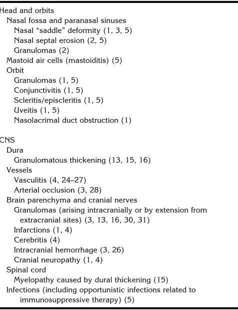
TABLE 2: Spectrum of reported manifestations of Wegener granu-lomatosis affecting the CNS, head, and orbits (reference no.)
Head and orbits
Nasal fossa and paranasal sinuses Nasal “saddle” deformity (1, 3, 5) Nasal septal erosion (2, 5) Granulomas (2)
Mastoid air cells (mastoiditis) (5) Orbit
Granulomas (1, 5) Conjunctivitis (1, 5) Scleritis/episcleritis (1, 5) Uveitis (1, 5)
Nasolacrimal duct obstruction (1)
CNS Dura
Granulomatous thickening (13, 15, 16) Vessels
Vasculitis (4, 24–27) Arterial occlusion (3, 28)
Brain parenchyma and cranial nerves
Granulomas (arising intracranially or by extension from extracranial sites) (3, 13, 16, 30, 31)
Infarctions (1, 4) Cerebritis (4)
Intracranial hemorrhage (3, 26) Cranial neuropathy (1, 4) Spinal cord
Myelopathy caused by dural thickening (15) Infections (including opportunistic infections related to
[image:5.612.316.554.421.731.2]ing and contrast enhancement in our patients 1 through 3 are due to granulomatous involve-ment of the dura. None of our patients had active paranasal sinus or orbital disease at the time of CNS signs and symptoms, and contig-uous extension is unlikely to be the cause of
their imaging findings. Unfortunately, the
causes of the MR imaging abnormalities in pa-tients 4 through 6 are not known with certainty. All three patients had end-stage renal disease and hypertension. It is possible that some or all of their MR findings may be related to hyperten-sion, although this is less likely in patients 5 and 6, in whom hypertension was well controlled at the time of CNS signs and symptoms. The CNS findings in patient 7, who had marantic endo-carditis and probable cardiogenic embolism, are also most likely the result of a complication from non-CNS disease. In addition to the three major means of production of CNS disease out-lined by Drachman, therefore, CNS complica-tions of non-CNS disease would appear to form a fourth category of CNS disease production (3). As survival is prolonged by more effective therapy, this fourth category may assume in-creasing importance.
Diffuse dural thickening and contrast en-hancement has previously been reported in connection with Wegener granulomatosis (13, 15, 16). Results of dural biopsy in previous cases have shown necrotizing granulomata, multinucleated giant cells, and lymphocytic in-filtration (13, 15, 16). Our cases of dural thick-ening differed from previously reported cases in two ways. First, abnormal imaging findings in our patients were confined to the dura, whereas in two previously reported cases, large focal regions of brain parenchyma adjacent to sites of dural thickening exhibited abnormal MR signal (13, 16). Second, in our patients, diffuse, sym-metric thickening of the entire dura was seen, whereas in previous reports focal, nodular, and plaquelike thickening was seen (13, 15, 16), which had mass effect on brain in one instance (16). As in our cases, previous reports noted that dural thickening and enhancement were present with little or no radiologic evidence of pial involvement (even though thickening and fibrosis of the pia-arachnoid were seen on bi-opsy specimens in one report) (16). Dural gran-ulomatous disease related to Wegener granulo-matosis can respond in a dramatic fashion to immunosuppressive therapy, manifested by rapid symptomatic improvement, as seen in
one of our patients, and a decrease in dural contrast enhancement, seen in a previously re-ported case (13).
Dural thickening caused by Wegener granu-lomatosis needs to be distinguished from other diseases that cause this finding. The differential diagnosis of dural thickening includes neurosar-coidosis, primary or secondary dural tumors,
infectious meningitis, hypertrophic cranial
pachymeningitis, neurosyphilis, and, in rare in-stances, intracranial fibromas. The dural dis-ease pattern in Wegener granulomatosis differs from that typically seen in neurosarcoidosis, in which a pial abnormality (with contrast en-hancement typically extending along the con-tour of the brain and within brain sulci) predom-inates (8). However, extraaxial granulomatous masses can be seen in neurosarcoidosis that may be similar in appearance to the regions of dural involvement in Wegener granulomatosis (17). Plaquelike or diffuse type dural enhance-ment and thickening can also be produced by primary tumors (eg, en plaque meningioma and, rarely, dural lymphoma) and metastatic tumor (9, 18, 19). In most cases, the distinction between Wegener granulomatosis and these tu-mors can be made on the basis of associated clinical or radiologic findings, for example, typ-ical non-CNS manifestations (present with We-gener granulomatosis but not with meningi-oma) and the presence of a primary neoplasm and elevated CSF protein, decreased CSF glu-cose levels, and tumor cells (in the case of metastatic disease). In cases in which the non-CNS features of Wegener granulomatosis are absent or subtle, however, the distinction be-tween tumor and Wegener granulomatosis can be made only by doing a biopsy.
con-trast enhancement, and appear hypointense on T2-weighted images. The distinction between the two entities can be made on clinical grounds, as well as on the basis of associated findings (eg, paranasal sinus opacification or other non-CNS disease manifestations, brain in-farction, or other parenchymal lesions) in We-gener granulomatosis. Diffuse or focal dural thickening can also be seen in some cases of neurosyphilis. Late syphilis can produce diffuse dural thickening similar to that seen in some of our patients with Wegener granulomatosis (22) and, rarely, intracranial gummas can produce focal masses that may appear to arise from the dura (23). The diagnosis can often (but not always) be reached by positive serologic VDRL or fluorescent treponema antibody test results and by positive CSF findings, response to pen-icillin, and, on occasion, dural biopsy findings (22).
Stroke directly attributable to Wegener gran-ulomatosis is uncommon (1, 4). When stroke does occur in patients with Wegener granulo-matosis it may be related to primary manifes-tations of the disease (eg, vasculitis or arterial occlusion due to granulomatous masses), sec-ondary effects (eg, the marantic endocarditis seen in patient 7), or causes unrelated to Wegener granulomatosis. In one series of 12 patients with Wegener granulomatosis who had cerebrovascular accidents, 5 had clinical fea-tures (seizures or encephalopathy) suggestive of vasculitis (4). However, despite the wide-spread necrotizing vasculitis involving the pe-ripheral and cranial nerves (4), cerebral vascu-litis in Wegener granulomatosis is uncommon (1, 3–5). In addition to infarction, cerebral vas-culitis related to Wegener granulomatosis has been reported to result in intraparenchymal or subarachnoid hemorrhage (3, 4, 24 –27). As in other vasculitides, findings at cerebral angiog-raphy can be negative in suspected cases of cerebral vasculitis related to Wegener granulo-matosis, possibly because of preferential in-volvement of small vessels (28). Clinical fea-tures strongly suggestive of cerebral vasculitis, however, were not seen in any of our patients. Although 3 patients had either cerebral or brain stem infarcts, angiography was not performed because the infarct could be explained on the basis of poorly controlled hypertension (patient 4), cardiogenic embolism (patient 7), or be-cause the Wegener granulomatosis was rela-tively inactive and a vasculitis was thought
un-likely. This suggests that other causes of infarction might be operative. In rare instances, cerebral infarction in Wegener granulomatosis can result from arterial occlusion caused by granulomatous masses extending from nasal or paranasal disease sites into the base of the skull (25, 29). Brain stem infarction, seen in one of our patients, has rarely been reported in con-nection with Wegener granulomatosis (28).
We did not encounter any CT or MR findings that were clearly caused by remote granuloma-tous lesions within brain parenchyma (as op-posed to remote lesions within dura). Remote granulomatous lesions arising within brain pa-renchyma are reported to be the least common form of CNS involvement in Wegener granulo-matosis (3). There are few actual reports of such lesions (3, 16, 30). These lesions can arise adjacent to sites of meningeal involvement or deep within brain tissue. On MR imaging, they can appear as homogeneously enhancing or ring-enhancing masses on T1-weighted images and as regions of hyperintense signal typically within white matter on T2-weighted images (16, 31).
Almost all of our patients had documented Wegener granulomatosis at the time of CT or MR imaging, and in many cases the neurologic symptoms could reasonably be attributed to the known disease. Because CNS symptoms and signs are rarely initial features of Wegener gran-ulomatosis, it seems likely that neuroradiologic findings, when present, will generally be seen only after clinical features of a systemic disease process are already evident. However, as this series demonstrates, the specific mechanism of CNS disease (ie, remote granuloma formation, vasculitis, or causes not related to the CNS) will not always be apparent from cross-sectional imaging studies.
References
1. Fauci AS, Haynes BF, Katz P, Wolff SM. Wegener’s granulomato-sis: prospective clinical and therapeutic experience with 85
pa-tients for 21 years.Ann Intern Med1983;98:76 – 85
2. Anderson JM, Jamieson DG, Jefferson JM. Non-healing
granu-loma and the nervous system.Q J Med1975;44:309 –323
3. Drachman DD. Neurological complications of Wegener’s
granu-lomatosis.Arch Neurol1963;8:145–155
4. Nishino H, Rubino FA, DeRemee RA, Swanson JW, Parisi JE. Neurological involvement in Wegener’s granulomatosis: an
anal-ysis of 324 consecutive patients at the Mayo Clinic.Ann Neurol
1993;33:4 –9
5. Hoffman GS, Kerr GS, Leavitt RY, et al. Wegener granulomatosis:
6. Asmus VR, Muhle C, Koltze H, et al. MRI features of Wegener’s
granuloma in the head.Fortschr Rontgenstr1992;157:11–14
7. Leavitt RY, Fauci AS, Bloch DA, et al. The American College of Rheumatology 1990 criteria for the classification of Wegener’s
granulomatosis.Arthritis Rheum1990;33:1101–1107
8. Sherman JL, Stern BJ. Sarcoidosis of the CNS: comparison of
unenhanced and enhanced MR images.AJNR Am J Neuroradiol
1990;11:915–923
9. Paakko E, Patronas NJ, Schellinger D. Meningeal Gd-DTPA
en-hancement in patients with malignancies.J Comput Assist
To-mogr1990;14:542–546
10. Wegener F. Uber generalisierte, septische Gefaesserkrankungen. Verh Dtsch Ges Pathol1936;29:202–210
11. Davies DJ, Moran JE, Niall JF, et al. Segmental necrotizing glo-merulonephritis with antineutrophil antibody: possible arbovirus
aetiology.Br Med J1982;285:606
12. Nolle B, Specks U, Ludemann J, Rohrbach MS, DeRemee RA, Gross WL. Anticytoplasmic autoantibodies: their
immunodiag-nostic value in Wegener’s granulomatosis.Ann Intern Med1989;
111:28 – 40
13. Weinberger LM, Cohen ML, Remler BF, Naheedy MH, Leigh RJ.
Intracranial Wegener’s granulomatosis. Neurology 1993;43:
1831–1834
14. Stumvoll M, Schnauder G, Overkamp D, Buettner UW, Grodd W, Eggstein M. Systemic vasculitis positive for circulating anti-neu-trophil cytoplasmic antibodies and with predominantly neurologic
presentation.Clin Invest1993;71:613– 615
15. Nishino H, Rubino FA, Parisi JE. The spectrum of neurologic
involvement in Wegener’s granulomatosis.Neurology1993;43:
1334 –1337
16. Tishler S, Williamson T, Mirra SS, Lichtman JB, Gismondi P, Kibble MB. Wegener’s granulomatosis with meningeal
involve-ment.AJNR Am J Neuroradiol1993;14:1248 –1252
17. Hayes WS, Sherman JL, Stern BJ, Citrin CM, Pulaski PD. MR and
CT evaluation of intracranial sarcoidosis.AJNR Am J Neuroradiol
1987;8:841– 847
18. Tyrrell RL II, Bundschuh CV, Modic MT. Dural carcinomatosis: MR
demonstration.J Comput Assist Tomogr1987;11:329 –332
19. Jazy FK, Shehata WM, Tew JM, Meyer RL, Boss HH. Primary
intracranial lymphoma of the dura.Arch Neurol1980;37:528 –
529
20. Phillips ME, Ryals TJ, Kambhu SA, Yuh WTC. Neoplastic vs
inflammatory meningeal enhancement with Gd-DTPA.J Comput
Assist Tomogr1990;14:536 –541
21. Martin N, Masson C, Henin D, Mompoint D, Marsault C, Nahum H. Hypertrophic cranial pachymeningitis: assessment with CT and
MR imaging.AJNR Am J Neuroradiol1988;10:477– 484
22. Moore AP, Rolfe EB, Jones EL. Pachymeningitis cranialis
hyper-trophica.J Neurol Neurosurg Psychiatry1985;48:942–944
23. Currie JN, Coppeto JR, Lessell S. Chronic syphilitic meningitis resulting in superior orbital fissure syndrome and posterior fossa
gumma: a report of two cases followed for 20 years.J Clin
Neu-roophthalmol1988;8:145–159
24. Yamashita Y, Takahashi M, Bussaka H, Miyawaki M, Tosaka K. Cerebral vasculitis secondary to Wegener’s granulomatosis:
com-puted tomography and angiographic findings.J Comput Tomogr
1986;10:115–120
25. Satoh J, Miyasaka N, Yamada T, et al. Extensive cerebral in-farction due to involvement of both anterior cerebral arteries by
Wegener’s granulomatosis.Ann Rheum Dis1988;47:606 – 611
26. Lucas FV, Benjamin SP, Steinberg MC. Cerebral vasculitis in
Wegener’s granulomatosis.Cleve Clin J Med1976;43:275–281
27. Payton CD, Jones JMB. Cortical blindness complicating
Wegen-er’s granulomatosis.Br Med J1985;290:676
28. Savitz JM, Young MA, Ratan RR. Basilar artery occlusion in a
young patient with Wegener’s granulomatosis.Stroke 1994;25:
214 –216
29. Goldberg AL, Tievsky AL, Jamshidi S. Wegener’s granulomatosis
invading the cavernous sinus: a CT demonstration. J Comput
Assist Tomogr1983;7:701–703
30. Oimomi M, Suehiro I, Mizuno N, Baba S, Okada S, Kanazawa Y. Wegener’s granulomatosis with intracerebral granuloma and
mammary manifestation.Arch Intern Med1980;140:853– 854
31. Miller KS, Miller JM. Wegener’s granulomatosis presenting as a primary seizure disorder with brain lesions demonstrated by