Forty years ago the Journal of Experimental Biology published Hans Lissmann’s paper ‘On the function and evolution of electric organs in fish’ (Lissmann, 1958) and his companion paper on ‘The mechanisms of object location in Gymnarchus niloticus and similar fish’ (Lissmann and Machin, 1958). It is fitting that the Journal should now publish a volume reviewing our current knowledge of the neuroethology of electroreception and electrogenesis, a field that can trace its origin to Lissmann’s original papers. Lissmann and before him Möhres (1957) suggested that social communication is an important function for weak electric discharges in fish. Möhres made some of the first playbacks of electric signals and evoked responses from mormyrid fish. In the 1960s, Szabo, Bullock, Bennett, Coates, Grundfest, Hagiwara and others explored these exciting new peripheral sense organs, electric organs and brain areas; but it was not until the work of Moller, Kramer, Black-Cleworth, Westby, Kirschbaum, Hopkins and others came out in the early 1970s that we started to glimpse the diversity of electric communication signals and functions (Black-Cleworth, 1970; Hopkins, 1972; Kirschbaum, 1995; Kramer, 1995; Moller, 1995; Westby, 1974, 1975). Since then, with more field studies, better information about sensory processing in the brain and additional studies of the motor control of electric signals (Kawasaki and Heiligenberg, 1988;
Kawasaki et al., 1988; Keller et al., 1991), we now know a great deal about the neuroethology of electric communication, perhaps more for electric fish than for any other sensory modality except vision.
Still, there is much to learn. In this paper, I briefly outline two design features of electric communication that make this modality unique. By understanding them, we may hope to understand more of how the electric modality has evolved and how selection has influenced the structure and function of signals and displays.
Design feature 1: limited signal range
The active space of an electric communication signal is determined by the amplitude of the signal at the source, the rate of signal decrease due to geometric spreading, the amplitude of masking background noise and the sensitivity of the receiver. From early recordings of the electric fields around captive gymnotiforms in large tanks (Knudsen, 1975), we learned that electric fish approximate dipole electric sources with electric fields that decline in field strength with the inverse cube of distance, as predicted from electrostatic theory. Using data from behavioral and physiological estimates of electric sensitivity, and from a series of calibrated playback JEB2093
How do the communication discharges produced by electric fish evolve to accommodate the unique design features for the modality? Two design features are considered: first, the limited range of signaling imposed on the electric modality by the physics of signal transmission from dipole sources; and second, the absence of signal echoes and reverberations for electric discharges, which are non-propagating electrostatic fields.
Electrostatic theory predicts that electric dicharges from fish will have a short range because of the inverse cube law of geometric spreading around an electrostatic dipole. From this, one predicts that the costs of signaling will be high when fish attempt to signal over a large distance. Electric fish may economize in signal production whenever possible. For example, some gymnotiform fish appear to be impedance-matched to the resistivity of the water; others
modulate the amplitude of their discharge seasonally and diurnally.
The fact that electric signals do not propagate, but exist as electrostatic fields, means that, unlike sound signals, electric organ discharges produce no echoes or reverberations. Because temporal information is preserved during signal transmission, receivers may pay close attention to the temporal details of electric signals. As a consequence, electric organs have evolved with mechanisms for controlling the fine structure of electric discharge waveforms.
Key words: electric fish, electrocommunication, gymnotiform, gymnotid, mormyriform, mormyrid, Mormyridae, electric organ, electrocyte, electrostatic field, conductivity, evolution, communication.
Summary
Introduction
DESIGN FEATURES FOR ELECTRIC COMMUNICATION
CARL D. HOPKINS*
Section of Neurobiology and Behavior, 263 Seeley G. Mudd Hall, Cornell University, Ithaca, NY 14853, USA *e-mail: [email protected]
experiments performed in the laboratory, we now estimate the active space of a 10–20 cm mormyrid or gymnotiform to be ellipsoidal with a maximum range of approximately 1 m.
Because the magnitude of the electric field decreases with the inverse cube of distance, one predicts that electric fish should be distance-limited by the cost of signal production. For a fish to double its active space will require an eightfold increase in the amplitude of its signal at the source. This restriction suggests the following consequences. (1) When a fish is broadcasting its signal at its normal amplitude, its output should be energetically efficient. (2) When a fish needs to signal at a greater distance, as in territorial defense or courtship calling, its energetic costs may become prohibitive as the fish tries to push the limits of signal range. Under these circumstances, we expect to see alternate bursts of energy with cost-saving economies. (3) If the costs of signaling are high, the ability to ‘display’ might be used to assess another’s health or fitness, because signal costs cannot be faked or used deceptively. We illustrate these three points with examples.
Costs of signal production
Surprisingly, we know practically nothing about the absolute caloric costs of signal production, except for Keynes’s early measurements of heat production by Electrophorus and
Malapterurus (Aubert and Keynes, 1968; Keynes, 1968). From
the simultaneous recordings of current and voltage from the electric organ of a mormyrid of Bell et al. (1976), we can roughly calculate the cost of a single electric organ discharge (EOD) as approximately 10−5J. When discharging continuously at 10 EODs s−1, this is equivalent to 8.64 J day−1. For a 10 g fish with an expected basal metabolic rate of 1006 J day−1(Schmidt-Nielsen, 1970), this represents only 1 % of its basal metabolic rate.
Rather than measuring the absolute costs of signaling, it might be more useful to compare the relative costs of electric signaling for different individuals, sexes or species. Do males expend more on EODs than females or adults more than juveniles, for example? How does one species compare with another, and does this influence signal design? How does the cost of generating EODs compare with the cost of generating a vocal signal that carries the same distance (see, for example, Crawford et al., 1997)? Are there costs and benefits associated with a particular habitat, season or other environmental factor? For these and other issues, we have yet to formulate the questions, much less have answers.
Economy in signaling by impedance matching If costs are high, fish might be expected to be efficient at signaling. We can illustrate this with some recent data on impedance matching among the gymnotiform fishes. The genus Brachyhypopomus (formerly Hypopomus) is one of the most diverse groups among the pulse-discharging Gymnotiformes, which is now known from molecular sequence data to be a monophyletic group (Sullivan, 1997). Because they are closely related, the kinds of close comparisons of costs mentioned above make sense. Several of
[image:2.609.311.560.205.560.2]the Brachyhypopomus exhibit seasonal sexual dimorphism in the caudal filament which carries the electric organ (Hagedorn, 1988; Hagedorn and Carr, 1985; Hopkins et al., 1990). Brachyhypopomus occidentalis males have short, but thick caudal filaments compared with females, which have short and slender filaments. Brachyhypopomus pinnicaudatus (Hopkins, 1991) males have long wide tails, while females have shorter, more slender tails (Hopkins et al., 1990); B. brevirostris and Brachyhypopomus sp. males have even longer tail filaments
Fig. 1. Sex differences in tail morphology of three species of
Brachyhypopomus (Hypopomidae, Gymnotiformes) from South
America tend to exaggerate trends that already characterize species differences. For example, Brachyhypopomus occidentalis (A) from Panama have short and thick caudal filaments. Breeding males (M) have even thicker filaments than females (F). Brachyhypopomus
pinnicaudatus (B) from French Guiana have caudal filaments of
intermediate length and width that are both longer and thicker in males than in females. The undescribed Brachyhypopomus sp. (C) from southern Venezuela and Guyana has a long and slender filament that is much longer in males. The caudal filament is composed largely of the electric organ. In all three of these species, the male electric organ discharge is longer in duration than that of the female. Figure sources: B. occidentalis from Hagedorn (1988); B.
pinnicaudatus from Hopkins et al. (1990); Brachyhypopomus sp.
than B. pinnicaudatus. These sex and species differences are illustrated in Fig. 1.
During fieldwork in Venezuela, we noticed that each species of Brachyhypopomus had very patchy distributions related to
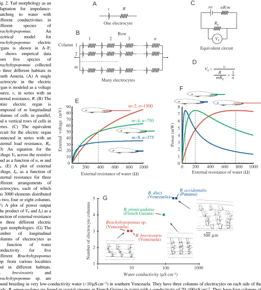
[image:3.609.54.566.136.704.2]the patchiness of water conductivity conditions in the environment (Fig. 2G). Correlated with this were differences among species in the structure of electric organs. Those species that live in water with very low conductivity (10µS cm−1or
Fig. 2. Tail morphology as an adaptation for impedance-matching to water with different conductivities in different species of
Brachyhypopomus. An
electrical model for
Brachyhypopomus electric
organs is shown in A–F; G shows empirical data from five species of
Brachyhypopomus collected
in three different habitats in South Ameria. (A) A single electrocyte in the electric organ is modeled as a voltage source, v, in series with an internal resistance, R. (B) The entire electric organ is composed of m longitudinal columns of cells in parallel, and n vertical rows of cells in series. (C) The equivalent circuit for the electric organ connected in series with an external load resistance, Re.
(D) An equation for the voltage Veacross the resistive load as a function of n, m and
Re. (E) A plot of external voltage, Ie, as a function of
external resistance for three different arrangements of electrocytes, each of which has 3000 elements distributed in two, four or eight columns. (F) A plot of power output (the product of Veand Ie) as a
function of external resistance for three different electric organ morphologies. (G) The number of longitudinal columns of electrocytes as a function of water conductivity for five different Brachyhypopomus
spp. from various localities and in different habitats.
B. brevirostris and
Brachyhypopomus sp. are
found breeding in very low-conductivity water (<10µS cm−1) in southern Venezuela. They have three columns of electrocytes on each side of the
body. B. pinnicaudatus are found in coastal streams in French Guiana in water with a conductivity of 70–100µS cm−1. They have four columns of
electrocytes on each side of the caudal filament. B. diazi and B. occidentalis, which are found in water of higher conductivity, in the Apuré River of Venezuela and in the Canal Zone of Panama, respectively, have five parallel columns. Sources: A–F from Sullivan et al. (1996), G from Comfort (1990), data on conductivity for B. occidentalis from M. Hagedorn (personal communication).
One electrocyte R v
A
B
C
D
Water conductivity (µS cm-1)
Number of electrocyte columns
1 2 3 4 5
1 1000
Brachyhypopomus sp.
(Venezuela)
B. brevirostris
(Venezuela) B. pinnicaudatus
(French Guiana)
B. diazi
(Venezuela)
B. occidentalis
(Panama)
500 µm
G
10 20 30 40 50 60 70 80
90 m=2, n=1500
m=4, n=750
m=8, n=375
0
External resistance of water (Ω)
E
External voltage (mV)
0 1000
0 1 2 3 4 5 6 7 8
Power (mW)
F
Many electrocytesColumn 1
2
m
Row
1 2 3 n
Equivalent circuit nR/m nv
Re
Ve
Ve= v
R mRe +
1 n
External resistance of water (Ω)
10 100
less) have long thin tail filaments that are exaggerated in length seasonally for males. Those species inhabiting water of intermediate conductivity (25–60µS cm−1) have thicker and shorter filaments, exaggerated in both length and thickness among males. Fish inhabiting high-conductivity water (above 80µS cm−1) have thick tail filaments, exaggerated in thickness in males (Fig. 2G).
The long and slender electric organs from the species inhabiting low-conductivity water typically have three longitudinal columns of electrocytes on each side of the body. The electric organs of intermediate length and width of fish from water of intermediate conductivity typically have four longitudinal columns of electrocytes on each side. The short and thick electric organs of fish from high-conductivity environments typically have five columns of cells on each side (Fig. 2G).
To understand better the economics of electric organ design, we have modeled the electric organ as a series of identical electrocytes arranged in series in longitudinal columns and in parallel as vertical rows (see Fig. 2B). Each electrocyte is represented as a voltage source, v, in series with a resistance, R (Fig. 2A). If we assume that the energetic cost of a single electrocyte is constant, a fish may deploy its electrocytes either into additional rows or into additional columns for the same energetic cost. We can then compare the output of different electric organs as a function of their geometry.
Fig. 2C shows an equivalent circuit for the electric organ modeled in Fig. 2B. For an electric organ with n rows in series and m columns in parallel, the total source voltage is nv, and the internal resistance is nR/m. The external resistance of the water is represented by Re. The equation in Fig. 2D is a solution for the external voltage, Ve, as a function of the numbers of rows and columns and the external resistance. If the electric organ were composed of 3000 electrocytes, they might be deployed in two columns of 1500 rows, four columns of 750 rows or eight columns of 375 rows, or in several other combinations. Those fish with eight columns and 375 rows do best at low external resistances (high conductivity). Fish with two columns of electrocytes and 1500 rows do best at high external resistance (low conductivity, Fig. 2E). A plot of the external power, IVe, where Veis the external voltage, and I is the current of the electric organ as a function of external resistance illustrates the impedance matching (Fig. 2F). The fish with a short, wide tail filament generates maximum external power in water with low external resistance (high conductivity); those with long thin tails generate highest power when the load resistance is high (low conductivity).
We have examined electric organs from many species of gymnotiform pulse fish, relating each observation to the conductivity of the water in which the individual was found. The trends in these data are especially clear when comparing species in a single clade, such as Brachyhypopomus. At higher water conductivity, the fish have a larger number of longitudinal columns of electrocytes and a reduced number of vertical rows of electrocytes. Fig. 2G shows the number of
electrocyte columns for five species of Brachyhypopomus collected in water of different conductivity. At lower conductivity, the fish tend to have longer, more slender tails, with fewer columns of electrocytes in parallel, but more rows in series (Sullivan et al., 1996). The best comparisons are those within a single genus, but several genera of pulse fish show the same trends. We expect to find the same phenomenon among wave species. From the tail morphology to the deployment of electrocytes into columns and rows, these fish have apparently evolved electric organs that are impedance-matched to the conductivity of their environment. Such electric organs would be expected to be operating at maximum efficiency in converting caloric costs into useful energy for signal production.
Once a fish has become developmentally committed to deploy its electrocytes in a given pattern of rows and columns, it may be more difficult for it to move from an area with one level of conductivity to an area where the conductivity is significantly different. In fact, conductivity changes may pose an ‘invisible barrier’ to the dispersal of some species, thereby adding to the patchiness of the environment and contributing to the establishment of geographical barriers within water systems. This may have been a factor in the process of speciation in Brachyhypopomus.
Economy in discharge amplitude and rate
For pulse fish such as Brachyhypopomus, the energy of signaling should be proportional to discharge rate and to the cost of producing a single pulse. This cost will be related to the product of the power and duration of a single EOD. Most pulse fish lower their discharge rates by day, when they are inactive, and elevate them at night. What can we say about the energy of the EOD? We had assumed that EOD amplitude and duration were fixed until Hagedorn (1995) observed in
Brachyhypopomus occidentalis and Franchina (1993, 1997)
and Franchina and Stoddard (1998) confirmed for
Brachyhypopomus pinnicaudatus that there are diurnal
Signal costs and sexual selection
Sexually dimorphic traits such as tail length in birds, color patterns and other forms of adornment and decoration often result from sexual selection, with females choosing males on the basis of signals that are costly for males to produce. Because these costly signals cannot be faked, they are honest indicators of male quality. This concept applies well to the electric fish already mentioned, Brachyhypopomus pinnicaudatus. Since male EODs are nearly twice the duration of female EODs, and since we expect the energetic cost to be proportional to the integral of voltage squared over the duration of the EOD, male EODs should cost more to produce than female EODs. When Hopkins et al. (1990) made field measurements of EOD amplitudes of males and females, they found that male EODs were weaker than female EODs when individuals of comparable total length were used. The amplitudes of male EODs were equal to those of female EODs if individuals of the same body size were compared (i.e. same length to the end of the anal fin), not including the extent of the long, sexually dimorphic tails. Paradoxically, males of this species do not produce a stronger signal with their much-exaggerated caudal filaments, they just match the amplitude of female EODs (or juvenile male EODs). Apparently, males that generate longer-duration EODs suffer a drop in the peak amplitude of the discharge. They appear to compensate for this drop by growing their tails to be longer and thicker so that they can generate greater voltage and current. In addition, during the breeding season, they may additionally boost the amplitude of the EOD at night.
Males pay an additional cost beyond the energetic cost of discharging long-duration pulses. That is the cost of the loss of their long, sexually dimorphic tails to predation. Males suffer a far greater frequency of tail damage than do females (Hopkins et al., 1990), apparently from predators. Sexual selection has set up a classical dynamic of competing selection pressures that result in a stable but sexually dimorphic signal system. Female Brachyhypopomus pinnicaudatus must be selecting their mates on the basis of longer-duration EODs over shorter ones because they ‘know’ that these pulses are more costly to produce and put the male at greater risk to produce them. In essence, the female knows that males that produce long pulses must be of exceptional quality. This model of signal evolution is simply a restatement of the handicap principle of Zahavi (1977), which has been well-studied for other modalities of signaling.
This principle of males paying a higher cost than females for signaling may apply equally well to African electric fish. In every case that has been reported (Bass, 1986b; Bass and Hopkins, 1983; Hopkins, 1986a; Landsman and Moller, 1988; Moller, 1995), male EODs are longer in duration than female EODs. We do not yet know how many times sex differences in EODs evolved in this group.
For the wave-discharging species, the same logic does not necessarily apply. For a sinusoidal waveform, the energy in the signal is independent of frequency so an increase in cost associated with elongating the discharge period would be offset by a decrease associated with the decline in repetition
frequency. Because wave species in the families Sternopygidae and Eigenmanniae control the repetition rate of the EOD in the pacemaker, and the duration of the pulses in the electric organ (Mills and Zakon, 1991; Zakon, 1996), it would be possible to change the duty cycle of the waveform independently of discharge frequency. The sex differences that do occur among wave species tend to result in both lower and higher frequencies for different species, however. For Sternopygus
macrurus, male EODs have a lower frequency and a longer
pulse duration (Hopkins, 1972; Mills and Zakon, 1991), while for Apteronotus leptorhynchus (Zakon, 1996), the male frequency is higher than that of the female. Hopkins (1974) and Hagedorn and Heiligenberg (1985) independently reported in field and laboratory studies that male Eigenmannia virescens
Petrocephalus sp. (large-eye)
Petrocephalus sp. (small-eye)
Pollimyrus isidori
Mormyrus rume
Hippopotamyrus psittacus
Mormyrops anguilloides
Pollimyrus petricolus
Marcusenius senegalensis
Brienomyrus niger
Hyperopisus bebe
Hippopotamyrus pictus
1 ms 1 ms
M
M F
F
M F II I
M F
M F
[image:5.609.314.569.257.618.2]M F
Fig. 3. Electric organ discharge (EOD) waveforms for eleven species of mormyrids co-existing in flooded swamps and in the main channel of the Niger River in the Central Delta of Mali. All EODs are shown with head-positivity upwards on the same time base. Sex differences, where present, are illustrated by typical examples of male (M) and female (F) EODs. The first three species have electric organs with non-penetrating stalks innervated on the posterior side; Pollimyrus
isidori has doubly penetrating stalks innervated on the posterior side;
had lower discharge frequencies than females with wide overlap. Kramer (1985) (see also Kramer, 1999) reports an isolated case of one male and one female of an Eigenmannia sp. that appear to be an exception to the general rule. Here, the male’s EOD pulse was shorter than the female’s although their repetition rates were approximately the same. Since the male’s duty cycle was lower than the female’s, his would require less energy to produce than hers if the amplitudes of their discharges were the same. If this case could be validated with additional specimens, it would merit further exploration from the viewpoint of energetics.
Design feature 2: non-propagating signals
Signal transmission
Electric signals are fundamentally different from acoustic
[image:6.609.73.528.285.670.2]signals because they are transmitted not as propagated waves, but as non-propagating electrostatic fields. This difference restricts the range of communication to the near-field electrostatic dipole. It also prevents signals from being corrupted by the reflections, refraction and reverberations that affect wave-propagating signals such as sound (Michelsen and Larsen, 1983; Wiley and Richards, 1982). As a consequence, the temporal fine structure of EODs is not affected by signal transmission except by the addition of electrical noise (for more details, see Hopkins, 1986b). Because temporal fine structure is more predictable in transmission for electric signals than for sound, we might expect cues encoded in pulse waveforms to have greater significance to receivers than would identical sound signals propagating as waves through air or water. This design feature has had a profound effect on the evolution of electric signals.
Time features and signal diversity
To illustrate the importance of temporal features in the electric modality, let us turn to the question of species recognition and the diversity of communication signals in natural communities of electric fish. Although we are still decades behind field studies on vocal signals in communities of birds and primates, there is now adequate information about electric signals for several communities of electric fish.
Fig. 3 shows the diversity of EODs of mormyrid fishes collected in the Niger River in Mali, central West Africa. The EOD waveforms show a wide range of diversity, from the shortest to the longest. Some EODs are biphasic, others are triphasic; some have one polarity, others have the reverse polarity. Sex difference in EODs are prevalent in all but the genus Petrocephalus. Within this population, species-specificity of EODs is most prominent among the male EODs, not the female EODs.
Fig. 4 shows the diversity of EODs from the mormyrid fishes from the Ivindo River in northeast Gabon of Central Africa. The EODs are highly diverse in waveform, wave duration and pulse polarity. While most species have biphasic EODs, a few have triphasic discharges and a few have tetraphasic waveforms. Electric organs composed of cells with very different anatomical and physiological properties produce the differences in the discharges.
The duration of these EODs varies over two orders of magnitude, from approximately 100µs in Petrocephalus to well over 10 ms in Paramormyrops gabonensis. The bandwidth of useful signaling ranges from 100 Hz in the species with the longest duration to over 20 kHz for the shortest. Both polarities of EODs are possible: normal and inverted. Mormyrops zanclirostris, for example, has an inverted polarity for the two main peaks (negative to positive) compared with all other species, where the main transition is positive to negative. This polarity inversion can be traced to the orientation of the cells in the electric organ. They are reversed in Mormyrops compared with all other species (Bass, 1986b).
Fig. 5 shows the diversity of EODs from different species belonging to the genus Campylomormyrus. Although these specimens were obtained through the aquarium trade rather than through fieldwork, they all originated in the Congo River basin. They represent only a fraction of the diversity of mormyrids from that river. Campylomormyrus exhibits a wide diversity of EOD waveform types, which we may use as an important character for discriminating among species that are otherwise cryptic. This information may help resolve differences in opinion regarding species identifications (see, for example, Poll et al., 1982; Roberts and Stewart, 1976).
C. tshokwe
C. sp. A
C. sp. B
C. sp. C
C. sp. D
C. sp. E
C. rhynchophorus
C. numenius
C. tamandua
[image:7.609.166.566.383.739.2]1 ms Fig. 5. Systematists disagree
The diversity of EODs in these communities of mormyrids suggests that the short and stereotyped EOD waveforms have a communicative significance of their own beyond that as a ‘carrier’ of signals encoded by the rhythm of the discharge or the sequences of pulse intervals. Indeed, the differences in EODs have permitted the discovery of new species previously overlooked by biologists who have used morphological characters only (Crawford and Hopkins, 1989; Roberts, 1989). One project currently under way is to describe eleven new species of Brienomyrus from the Ogooué River basin of Gabon, most of which were uncovered by the distinctive EOD waveforms they produce (C. D. Hopkins, G. Teugels, and R. Rundell, unpublished).
Evolution of electrogenic diversity
The diversity of EOD waveforms that we see in both gymnotiforms and mormyriforms can be traced to the anatomical and physiological characteristics of the electric organs of these fish. This diversity has been explored in both groups and is reviewed in several earlier publications (Bass, 1986a; Bennett, 1971). There is generally a good
understanding of the relationship between the anatomy of electrocytes and the physiology of the EOD. If we are to understand the evolution of electric signals, however, we must attempt to understand more than just the adaptive principles of signal design and the physiology of different types of electrocytes. We must attempt to integrate this knowledge with new understanding of evolutionary relationships among the animals derived from molecular sequencing techniques and modern cladistics.
Mormyrid electrocyte evolution
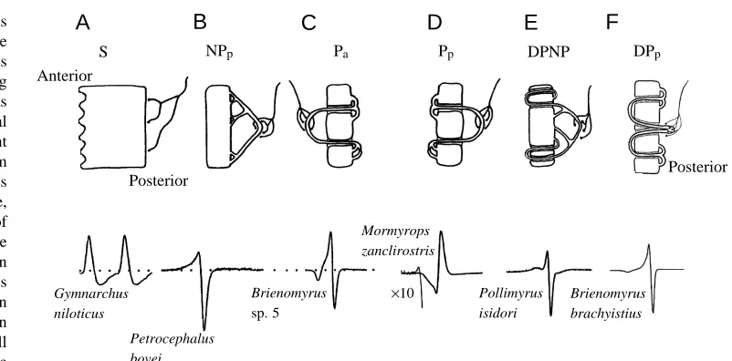
Mormyrid electric organs are made up of ‘electrocytes’ that are flattened, multinucleated disks several millimeters in diameter. There is a stilt-root-like stalk system that emerges as stalklets from one of the flattened faces of the electrocyte (usually posterior); these stalklets fuse with other stalklets, until they finally fuse to make up a large trunk of stalks which receives innervation from the electromotor nerve. Four parallel columns of electrocytes, with over a 100 in series, make up the electric organ in the caudal peduncle. In cross section, one sees how each electrocyte is designed (Fig. 6). In Gymnarchus, innervation is on the posterior face of the electrocyte, which
Fig. 6. The electric organs of mormyriform fishes are composed of electrogenic cells called ‘electrocytes’ of varying degrees of complexity, as illustrated in cross-sectional diagrams for six different morphological classes from different species. Each species has electrocytes of a single type, each of which is capable of generating the waveform of the whole-animal electric organ discharge (EOD). (A) Stalkless electrocytes (S) are found in
Gymnarchus niloticus and also in
the larval electric organs of all mormyrids. The electrocytes are
cylindrical in shape and are innervated only on their posterior surface. Only the posterior face fires an action potential. (B) Non-penetrating stalk electrocytes with posterior innervation (NPp) have a well-developed series of stalklets emerging from the posterior face of the electrocyte,
which fuse repeatedly to form stalks of greater and greater diameter. Innervation is on the largest stalks on the posterior side. Action potentials originating in the stalk propagate to excite the posterior face, which generates an inward current to produce a head-positive potential in the water. It also depolarizes the anterior face, which generates the head-negative phase after a delay. (C) Penetrating stalk electrocytes with anterior innervation (type Pa) have stalklets on the posterior face that penetrate through the cell to receive innervation on the anterior side. The
stalk potential generates no observable effect until it reaches the point of penetration. Here, inward current in the stalk is directed through the electrocyte and posteriorly to create the first weak head-negative pre-pulse. When the spike reaches the posterior face, it spreads across the face, causing a head-positive peak in the EOD. This is followed by firing of the anterior face, which generates the final head-negative phase. (D) When the electrocyte is anatomically reversed, and the innervation is on the posterior side (Pp), the EOD is inverted in polarity compared
with type Pa electrocytes. The light line shows a 10-fold gain expansion for Mormyrops zanclirostris. (E) Doubly penetrating and
non-penetrating stalked electrocytes (type DPNP) have stalks that either make a double penetration through the face of the electrocyte or contact the posterior face directly. The stalk potential generates a complex pre-pulse, which may be head-negative or head-positive. (F) Doubly penetrating stalked electrocytes with posterior innervation (type DPp) are innervated on the posterior side, and the stalk penetrates immediately and
completely through the cell to the anterior side, spreads out across the anterior surface and then makes numerous penetrations to return to the posterior side. This cell type is nearly identical to type Pamentioned above, as is the EOD, which is triphasic (adapted from Alves-Gomes and
Hopkins, 1997; Hopkins, 1999).
A
B
C
D
E
F
S NPp Pa Pp DPNP DPp
×10
Anterior
Posterior Posterior
Gymnarchus niloticus
Petrocephalus bovei
Mormyrops zanclirostris
Pollimyrus isidori Brienomyrus
sp. 5
[image:8.609.161.562.67.265.2]entirely lacks a stalk system (type S, ‘stalkless’). For the remaining 200 or more species of mormyrids, the innervation is on a stalk system that receives innervation either on the anterior or posterior side and which may or may not penetrate once or twice through the electrocyte. Six different electrocyte types are depicted in Fig. 6.
Molecular sequence data and the evolution of electric organs
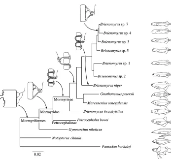
The molecular sequence data of Alves-Gomes and Hopkins (1997) derived from mitochondrial rRNA genes (12S and 16S) places Gymnarchus niloticus at the base of the phylogenetic tree for Mormyriformes (Fig. 7), thereby confirming an earlier osteological analysis by Taverne (1972). The ancestral position of Gymnarchus suggests that both wave discharges and the relatively simple box-like, stalkless electrocytes are the primitive condition for the group. The sequence data places
Petrocephalus at the base of the Mormyridae, also confirming
the osteology. Its stalked electrocytes and the caudal position of the electric organ are substantially different from those of
Gymnarchus. Action potentials arising in the stalk travel to the
posterior surface of the cell. When the cell fires, it causes the first, head-positive phase of the EOD. Since both faces of the electrocyte are electrically excitable, the EOD is a biphasic discharge. Sequence data suggest that penetrating stalks are a more derived condition for mormyrids. For some groups, such as the genus Brienomyrus shown in Fig. 7, there may be both penetrating and non-penetrating stalked species. This demonstrates the possibility that reversions to the primitive,
non-penetrating stalked condition may occur within a clade. Alves-Gomes and Hopkins (1997) suggest that it may have happened in at least three different genera within the family (see their Table 6).
A highly discordant phylogeny to the one presented here and to a second one of Taverne (1972) was proposed by van der Bank and Kramer (1996) on the basis of an allozyme data set, of recordings of electric organ discharges and of habitats and food preferences. However, since EOD waveforms can be highly variable, even within a class of anatomically similar electric organ types, and because competition between closely related taxa can drive habitat and food preferences apart, their data set may not be a good source for synapomorphies. The phylogeny of Alves-Gomes and Hopkins has recently been confirmed using molecular sequence data derived from cytochrome b (Lavoué et al., 1999).
Model for electrocyte development
Alves-Gomes and Hopkins (1997) and Hopkins (1999) suggest that the reversions from type Pa(anterior innervation) electrocytes to NPp(posterior innervation) electrocytes within
the genus Brienomyrus might have evolved by
[image:9.609.208.555.66.389.2]paedomorphosis – the reversion to a primitive state by arrested development. The actual mechanism by which stalks penetrate through electrocytes is unknown, although it has fascinated microscopists ever since Fritsch (1891). Alves-Gomes and Hopkins (1997) suggested two possible mechanisms to explain the ontogeny of penetrating stalks. In the first, the developing
stalk, already innervated on the posterior side, migrates medially to the edge of the electrocyte and then moves rostrally, to pull the smaller rootlets of the stalk through the edge of the cell. In the second, the thickened middle part of the developing stalk pushes through the face of the electrocyte from the posterior side to create a doubly penetrating stalk system which is still innervated on the posterior side. The proximal end of the stalk then migrates rostrally through the edge of the cell, pulling the innervated portion of the stalk entirely through to the anterior side (Fig. 8).
According to these models, arrested development may occur at two stages. There may be failure to undergo any type of stalk migration at all, and this causes a reversion to NPpelectrocytes. Alternatively, there may be a failure to undergo a complete migration of the large proximal ends of the stalks, and this would result in the DPpmorphology as seen in B. niger.
Larval electric organs in mormyrids
Larval organs in mormyrids are probably homologous to adult electric organs in Gymnarchus. Mormyrids have a larval electric organ that is active well before the functioning of the main adult electric organ. Larval electric organs have been described for Pollimyrus adspersus (Westby and Kirschbaum, 1977, 1978) and in several other species (for a review, see Kirschbaum, 1995). In the case of mormyrids, the larval electric organ lies deep in the lateral body musculature in four longitudinal columns on each side of the body, but in a position far anterior to the caudal peduncle where the adult organ develops. Larval electrocytes bear a close resemblance to the
adult organ in Gymnarchus in the number of columns, in the box-like electrocyte morphology which is ‘stalkless’, in the position within the body and in physiology. Both larval electric organs and the electric organs in Gymnarchus are electrically active only on the posterior (innervated) face. The non-innervated face is inexcitable. Kirschbaum (1995) has proposed that the larval organ and the adult organ are homologous, with the larval electric organ as plesiomorphic and the adult organ as apomorphic, whereas Fessard (1958) thought that the electric organs in Gymnarchus and in adult mormyrids were distinct and had evolved independently. It is perhaps now more correct to state that the Gymnarchus electric organ is homologous to the larval electric organ in mormyrids, and that the adult organ is an independently derived structure. In a similar fashion, the adult electric organ with non-penetrating stalks is the plesiomorphic condition, whereas the penetrating stalked electrocytes are apomorphic.
Conclusions
[image:10.609.206.561.74.339.2]Forty years ago Lissmann attempted to understand the function and evolution of electric organs in fish, a problem that Darwin had seen as a particular difficulty in understanding the remarkable evolution of powerful electric organs in electric eels, rays and skates. The weakly electric fish provided an answer to the mystery of how these electric organs came about. Now, with insights into the mechanisms of communication in electric fish and better techniques for analysis of relatedness, we are gaining new insights into the features of the modality
Fig. 8. Artist’s three-dimensional drawing illustrating a hypothesis for the development of electrocytes among the Mormyridae with penetrating stalks from non-penetrating electrocyte precursors. According to the hypothesis, the electrocyte first develops a non-penetrating stalk that is innervated on the posterior side (A). It is proposed that the stalk system migrates, pulling the stalks through the edge of the electrocyte. Two possibilities are suggested: in the first (B), the proximal, thickened end of the stalk pulls through the edge of the electrocyte first, sealing around the stalk as it continues to migrate rostrally to make the penetrating electrocyte shown in D. In the second, the mid region of the stalk pushes through the electrocyte first, making a double penetration (C), and the proximal or thick end pulls through the edge later to make the penetrating electrocyte shown in D. Observations of larval electric organs indicate that Hyperopisus occidentalis and
Mormyrops deliciosus, which have Pa
that have guided the evolution of signals and the constraints on evolution imposed on their phylogenetic history.
The research was supported in part by grants from the National Institute of Mental Health (Grant MH37972), the National Geographic Society (5801-96) and the National Science Foundation (INT-9605176). I thank R. W. Turner, L. Maler and M. Burrows for organizing this volume, G. Harned for technical assistance, and M. Friedman, J. P. Sullivan, N. Comfort, W. Crampton, C. Franchina, P. Stoddard, and P. Lovell for fruitful discussion, and M. Nelson for artwork.
References
Alves-Gomes, J. and Hopkins, C. D. (1997). Molecular insights into
the phylogeny of mormyriform fishes and the evolution of their electric organs. Brain Behav. Evol. 49, 324–351.
Aubert, X. and Keynes, R. D. (1968). Temperature changes during
and after the discharge of the electric organ in Electrophorus
electricus. Proc. R. Soc. B 169, 241–263.
Bass, A. H. (1986a). Electric organs revisited: evolution of a
vertebrate communication and orientation organ. In
Electroreception (ed. T. H. Bullock and W. Heiligenberg), pp.
13–70. New York: Wiley.
Bass, A. H. (1986b). Species differences in electric organs of
mormyrids: substrates for species-typical electric organ discharge waveforms. J. Comp. Neurol. 244, 313–330.
Bass, A. H. and Hopkins, C. D. (1983). Hormonal control of sexual
differentiation: Changes in electric organ discharge waveform.
Science 220, 971–974.
Bell, C. C., Bradbury, J. and Russell, C. J. (1976). The electric
organ of a mormyrid as a current and voltage source. J. Comp.
Physiol. 110, 65–88.
Bennett, M. V. L. (1971). Electric organs. In Fish Physiology, vol.
V (ed. W. Hoar and D. J. Randall), pp. 347–491. New York: Academic Press.
Black-Cleworth, P. (1970). The role of electric discharges in the
non-reproductive social behaviour of Gymnotus carapo. Anim. Behav.
Monogr. 3, 1–77.
Comfort, N. C. (1990). Functional analysis of sexual dimorphism in
a pulse-type electric fish, Hypopomus. In Neurobiology and
Behavior. MSc Thesis, Cornell University. Ithaca, NY, USA. Crawford, J. D. and Hopkins, C. D. (1989). Detection of
a previously unrecognized mormyrid fish (Mormyrus
subundulatus) by electric discharge characteristics. Cybium 13,
319–326.
Crawford, J. D., Jacob, P. and Bénech, V. (1997). Sound
production and reproductive ecology of strongly acoustic fish in Africa: Pollimyrus isidori, Mormyridae. Behaviour 134, 677–725.
Dunlap, K. D., McAnelly, M. L. and Zakon, H. H. (1997). Estrogen
modifies an electrocommunication signal by altering the electrocyte sodium current in an electric fish, Sternopygus. J. Neurosci. 17, 2869–2875.
Dunlap, K. D., Thomas, P. and Zakon, H. H. (1998). Diversity of
sexual dimorphism in electrocommunication signals and its androgen regulation in a genus of electric fish, Apteronotus. J.
Comp. Physiol. A 183, 77–86.
Dunlap, K. D. and Zakon, H. (1998). Behavioral actions of
androgens and androgen receptor expression in the
electrocommunication systems of an electric fish, Eigenmannia
virescens. Hormones Behav. 34, 30–38.
Fessard, A. (1958). Les organes électrique. In Traité de Zoologie, vol.
13 (ed. P. P. Grassé), pp. 1143–1238. Paris: Masson.
Franchina, C. R. (1993). The waveform of the weakly electric fish Hypopomus pinnicaudatus changes daily in the male. J. Comp. Physiol. A 173, 742.
Franchina, C. R. (1997). Ontogenetic, day–night and socially
mediated changes in the electric organ discharge waveform of a weakly electric fish (Gymnotiformes, Hypopomidae). PhD Thesis, Cornell University. Ithaca, NY, USA.
Franchina, C. R. and Stoddard, P. K. (1998). Plasticity of electric
organ discharge waveform of the electric fish, Brachyhypopomus
pinnicaudatus. I. Quantification of day–night differences. J. Comp. Physiol. A 183, 759–768.
Franchina, C. R., Stoddard, P. K., Volmar, C. H. and Salazar, V.
(1998). Social stimulation elicits both sudden and gradual increases in duration and amplitude of the elctric organ discharge of male gymnotiform electric fish. In International Congress of
Neuroethology V. San Diego, California.
Fritsch, G. (1891). Zweiter Berich über neuere Untersuchungen an
elektrischen Fischen. Sitzber. K. Preuss. Akad. Wiss. Berlin, Phys.
Math. Kl. 1891, 601–602.
Hagedorn, M. (1988). Ecology and behavior of a pulse-type electric
fish Hypopomus occidentalis, Gymnotiformes Hypopomidae, in a fresh-water stream in Panama. Copeia 1988, 324–335.
Hagedorn, M. (1995). The electric fish Hypopomus occidentalis can
rapidly modulate the amplitude and duration of its electric organ discharges. Anim. Behav. 49, 1409–1413.
Hagedorn, M. and Carr, C. (1985). Single electrocytes produce a
sexually dimorphic signal in South American electric fish,
Hypopomus occidentalis (Gymnotiformes, Hypopomidae). J. Comp. Physiol. A 156, 511–523.
Hagedorn, M. and Heiligenberg, W. (1985). Court and spark:
electric signals in the courtship and mating of gymnotoid fish.
Anim. Behav. 33, 254–265.
Hopkins, C. D. (1972). Sex differences in electric signalling in an
electric fish. Science 176, 1035–1037.
Hopkins, C. D. (1974). Electric communication: functions in the
social behavior of Eigenmannia virescens. Behaviour 50, 270–305.
Hopkins, C. D. (1986a). Behavior of Mormyridae. In
Electroreception (ed. T. H. Bullock and W. F. Heiligenberg), pp.
527–576. New York: John Wiley & Sons.
Hopkins, C. D. (1986b). Temporal structure of non-propagated
electric communication signals. Brain Behav. Evol. 28, 43–59.
Hopkins, C. D. (1991). Hypopomus pinnicaudatus (Hypopomidae) a
new species of gymnotiform fish from South America. Copeia 1, 151–161.
Hopkins, C. D. (1999). Signal evolution in electric communication.
In Neural Mechanisms of Communication (ed. M. Hauser and M. Konishi). Cambridge, MA: MIT Press (in press).
Hopkins, C. D., Comfort, N. C., Bastian, J. and Bass, A. H. (1990).
A functional analysis of sexual dimorphism in an electric fish,
Hypopomus pinnicaudatus, order Gymnotiformes. Brain Behav. Evol. 35, 350–367.
Kawasaki, M. and Heiligenberg, W. (1988). Distinct mechanisms
of modulation in a neuronal oscillator generate different social signals in the electric fish Hypopomus. J. Comp. Physiol. A 165, 731–741.
Kawasaki, M., Maler, L., Rose, G. J. and Heiligenberg, W. (1988).
nucleus in gymnotiform electric fish: The accommodation of two behaviors in one nucleus. J. Comp. Neurol. 276, 113–131.
Keller, C. H., Kawasaki, M. and Heiligenberg, W. (1991). The
control of pacemaker modulations for social communication in the weakly electric fish Sternopygus. J. Comp. Physiol. A 169, 441–450.
Keynes, R. D. (1968). The temperature changes during and after the
discharge of the electric organ in Malapterurus electricus. Proc. R.
Soc. B 169, 265–274.
Kirschbaum, F. (1995). Reproduction and development in mormyriform and gymnotiform fishes. In Electric Fishes: History
and Behavior (ed. P. Moller), pp. 267–301. London: Chapman &
Hall.
Knudsen, E. I. (1975). Spatial aspects of electric fields generated by
weakly electric fish. J. Comp. Physiol. 99, 193–118.
Kramer, B. (1985). Jamming avoidance in the electric fish Eigenmannia: harmonic analysis of sexually dimorphic waves. J. Exp. Biol. 119, 41–69.
Kramer, B. (1995). Electroreception and Communication in Fishes.
Stuttgart: Georg Fischer Verlag.
Kramer, B. (1999). Waveform discrimination, phase sensitivity and
jamming avoidance in wave-type electric fish. J. Exp. Biol. 202, 1387–1398.
Landsman, R. E. and Moller, P. (1988). Testosterone changes the
electric organ discharge and external morphology of the mormyrid fish Gnathonemus petersii Mormyriformes. Experientia 44, 900–903.
Lavoué, S., Bigorne, R., Lecointre, G. and Agnèse, J. F. (1999).
Phylogenetic relationships of electric elephant-fishes (Mormyridae: Teleostei) inferred from cytochrome b sequences. Molec. Phylog.
Evol. (in press).
Lissmann, H. W. (1958). On the function and evolution of electric
organs in fish. J. Exp. Biol. 35, 156–191.
Lissmann, H. W. and Machin, K. E. (1958). The mechanisms of
object location in Gymnarchus niloticus and similar fish. J. Exp.
Biol. 35, 457–486.
Lovell, P. V., Hopkins, C. D. and Harned, G. D. (1997). The
diversity of electric organ discharges found in eight species of
Campylomormyrus electric fish (Mormyridae). In 27th Annual Meeting of the Society for Neuroscience, Part 1, Society for Neuroscience Abstracts., vol. 23, pp. 249. New Orleans, USA. Michelsen, A. and Larsen, O. N. (1983). Strategies for acoustic
communication in complex environments. In Neuroethology and
Behavioral Physiology (ed. F. Huber and H. Markl), pp. 321–331.
Berlin: Springer.
Mills, A. and Zakon, H. H. (1991). Chronic androgen treatment
increases action potential duration in the electric organ of
Sternopygus. J. Neurosci. 11, 2349–2361.
Möhres, F. P. (1957). Elektrische Entladungen im Dienste der
Revierabgrenzung bel Fischen. Naturwissenschaften 44, 431–432.
Moller, P. (1995). Electric fishes: history and behavior. In Chapman & Hall Fish and Fisheries Series; 17, pp. xxiv, 584. London, New
York: Chapman & Hall.
Poll, M., Gosse, J. P. and Orts, S. (1982). Le genre
Campylomormyrus Bleeker 1874, étude systématique et description
d’une espèce nouvelle (Pisces, Mormyridae). Bull. Inst. R. Sci. Nat.
Belg. 54, 1–34.
Roberts, T. (1989). Mormyrus subundulatus, a new species of
mormyrid fish with a tubular snout from West Africa. Cybium 13, 51–54.
Roberts, T. and Stewart, D. J. (1976). An ecological and systematic
survey of fishes in the rapids of the lower Zaire or Congo river.
Bull. Mus. Comp. Zool. 147, 239–317.
Schmidt-Nielsen, K. (1970). Animal Physiology. Englewood Cliffs,
NJ: Prentice-Hall.
Sullivan, J. P. (1997). A phylogenetic study of the neotropical
hypopomid electric fishes (Gymnotiformes, Rhamphichthyoidea). In Department of Zoology, pp. 335. Durham, NC: Duke University.
Sullivan, J., Hopkins, C. D. and Comfort, N. C. (1996). Water
conductivity as an invisible barrier to dispersal in South American freshwater for hypopomid electric fishes. In American Society for
Ichthyology and Herpetology, vol. 1995. New Orleans: American
Society for Ichthyology and Herpetology.
Szabo, T. (1960). Development of the electric organ of Mormyridae. Nature 188, 760–762.
Taverne, L. (1972). Ostéologie des genres Mormyrus Linné, Mormyrops Müller, Hyperopisus Gill, Isichthys Gill, Myomyrus
Boulenger, Stomatorhinus Boulenger et Gymnarchus Cuvier. Considérations générales sur la systématique des poissons de l’ordre des mormyriformes. Musée R. l’Afrique Centrale, Sci. Zool.
Tervuren, Belgium 200, 1–194.
van der Bank, F. H. and Kramer, B. (1996). Phylogenetic
relationships between eight African species of Mormyriform fish (Teleostei, Osteichthyes): Resolution of a cryptic species and reinstatement of Cyphomyrus Myers, 1960. Biochem. Systematics
Ecol. 24, 275–290.
Westby, G. W. M. (1974). Assessment of the signal value of certain
discharge patterns in the electric fish, Gymnotus carapo by means of playback. J. Comp. Physiol. A 92, 327–341.
Westby, G. W. M. (1975). Comparative studies of the agressive
behaviour of two gymnotid electric fish (Gymnotus carapo and
Hypopomus artedi). Anim. Behav. 23, 192–213.
Westby, G. W. M. and Kirschbaum, F. (1977). Emergence and
development of the electric organ discharge in the mormyrid fish,
Pollimyrus isidori. I. The larval discharge. J. Comp. Physiol. A 122,
251–271.
Westby, G. W. M. and Kirschbaum, F. (1978). Emergence and
development of the electric organ discharge in the mormyrid fish,
Pollimyrus isidori. II. Replacement of the larval by the adult
discharge. J. Comp. Physiol. A 127, 45–59.
Wiley, R. H. and Richards, D. G. (1982). Adaptations for acoustic
communication in birds. In Acoustic Communication in Birds, vol. 1 (ed. D. E. Kroodsma and E. H. Miller), pp. 131–181. New York: Academic Press.
Zahavi, A. (1977). The cost of honesty (further remarks on the
handicap principle). J. Theor. Biol. 67, 603–605.
Zakon, H. H. (1996). Hormonal modulation of communication