EXERCISE IN BIRDS
BY P. J. BUTLER
School of Biological Sciences, University of Birmingham, Birmingham, B15 2TT, United Kingdom
Summary
Birds have two independent locomotor systems: the forelimbs (wings) are used predominantly for aerial flight, but may be used for underwater propulsion, e.g. in penguins; the hindlimbs (legs) are used for running, surface swimming and diving. In birds of similar mass, energy consumption during flight is approximately 2.5 times greater than that when running or swimming at maximum speed. This difference is the result not only of the larger mass of the flight muscles compared with that of the leg muscles, but also of their greater oxidative capacity. Interestingly, the relationship of energy consumption to body mass in cursorial birds when running is similar to that of volant birds when flying. Energy consumption during diving may be as high in some birds (e.g. tufted duck) as when they are swimming at maximum sustainable speed, and this is not influenced by water temperature.
The composition of the flight and leg muscles is different. The muscles of the leg consist of deeply situated slow oxidative (SO) fibres, which are active during quiet standing and walking, fast oxidative glycolytic (FOG) fibres, which are recruited during walking and sustained running or swimming, and peripherally located fast glycolytic (FG) fibres, which are recruited at the highest running or swimming speeds. In most volant birds, the pectoralis muscle consists predominantly of FOG fibres with a smaller percentage of FG fibres. There is some controversy over the occurrence of SO fibres in some species, although they are most probably present in those that glide. The FOG fibres are highly oxidative, with a high capillary density.
The respiratory and cardiovascular adjustments that occur during flying, running and diving are described, and the ability of some species of birds to fly at extremely high altitudes, where the partial pressure is one-third of the sea level value, is discussed.
Introduction
An important feature of exercise in birds is the fact that most of them have two independent locomotor systems. The forelimbs (wings) are used predominantly for aerial flight, but in some species, most notably the penguins, they are used for underwater propulsion. The hindlimbs (legs) are used for running, surface
swimming and diving. This review will therefore look at oxygen uptake during various forms of locomotion performed by a range of species of birds, the muscles of the legs and breast that generate the extra demand for oxygen during locomotion, and the role of the respiratory and cardiovascular systems combined in presenting the necessary oxygen to the working muscles.
Exercise in air Oxygen uptake
It has been noted previously (Butler, 1981, 1982a; Butler and Woakes, 1985) that maximum oxygen uptake (VO2max) in volant birds when running or swimming
is similar to that in mammals of similar body mass (Pasquis etal. 1970), and less than half of the minimum oxygen uptake (VO2mw) in birds during level flapping
flight. What is more, birds that are exclusively cursorial, such as the domestic cockerel, rhea and emu, have a V^max when running that is similar to V'o^min of birds during forward flapping flight.
When comparing maximum oxygen consumption during exercise in different species of bird and mammal, values are often given as a multiple of the resting value. This is only really of any use if the resting values are obtained under standard conditions, which is not always the case. Making direct comparisons between values of VOl during exercise is complicated by the fact that the animals to
be compared are of different body mass and VOl is not linearly related to mass
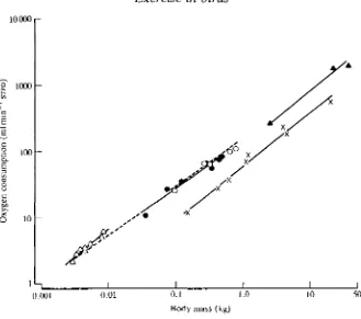
(Fig. 1). Using data from seven species of birds during forward flapping flight in wind tunnels (Tucker, 1968, 1972; Bernstein etal. 1973; Butler etal. 1977; Torre Bueno and Larochelle, 1978; Gessaman, 1980; Hudson and Bernstein, 1983), ^o2min (mlrnin"1 STPD) was found to be 149M072 (where M is body mass in kg).
Using data for six species of volant birds (Prange and Schmidt-Nielsen, 1970; Fedak etal. 1974; Bamford and Maloiy, 1980; Grubb, 1982; Woakes and Butler, 1983; Warncke et al. 1988)and two species of penguins (Pinshow et al. 1976, 1977) while walking, running or swimming, Vo2max was found to be 60.5M081 and for three species of cursorial birds (Taylor et al. 1971; Brackenbury and Avery, 1980; Grubb etal. 1983) Vo2max was found to be 145M074. Thus, although the mass exponents are not exactly the same for the three groups, these equations (see Fig. 1) illustrate the large (2.4 times for a 1 kg animal) difference in aerobic metabolism in different species of birds engaged in the same type of exercise (running) and, it is assumed, between different types of exercise (running vs flying) in the same species. The only species, in fact, for which data have been obtained during forward flapping flight and while running is the pigeon (Butler etal. 1977; Grubb, 1982), in which VOl while flying at 10ms"1 was 77.8mlmin"1
10000
S 1000
I I
0.001 0.01 0.1 1.0
Body mass (kg)
[image:3.451.60.389.59.350.2]10 50
Fig. 1. Double logarithmic plots of oxygen consumption (ml min ' STPD) against body mass (kg) for hovering hummingbirds (A A), birds during forward flapping flight ( • • ) , cursorial birds running (A A), volant birds and penguins running or swimming (x x) and bats during forward flapping flight (O O ) . For further details, see text.
Hovering flight is, at least theoretically (Rayner, 1979), energetically very costly and data from eight studies on six species of hummingbirds (Lasiewski, 1963; Berger and Hart, 1972; Berger, 1974, 1985; Epting, 1980; Bartholomew and Lighton, 1986) tend to support this; VOl during hovering is 371M087. However, if
the data from these six species of hummingbirds are added to those from the seven species of birds during forward flapping flight, the relationship is VOl=\A5M°i69,
60 i—
50
g 40
I 30
ex
B
C O -v, o 2U
10
01- I I I I J
F, off F, on 0.25 0.35 0.45 0.55
Water velocity ( m s "1)
[image:4.451.72.373.53.306.2]0.65 0.75 0.85
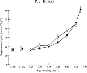
Fig. 2. The relationship between mean ( ± S . E . , N=6) oxygen consumption and
swimming speed in trained (•) and untrained (D) tufted ducks. F, off, F, on, flume motor turned off or on. (From Butler and Turner, 1988.)
influence on VOl during hovering (Schuchman, 1979), whereas in other species
there is only a slight effect (Berger and Hart, 1972).
It is interesting to note, in passing, that for five species of bats flying on the level in wind tunnels, V^mm was found to be 151A/0'71 (Thomas, 1975; Carpenter, 1985, 1986) and that this is similar to that for birds during forward flapping flight (Fig. 1).
It is clear from studies on the tufted duck, Ay thy a fuligula, that the level of fitness of an animal can have a large influence on VO2max (Butler and Turner,
1988). Training resulted in a 27% increase in VOimax in swimming ducks (Fig. 2)
or, looking at it the other way round, VO2max w a s approximately 22 % lower in inactive birds compared with that in active ones. It would be of great interest to know to what extent flight performance can be influenced by training (see below).
Exercise under water (diving)
Aerobic metabolism
14.4s (water temperature 13.5°C), mean oxygen uptake (VOl, STPD) was
34.0mlmin~1 (57mlmin~1kg~1) and not significantly different from the value (SS^mlmin"1, 63mlmin~1kg~1) obtained from the same ducks swimming at maximum sustainable speed on the surface. These values are some 3.5-3.8 times resting oxygen consumption, respectively, and indicate that feeding under water is, in energetic terms, a very costly business for these animals.
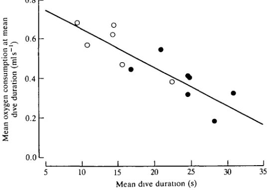
On the basis of the usable oxygen stores in the tufted duck (Keijer and Butler, 1982), this species should be able to remain submerged and metabolise aerobically at the level given above for approximately 50 s. This is some 2.5 times longer than the preferred dive duration of these animals on a 1.9-2.8 m deep pond (Stephen-son et al. 1986), and would suggest that they metabolise completely aerobically during most voluntary dives, using the oxygen stored in the body and replacing it upon surfacing. It could be, however, that oxygen consumption actually decreases as dive duration progresses beyond 15 s (Bevan et al. 1991). Certainly, obser-vations on individual ducks indicate that mean oxygen consumption at mean dive duration is lower in those that perform longer dives (Fig. 3). This could result from reduced buoyancy as the dive progresses [in those birds that perform longer dives it could be the result of the reduced volume of the respiratory system (Stephenson et al. 1989a,fr)] and/or it could indicate that aerobic metabolism declines as a dive proceeds with, perhaps, increasing anaerobiosis. This latter suggestion would imply associated cardiovascular adjustments (see below).
As the energetic cost of feeding under water is so high for aquatic birds such as
0.8
r-0.6
o •«
1
.9 0.4
H -5
0.2
0.0 L
I I
10 15 20 25 Mean dive duration (s)
[image:5.451.91.361.376.567.2]30 35
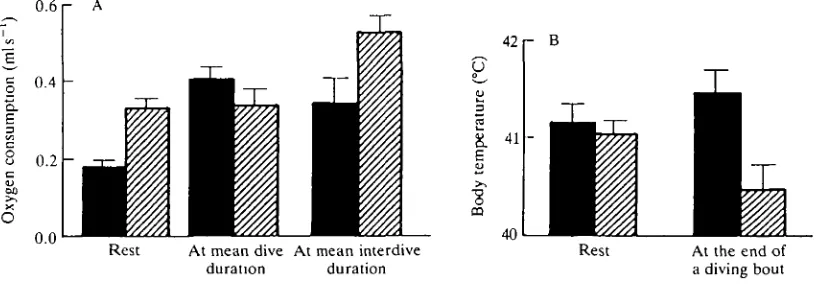
tufted ducks, an important question is what happens in winter when they are cold water? When at rest on water under winter conditions (air temperature 6°C, water temperature 7.5 °C), oxygen consumption in tufted ducks is twice that under summer conditions (26°C and 23°C, respectively), and deep body temperatures are similar (Fig. 4). However, mean oxygen consumptions at mean dive durations are not significantly different under the two conditions (Bevan and Butler, 1989), whereas mean oxygen consumption at mean duration at the surface is some 50 % higher under winter conditions. Deep body temperature is a full 1°C lower after a bout of diving in winter conditions compared with that at summer temperatures (Fig. 4B). It would appear that while actually feeding under water the heat from locomotor activity is insufficient to maintain core temperature in winter, but that the ducks do not enhance metabolic heat production during this period. In contrast, while at the surface between dives, aerobic metabolism is higher during winter but is still insufficient to prevent a reduction in body temperature over a complete bout of diving. The outcome is that, overall, feeding in winter is metabolically more expensive (approximately 40 % in the present study) for tufted ducks than it is in summer, but not as great as might be expected from the elevation of oxygen uptake seen in ducks resting on water in winter conditions.
Penguins, which are much better adapted than tufted ducks to an aquatic existence, may be able to feed much more efficiently. From direct measurements of oxygen uptake in Humboldt penguins, Spheniscus humboldti, diving for food in a pool and in little penguins, Eudyptula minor, swimming at 0.7 m s "1 in a water channel, it appears that aerobic energy expenditure during underwater swimming in penguins is approximately 30% greater than, but not significantly different
o. &
0.6
0.4
0.2
0.0
Rest At mean dive At mean interdive duration duration
U
03
42
41
40 r B
Rest At the end of a diving bout
Fig. 4. (A) Histogram showing mean oxygen uptake (+S.E., ^=6) in tufted ducks at rest, at mean dive duration and at mean interdive duration. The birds were acclimated to summer (air temperature 26°C, water temperature 23°C; filled columns) or winter (air temperature 6°C, water temperature 7.5°C; hatched columns) conditions. (B) Histogram showing mean deep body temperature (+S.E., N=6) in tufted ducks at
[image:6.451.29.437.405.547.2]Pbm, that in resting birds (Butler and Woakes, 1984; Baudinette and Gill, 1985).
Clearly, these experimental conditions are somewhat different from those that exist at the feeding grounds. However, from the measurements of oxygen consumption that have been made for Humboldt penguins swimming under water and from the usable oxygen stores of a similar-sized penguin, calculated from the data of Kooyman (1975), these birds should be able to remain submerged and completely aerobic for approximately 2.3 min. Data from chinstrap penguins, Pygoscelis antarctica (which are of a similar size to Humboldt penguins), diving freely off Signy Island, South Orkneys, indicate that at night these birds dive for an average of approximately 1.6 min (Lishman and Croxall, 1983). It would appear, therefore, that these medium-sized penguins, like diving ducks, remain com-pletely aerobic during normal feeding dives.
Jones and Furilla (1987) point out that the average daily metabolic rate (ADMR) measured with labelled water in a number of species of penguins when foraging is between 40 and 60% greater than the fasting rate. Unfortunately, in some of the field studies it was not possible to estimate metabolic rate when the birds were actually diving, so the value for ADMR probably includes periods of rest (Kooyman et al. 1982; Davis et al. 1983). By incorporating information on time budgets, Nagy et al. (1984) estimated that, when swimming under water at sea at an average velocity of 1.75 m s "1, energy expenditure in jackass penguins, Spheniscus demersus, is six times the resting value. This can be compared with the metabolic rate of 4.3 times the lowest resting value in Humboldt penguins swimming at 1.25ms"1 (Hui, 1988). Also, estimated metabolic rate for gentoo penguins, Pygoselis papua, foraging at sea is some 6-7 times the calculated standard metabolic rate (Davis et al. 1989). It would appear, therefore, that aerobic metabolism during diving in penguins can be substantially elevated above the resting value, as it is in ducks; the difference is that penguins can swim faster.
Locomotory muscles
which is probably related to the fact that they spend much of their time floating water.
Studies on mammals also indicate that the deep extensor muscles (mainly SO fibres) are active during quiet standing as well as during locomotion. During walking, deep SO and FOG fibres are recruited in extensor muscle groups, and with increasing speed there is progressive recruitment of the more peripheral FG fibres (Armstrong et al. 1977; Walmsley et al. 1978). Suzuki etal. (1985) propose that a similar recruitment pattern exists in the leg muscles of birds. Laughlin and Armstrong (1982) have demonstrated for the rat that blood flow distribution within and among different muscles is related to their recruitment patterns during quiet standing and exercise. Again, a similar situation is assumed to exist in the legs of birds.
The major flight muscles in birds are the pectoralis and supracoracoideus and together they constitute, on average, 17 % of body mass for a range of birds, with the pectoralis being approximately 10 times the mass of the supracoracoideus (Greenewalt, 1962). This reflects the more important role of the former in causing depression of the wing and therefore in generating lift and thrust. The upstroke, which is effected by the supracoracoideus, is merely a recovery stroke. In hummingbirds and penguins, in contrast, the ratio of the masses of the two muscles is approximately 2 and in the former their combined mass accounts for 20-30 % of total body mass (Greenewalt, 1962; Baldwin et al. 1984). Lift is generated during both phases of the wingbeat during hovering in hummingbirds and during swimming in penguins.
In volant birds, the pectoralis muscle consists predominantly of FOG fibres together with a smaller (usually) percentage of FG fibres. In a few species of birds there are also some SO fibres, which are normally involved in postural support. Their presence was thought to be associated with the gliding or soaring behaviour of some birds, e.g. herring gull (Talesara and Goldspink, 1978). This appears to be the case for the turkey vulture, Cathartes aura (Rosser and George, 1986a), although Rosser and George (19866) found SO fibres in the pectoralis muscle of only two out of 42 volant species, and this did not include the herring or ring-billed gulls. The reported presence of a large percentage (12 %) of SO fibres in the dorsal areas of the pectoralis of the grey catbird, Dumetella carolinensls (Marsh, 1984), which is a migrating species, is perplexing in view of the fact that Rosser and George (19866) found no indication of such fibres in the pectoralis muscles of 17 other species of passerines. Thus, the functional significance of SO fibres in the pectoralis of volant birds, when they are present, is not clear in all cases.
The FG fibres are relatively large and make up anything from 0 % to approximately 50 % of the fibres in the pectoralis of the volant species (Rosser and George, 19866). They metabolize glycogen anaerobically and are used predomi-nantly during take-off and landing and when the birds undergo sudden changes in direction (Parker and George, 1975; Dial etal. 1987).
Piaerobically, are nonetheless highly oxidative. In free-living tufted ducks, the mass-specific activity of citrate synthase (CS), an indicator of the capacity for oxidation of acetyl Co A in the citric acid cycle, is approximately 20 % higher in the pectoralis muscle than in the heart (Butler and Turner, 1988). CS activity in the leg muscles of volant birds may be less than half of that in the pectoralis muscles (Marsh and Dawson, 1982; Butler and Turner, 1988). Marsh (1981) reported a very high activity of CS (200 /imolmin^g"1 fresh mass) in the pectoralis of the grey catbird just prior to migration and claimed that it was 'the highest reported for any vertebrate skeletal muscle' (it is approximately twice that for the supracora-coideus muscles of the same species). An even higher activity of CS (343jumol m i n ^ g "1 wet mass) has been reported in the flight muscle of the hummingbird, Selasphorus rufus (Suarez et al. 1986). It must be remembered, however, that CS activity most likely scales with body mass in a similar fashion to oxygen uptake (see above, and Somero and Childress, 1980). Nonetheless, the FOG fibres in the pectoralis muscle are so oxidative that Talesara and Goldspink (1978) have suggested that they should be designated FO fibres.
The proportion of FO(G) fibres varies throughout the pectoralis muscle of any one species, being greater in the deeper regions, and also between volant species, from 100% in starlings, owls, hawks and kingfishers (and, it is assumed, hummingbirds) to approximately 50% in the ruffed grouse (Rosser and George, 1986£>). Thus, the pectoralis muscle normally consists of a preponderance of fast contracting but highly oxidative fibres, which are required to beat the wings at high frequencies, sometimes for many hours.
Related to the greater potential oxygen demand in the pectoralis of flying birds is a greater capillary/fibre ratio than that in the leg muscles of birds and mammals (Pietschmann et al. 1982; Turner and Butler, 1988). Along with the smaller cross-sectional area of the FO(G) fibres in the pectoralis, there are, therefore, shorter diffusion distances from capillaries to fibres. In fact, the smallest fibres and the greatest capillary densities are found in birds that migrate the longest distances (Lundgren and Kiessling, 1988). Also, in the tufted duck, the volume density of mitochondria is greater in the pectoralis muscle than in the muscles of the leg (Turner and Butler, 1988). In fact, it is evident from this last study that the difference in Vo2max during running in volant birds and V^mm during forward flapping flight is not merely the result of the different muscle masses, as suggested by Prange and Schmidt-Nielsen (1970), but also of the different oxidative capacities of the muscles.
o 09
2 0.8
0.7
I
10 20 30
Flying time (min)
[image:10.451.75.375.50.289.2]40 50 60
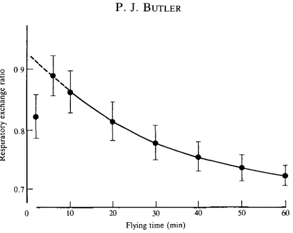
Fig. 5. Mean values ( ± S . D . , N=5) of the respiratory exchange ratio of pigeons at rest
and when flying for up to 60 min in a wind tunnel. (After Rothe et al. 1987.)
approximately 40kJ of energy, whereas for carbohydrate the value is 17.5 kJg \ Also, it can be stored in adequate abundance for long-term or migratory flight (Blem, 1976). However, the maximum theoretical rate of catabolism of a particular fuel for muscle contraction is determined by the activities of the relevant enzymes, but factors such as transport of substrates, particularly free fatty acids (FFA), and availability of cofactors will also limit its rate of utilization (Hultman and Harris, 1988). These authors quote calculated values, based on data from humans, which indicate that the maximum rate of ATP synthesis from the complete oxidation of carbohydrate is 0.51-0.68 mmol s"1 kg"1, whereas the value for the oxidation of FFA is 0.24 mmol s"1 kg"1. This relatively low rate indicates a rate-limiting step before the formation of acetyl CoA, as succeeding steps are also involved in oxidation of carbohydrates. If birds do metabolize at the rates indicated in Fig. 1 during long-distance flight, while oxidizing fats, it must be assumed that they do not possess the constraints associated with oxidation of FFA that are present in humans.
Associated with pre-migratory fattening is an increase in the mass of the pectoralis muscle (Marsh, 1984). In the grey catbird, there is no associated change in the specific activity of CS, although there is an increase in activity of 3-hydroxyacyl-CoA dehydrogenase (HAD), which is a key enzyme in the /S-oxidation of fatty acids (Marsh, 1981). There is, however, an increase in CS and cytochrome oxidase (COX) activities in a number of other species prior to migration (Lundgren and Kiessling, 1985, 1986). ^
^comotor activity (training) just prior to migration effect the adaptations seen in the pectoralis muscle. It would seem that hypertrophy of the pectoralis is a direct result of the increase in body mass associated with pre-migratory fattening (Marsh, 1984). Endurance training can increase muscle capillarity and mass-specific activity of CS and cause a transformation of muscle fibres from FG to FOG in the leg muscles of ducks (Butler and Turner, 1988). It has been proposed that the increased oxidative capacity in migratory reed warblers Acrocephalus scirpaceus is a training effect (Lundgren and Kiessling, 1986). These authors do point out, however, that both increased locomotor activity and hyperphagia are initiated by the hormones corticosterone and prolactin (Meier and Martin, 1971). Nonethe-less, studies on the levels of thyroid hormones (T3 and T4) and growth hormone in migratory Canada geese have suggested that they are directly involved in the hypertrophy of the pectoral muscles prior to migration (John and George, 1978; John et al. 1983). Also, high levels of T4 and T3 can cause increases in activities of CS and cytochrome c in the soleus muscle of rats (Winder, 1979).
'Conditioning' of the pectoral muscles before migration takes on even greater significance when it is realised that periods of relative inactivity, such as those occurring during moulting and incubation, cause significant reductions in muscle mass and myoglobin concentration and in CS and HAD activities (Pages and Planas, 1983; George et al. 1987; Butler and Turner, 1988; Piersma, 1988). In contrast, the relative mass of the leg muscles may increase during the moulting period, which could be an undesirable burden during migration.
With the possible exception of the larger and deeper-diving penguins (emperor, king, AdeTie), the anaerobic capabilities of the locomotory muscles of diving birds (pectoralis in penguins and alcids) are relatively low (Baldwin et al. 1984; Davis and Guderley, 1990). In fact, the latter authors conclude that there are no special biochemical adaptations in the locomotory muscles of diving birds, although there is a tendency, particularly among the penguins, for them to have a higher concentration of myoglobin (Butler, 1990).
Respiratory and cardiovascular adjustments during exercise in air
It is the combined functions of the respiratory and cardiovascular systems to supply the required oxygen and metabolic substrates to the locomotor muscles and to remove the waste products of metabolism (e.g. CO2 and heat). It is clear from the earlier discussion that the respiratory and central cardiovascular systems do not impose any limit on oxygen consumption during running or swimming in volant birds. Thus, this part of the review will concentrate mainly on those studies performed on flight and on running in cursorial species.
flight (cf. VQ2 of birds and bats during flight) or with respect to their
tolerance of high altitude (see below).
Ventilation
Respiratory data are given for six species of birds in Table 1. Two of these are hovering hummingbirds and four are birds during forward flapping flight. It can be seen that, for the flying birds at relatively low ambient temperatures (<23°C), ventilation volume (Vi) increases by a similar proportion as oxygen uptake (Vo2) and that the proportion of oxygen extracted from the inspired gas (O2ext) is similar to that in resting birds. Oxygen extraction during hovering for one of the hummingbirds, Colibri coruscans, is similar to that for the flying birds, whereas for the other, Amazilia fimbriata, it is somewhat lower. The respiratory frequencies (/resp) a r e v e rY high m the hovering hummingbirds. The relative contributions of respiratory frequency and tidal volume (Vt) to the increase in Vi during forward flapping flight vary among species. In white-necked ravens, VT does not change at all, in fish crows it almost doubles and in starlings there is a fourfold increase. Thus, in the first two species, frequency makes the greater contribution, whereas in the third, volume predominates.
Despite the apparent matching between ventilation volume and oxygen uptake in the four species of flying birds listed in Table 1, there does appear to be an increase in effective lung ventilation above that required by metabolic rate (hyperventilation) during flight in starlings, as partial pressure of oxygen (-PoJ in the air sacs increases and Pco2 decreases (Torre-Bueno, 1978a). Such
hyperventi-lation is even more apparent in ducks when running or swimming (Kiley et al. 1979; Woakes and Butler, 1986) and in running chickens (Brackenbury et al. 1982). An explanation could be that hyperventilation results from the increase in body temperature that occurs in all exercising birds, even during flight at low (0°C) environmental temperatures (see, for example, Torre-Bueno, 1976; Hudson and Bernstein, 1981; Hirth et al. 1987). However, when the usual elevation in body temperature during running is prevented in ducks and domestic fowl, there are still signs of hyperventilation (Kiley et al. 1982; Gleeson and Brackenbury, 1984). In both cases there was a slight acidosis which could have stimulated ventilation. Thus, it does appear as if factors other than hyperthermia contribute to the overall hyperventilation in exercising birds.
Table 1. Values of respiratory frequency, f,,, (min-'); tidal volume, VT (ml); ventilation volume, VI (1 min-' BTPS, except for fish crow where it is 1 min-' STPD), oxygen extraction, 02ext, and oxygen uptake,
vO,
(ml O2 min-' STDP) during rest and while ho~wing in two species of hummingbirds and while flying in a wind tunnel for four other species of birds Rest FlightMass (kg)
43 I
-42
O
S 41
O-E
40
39 I I I
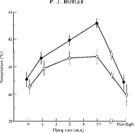
2 3 4 Flying time (min)
[image:14.451.94.358.55.316.2]5 + Post-flight
Fig. 6. Mean values (±2s.E., N=5) of brain (O) and colonic ( • ) temperatures in kestrels before, during and after flapping flight at 10 ms"1 at an ambient temperature of 23°C. Data at 5+ min were obtained between 5 and 15 min after the onset of flight and represent steady-state values. (After Bernstein et al. 1979.)
opthalmicum (RMO). It is closely associated with the circulatory system of the eye, and warm arterial blood from the body is thought to be cooled by the counter-current exchange with venous blood returning from the relatively cool beak, evaporative surfaces of the upper respiratory tract and the eye (Mitgard, 1983).
The hyperventilation during flight no doubt serves a thermoregulatory function and as ambient temperature increases, a greater proportion of total heat loss during flight is by respiratory evaporation. However, even at an ambient temperature of 30°C this is only approximately 20% for the budgerigar (Tucker, 1968) and 30 % for the fish crow, white-necked raven and pigeon (Hudson and Bernstein, 1981; Biesel and Nachtigall, 1987). So, the majority of metabolic heat must be dissipated by means other than respiratory evaporation. Indeed, herring gulls can lose up to 80 % of total heat production through their feet during flight (Baudinette et al. 1976). In pigeons, the value is probably slightly less, at 50-65 % (Martineau and Larochelle, 1988).
Biough to enable a greater proportion of heat to be dissipated by non-evaporative means, thus keeping them in water balance.
Studies on ventilatory control in running Pekin ducks have indicated that neither the carotid bodies nor the pulmonary CO2 receptors are of any importance and that input from muscle afferents may play a major role (Kiley and Fedde, I983a,b; Faraci et al. 1984).
Circulation
The role of the various components of the circulatory system in presenting oxygen to (and removing CO2 from) the exercising muscles can best be described by Fick's formula: FO 2= / H X Vs(CaO2-CvO2), where VOz is the rate of oxygen
consumption, / H is heart rate, Vs is cardiac stroke volume, CaO2 is the oxygen content of arterial blood and CvO2 is the oxygen content of mixed venous blood.
There are two studies on flying or cursorial birds in which data have been collected on all but one of these variables (thus allowing the absent one to be calculated), Butler et al. (1977) working with pigeons and Grubb et al. (1983) working with emus. When flying in a wind tunnel at 10 m s"1, oxygen uptake in the pigeons was 10 times the resting value (Table 2). The respiratory system maintained CaO2 at slightly below the resting value but CvO2 was halved, giving a 1.8-fold increase in (CaO2—CvO2). There was no significant change in Vs, so the major factor in transporting the extra oxygen to the muscles was the sixfold increase in heart rate. When running on a treadmill at a 6° incline and a speed of 1.33 m s"1, emus had an oxygen uptake that was 11.4 times the resting value and, although an increase in heart rate to 3.9 times the resting value was the major factor, a 1.8-fold increase in cardiac stroke volume and a similar increase in (CaO2—Cvch) both made significant contributions to the enhanced delivery of oxygen to the exercising muscles. As mean arterial blood pressure did not change in either species during exercise, total peripheral vascular resistance fell by the same proportion as cardiac output (faxVs) increased. It is interesting to note that
Table 2. Mean values of oxygen uptake and cardiovascular variables measured in pigeons (Butler et al. 1977) and emu (Grubb et al. 1983) at rest and after 6min of steady level flight in a wind tunnel at a speed of 10ms~' for the former and after 20min running on a treadmill at a 6° incline and a speed of 1.33 ms'1 for the latter
Pigeon (0.442 kg) Emu (37.5 kg)
Oxygen uptake (ml min i STPD) Heart rate (beats min"1) Cardiac stroke volume (ml) Oxygen content of arterial blood
Oxygen content of mixed venous blood (vol%)
Rest 9.0 115
1.44 15.1
10.5
Flying 88.4 670
1.58 13.7
5.4
Rest 156.7 45.8 57 15.2
9.0
Running 1807
180 102.7
15.2
heart rate in hovering hummingbirds may exceed 1200 beats min 1 (Berger et afl 1979).
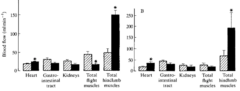
During swimming in both tufted ducks and barnacle geese, an approximately twofold increase in oxygen consumption above the resting value is accompanied by a threefold increase in blood flow to the muscles of the legs (Butler et al. 1988; R. M. Bevan and P. J. Butler, in preparation). In the ducks, there is also a significant reduction in blood flow to the flight muscles, whereas this is not the case for the barnacle geese (Fig. 7). As the cardiovascular system is able to cope with the greater demand for oxygen during flapping flight, redistribution of blood away from the inactive pectoral muscles during swimming may not be important to volant birds. It is possible that the difference in response between the tufted duck and barnacle goose is related to the diving behaviour of the duck, when reduced blood flow to the flight muscles is a likely mechanism for conserving oxygen for the central nervous system (CNS), heart and leg muscles (Butler, 1988). It should be noted, however, that 'resting' blood flow to the flight muscles is higher in the tufted duck than in the barnacle goose. Whether this is an artefact remains to be seen, but it could be an explanation for the different responses in the two species. Nonetheless, it would certainly be interesting to see what happens to blood flow to the leg muscles during forward flapping flight.
Birds have larger hearts and lower resting heart rates than mammals of similar body mass (Lasiewski and Calder, 1971; Grubb, 1983). They also have a greater cardiac output for a given oxygen consumption than similar-sized mammals (Grubb, 1983). In other words, (Cao^-Cvo,) is lower in birds than in similar-sized mammals, because CvOl is not normally reduced to such a low level. Thus, the
200
7 150 c
100
5 5°
Heart Gastro- Kidneys Total Total intestinal flight hindhmb
tract muscles muscles
Heart Gastro- Kidneys Total Total intestinal flight hindhmb
[image:16.451.20.434.412.569.2]tract muscles muscles
cardiac output in birds may be an important factor in their attaining a higher maximum oxygen uptake (V^max) during flight than similar-sized mammals when running. It is certainly interesting to note that bats have larger hearts and a higher blood oxygen-carrying capacity than other mammals of similar size (Jiirgens etal. 1981) and that, as mentioned earlier, their VOl during flight is
similar to that of birds of similar mass.
High altitude
Most passerines migrating at night fly below 2000 m, although some birds have been observed at extremely high altitudes. A flock of 30 swans (probably whooper: Cygnus cygnus) was located by radar off the west coast of Scotland at an altitude of 8000-8500 m (Stewart, 1978). Bar-headed geese Anser indicus have been observed flying at altitudes of approximately 9000 m (where the PO2 is, at 6.9 kPa,
approximately one-third of the sea-level value) during their migration across the Himalayas (Swan, 1961). Unfortunately, there have been no physiological studies on birds flying at high altitude (real or simulated), but work on inactive animals has indicated the adaptations that may be of great importance in the altitude performance of birds.
It is the cardiovascular system that shows most adaptations to the conditions of high altitude. Unlike Pekin ducks, bar-headed geese do not increase their haematocrit (packed cell volume of the blood) and haemoglobin (Hb) concen-tration when exposed to simulated high altitude (Black and Tenney, 1980). This means that there is no increase in blood viscosity, thus preventing a possible reduction in circulation of the blood. It also means that there is no increase in the oxygen-carrying capacity of the blood. This is more than counterbalanced by the higher affinity for oxygen (low P^) of the Hb of the goose (P50 for goose blood is approximately 5kPa at pH7.5 compared with 7.5kPa in the duck), which allows the maintenance of a higher CaO2 (and hence CaO2-CvO2) at high altitudes than in the duck. A comparison between the Hb of greylag and bar-headed geese indicated that the difference in oxygen affinity is the result of a very small intrinsic difference that is magnified by the presence of inositol pentaphosphate (Rollema and Bauer, 1979).
It is also clear from the data of Black and Tenney (1980) that the respiratory system of the bar-headed goose is able to maintain a very small difference between the Po2 >n inspired air (PioJ a n^t n a t in arterial blood (Pao2) when at high altitude. At sea level, P\O2—Pa.O2 is 7 kPa, whereas at a simulated altitude of 10 668 m it is a
mere 0.5 kPa. The large increase in ventilation that is required to maintain such a small difference between PiO2 and P ao, also causes a decline in PaCOz and an
hypoxia) and hypocapnia attenuates the increase in cerebral blood flow induced rj^ hypoxia (Grubb etal. 1979). However, these authors found that blood flow is similar at a given O2 content in both normocapic and hypocapnic ducks. This is because during hypocapnia (and alkalosis) there is a leftward shift of the oxygen equilibrium curve so that a given oxygen content is achieved at a lower POl. In fact,
in bar-headed geese, the alkalosis during severe hypoxia is greater than that in Pekin ducks which, together with the higher affinity of their Hb for oxygen, means that at a given (low) PaOl, Ca^ is much (two times) greater in the geese (Faraci
etal. 1984). Thus, similar (or even greater) oxygen deliveries to the brain and heart can be achieved in hypoxic bar-headed geese at lower cerebral and coronary blood flows than in hypoxic ducks. This may be a very important feature when the geese are flying over the Himalayas.
It has been suggested that the more effective lungs of birds, compared with those of mammals, may contribute significantly to the ability of birds better to tolerate high altitude (Scheid, 1985). A more recent analysis, however, has dismissed this notion (Shams and Scheid, 1989). The difference between birds and mammals appears to be the ability of the former to tolerate lower Paco2> thus
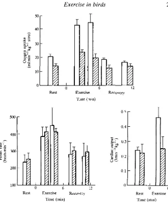
enabling the respiratory system to maintain PaO:, at as high a level as possible. Despite all of these apparent adaptations to life at high altitude, experiments on bar-headed geese running on a treadmill under hypoxic conditions, similar to those at the top of Mount Everest, indicate severe limitations to oxygen uptake (Fedde et al. 1989). Under normoxic conditions, running at 0.6ms"1 at a 2° incline caused a doubling in VOl and cardiac output from approximately 20 ml min"1 kg"1
and 0.241 min"1 kg"1, respectively (Fig. 8). Cardiac stroke volume was constant at approximately 2.5 ml. Under hypoxic conditions, resting oxygen uptake fell to approximately 15 ml min"1 kg"1 and was not significantly different after 6 min of exercise. Cardiac output also remained unchanged during exercise at approxi-mately 0.251 min"1 kg"1, whereas cardiac stroke volume declined significantly from 1.9ml at rest to 1.4ml during exercise. The authors argue that diffusion of oxygen from the muscle capillaries to the mitochondria may limit the aerobic capacity of the exercising leg muscles during hypoxia. If this is the case, the situation could be different in the pectoral muscles during flight at high altitude. The question is, how does the heart manage to cope with the extra demands of flight at high altitude when it appears to be at its limit during running under hypoxic conditions similar to those at the top of Mount Everest? The resting heart rate was rather high in the birds used in this study, which probably means that they were stressed (see Woakes and Butler, 1986). Whether this could explain the results remains to be seen. It is also possible that birds of this species routinely migrate across the Himalayas at altitudes substantially below that at the top of Mount Everest.
5O
40
Q
I
6
D.I
Of e
20
10
I
I
i
1
1
Rest
CO J 2
U CO
X «
500r
400
300
200
100
Jft
6 12 Exercise Recovery
Time (min)
0.5
0.4
f-f" 0.3
•2'g 0.2
CO
0.1
I
12
Rest Exercise Time (min)
Recovery
0 6 Rest Exercise
[image:19.451.54.400.48.466.2]Time (min) Fig. 8. Mean values (+S.E.) of oxygen uptake, heart rate and cardiac output in bar-headed geese at rest and while running at 0.6 ms"1 on a 2° incline (exercise) during normoxia (D) and hypoxia simulating that at the top of Mount Everest (0). (Modified from Fedde et al. 1989.)
Cardiovascular adjustments during diving
grebes, divers, cormorants and a range of auks, are, to a greater or lesser exten™ active predators of fish and remain submerged for an average of 20-70 s, with the guillemot, Uria aalge, having a reported maximum dive duration of 202 s (Wanless et al. 1988) and the imperial cormorant (blue-eyed shag), P. atriceps, of 312s (Naito et al. 1991). The latter species has been monitored diving to a maximum depth of 116 m, whereas other birds have been trapped in nets at 180 m (guillemots), 120m (razorbills Alca torda) and 60m (great northern diver, Gavia immer) (Schorger, 1947; Piatt and Nettleship, 1985). These dive durations and depths compare very favourably with those of the smaller penguins. When inshore, the little penguin, Eudyptula minor, remains submerged for very short periods of 10-15 s and the greatest recorded depth is 60m (Mill and Baldwin, 1983), whereas the larger chinstrap and gentoo penguins have mean dive durations of 91 and 128 s and maxima of 130 and 190 s, respectively (Trivelpiece et al. 1986). The greatest recorded depths for these birds are 70 and 100 m, respectively (Lishman and Croxall, 1983; Conroy and Twelves, 1972).
It is the larger penguins, the kings and emperors, Aptenodytes patagonica and A. forsteri, that are the most impressive avian divers. When diving naturally and presumably feeding, the emperors remain submerged for 2.5-9min with a maximum recorded depth of 265 m (Kooyman et al. 1971). King penguins can dive to between 240 and 290 m, although 50% of the dives are probably to less than 50 m (Kooyman et al. 1982).
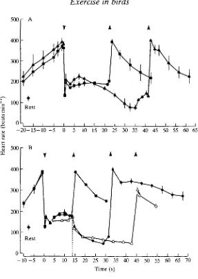
With oxygen consumption of tufted ducks during dives of approximately 15 s duration being similar to that at maximum sustained swimming speed, similar cardiovascular adjustments might also be expected. However, Woakes and Butler (1983) discovered that mean heart rate just before surfacing from dives of approximately 15 s duration is significantly lower than that recorded from the same animals w^ien they are swimming at the surface and consuming oxygen at the same rate (Fig. 9). Assuming that heart rate is an indicator of the degree of peripheral vasoconstriction (and thus, of anaerobic metabolism), it has been suggested (Butler, 19826) that, during most dives, perfusion (and hence aerobic metabolism) of the active muscles (heart, legs) and of the central nervous system is similar to that during exercise in air, whereas the viscera and inactive muscles (which, of course, includes the large pectorals in these birds) may be perfused less than during exercise in air in order to conserve oxygen for the active muscles, heart and CNS. This hypothesis has gained some support from qualitative studies using micro-aggregated albumin labelled with "mT c (Jones et al. 1988), and from direct measurements of blood flow through major arteries (R. M. Bevan and P. J. Butler, in preparation).
300r
Fig. 9. Mean heart rate ( ± S . E . , N=6) for tufted ducks at rest, during voluntary dives of 14.4 s mean duration and while swimming. Oxygen consumptions (VOi) at mean dive duration and while swimming were the same: 34mlmin~'. (From Woakes and Butler, 1983.)
ducks were swimming at 0.75 m s"1 and by 33 % during the last 5 s (excluding data over the final second) of dives with a mean duration of 15.5s. Under similar conditions, blood flow through both ischiadic arteries was SOmlmin"1 at rest, increasing 4.8 times during swimming and 5.1 times during diving. Thus, the proportion of cardiac output flowing through the ischiadic arteries was greater during diving than during swimming and this inevitably means that the rest of the body was perfused by less blood during diving than during swimming. This is, unfortunately, not clearly reflected in the estimated blood flow through the brachial artery. Although flow through the brachial arteries, at 26 ml min"1, was significantly lower during diving than that in the resting birds (66 ml min"1), it was not, however, significantly lower than that during swimming (42ml min"1). A region of the body that could well receive less blood during diving than during swimming is, of course, the respiratory muscles.
It would appear, therefore, from the evidence presented so far, that Butler's (1982b) hypothesis is correct, i.e. during diving in ducks the cardiovascular system exhibits a modified exercise response, with the legs, as well as the heart and CNS, being perfused adequately to maintain overall aerobic metabolism. Other, inactive, parts of the body are less well perfused than during surface swimming. Under certain circumstances, however, the balance may tip towards more of an ^xygen-conserving response.
distances under water for their food (e.g. under ice in winter), heart r a i l progressively declines after approximately 10 s, so that by approximately 30 s it is significantly below the resting value (Fig. 10A). During these dives the birds actively swim to and from the food. However, during normal vertical dives of similar duration and when the birds surface passively, heart rate remains elevated above the resting level. If, as indicated by the sub-resting heart rate, blood lactate does accumulate during a long horizontal dive, it does not have a large inhibitory effect on exercise, because the birds are still able to perform a number of dives in succession. There may, however, be a cumulative effect since the number of dives in a bout is lower than normal. If a duck is temporarily unable to surface from a voluntary dive, e.g. if it is disoriented under ice, there is an immediate reduction in heart rate as soon as the bird becomes aware of the situation. Heart rate follows the same time course and reaches a similar level to that seen during involuntary submersion (Fig. 10B), when selective vasoconstriction, a reduction in aerobic metabolism and increased lactate production are known to occur (Butler and Jones, 1982). This intense bradycardia occurs in 'trapped' ducks, despite the fact that they are still active under water.
It is clear that the cardiovascular response to diving in ducks is highly labile. On the basis of the cardiac response, it does appear that, under certain circumstances, the cardiovascular and metabolic responses during voluntary dives can shift progressively or more immediately to an oxygen-conserving response, i.e. relatively more intense peripheral vasoconstriction and increased lactate pro-duction in some tissues and organs, maybe even in the legs themselves in 'trapped' ducks.
Although physiologically expedient, in as much as oxygen is conserved, it appears that such a shift affects the feeding behaviour of the birds. At one extreme, it is usual for a bird to remain at the surface for an hour or more before making another dive after temporarily being unable to surface from a voluntary dive. Also, the time spent feeding during each dive decreases as horizontal distance to the food increases (Stephenson etal. 1986). This is contrary to the predictions of optimal foraging theory, which suggest that if more energy is expended in reaching a source of food, more time would be spent in obtaining (more) food (Charnov, 1976). This apparent contradiction in aquatic birds may result from the opposing time constraint imposed by physiological factors associated with the maintenance of aerobic metabolism, at least in the active muscles, CNS and heart. It appears that, under such conditions, the physiological adjustments allow optimization of feeding behaviour, but that these adjustments impose their own constraints upon that behaviour.
500
400
300
200
100
o
r- A
I I I I I I I t I I I I I I I I I
-20-15-10 -5 0 5 10 15 20 25 30 35 40 45 50 55 60 65
500 400 300 200 100 0
r
BA
f
_ i
Rest1 A
-10-5 0 5 10 15 20 25 30 35 40 45 50 55 60 65 70 Time (s)
[image:23.451.73.360.70.473.2]responsible for, the bradycardia seen during extended horizontal dives and when tufted ducks are temporarily unable to surface from a voluntary dive (Fig. 10, Butler and Stephenson, 1988). They also have a slight inhibitory effect on heart rate towards the end of 'normal' vertical dives of approximately 20 s duration (Butler and Woakes, 19826). In the latter study, there was also a significant increase in dive duration following bilateral denervation of the carotid bodies. Thus, these sense organs do not appear to play a dominant role in cardiac control during voluntary diving in ducks and are not involved in the immediate reduction in heart rate seen when 'trapped' ducks become aware of their predicament (Fig. 10B). Inactivation of receptors in the nasal passages with local anaesthesia prevented voluntary diving in some redhead ducks and heart rate was 10-30 % higher than in untreated ducks during the first 12-5 s of submersion when the animals dived in response to being chased (Furilla and Jones, 1986).
Thus, it seems that receptors in the nasal passages have an inhibitory effect during the first few seconds of voluntary submersion and that the carotid body chemoreceptors have a similar influence after 10-15 s of submersion. However, it is clear that the dramatic reduction in heart rate, from the elevated pre-dive level, occurs before the nasal areas contact the water (Butler and Woakes, 1982a) and is probably centrally mediated. The motor side of the cardiac response resides entirely in the vagal branches to the heart (Butler and Woakes, 1982a; Furilla and Jones, 1987). It is clear from this brief discussion that the neural control of the cardiac response in freely diving birds is very complex and well worthy of further study.
References
ARMSTRONG, R. B., MARUM, P., SAUBERT, C. W., SEEHERMAN, H. J. AND TAYLOR, C. R. (1977). Muscle fiber activity as a function of speed and gait. J. appl. Physiol. 43, 672-677'.
ARMSTRONG, R. B. AND LAUGHLIN, M. H. (1985). Metabolic indicators of fibre recruitment in mammalian muscles during locomotion. J. exp. Biol. 115, 201-213.
BALDWIN, J., JARDEL, J.-P., MONTAGUE, T. AND TOMKIN, R. (1984). Energy metabolism in
penguin swimming muscles. Molec. Physiol. 6, 33-42.
BAMFORD, O. S. AND MALOIY, G. M. O. (1980). Energy metabolism and heart rate during treadmill exercise in the Marabou stork. J. appl. Physiol. 49, 491-4%.
BARTHOLOMEW, G. A. AND LIGHTON, J. R. B. (1986). Oxygen consumption during hover-feeding in free-ranging anna hummingbirds. J. exp. Biol. 123, 191-199.
BAUDINETTE, R. V. AND GILL, P. (1985). The energetics of 'flying' and 'paddling' in water: locomotion in penguins and ducks. J. comp. Physiol. 155, 373-380.
BAUDINETTE, R. V., LOVERIDGE, J. P., WILSON, K. J., MILLS, C. D. AND SCHMIDT-NIELSEN, K. (1976). Heat loss from feet of herring gulls at rest and during flight. Am. J. Physiol. 230, 920-924.
BAUDINETTE, R. V. AND SCHMIDT-NIELSEN, K. (1974). Energy cost of gliding flight in herring gulls. Nature 248, 83-84.
BERGER, M. (1974). Energiewechsel von Kolibris beim Schwirrflug unter Hohenbedingungen. /. Orn. 115, 273-288.
BERGER, M. (1978). Ventilation in the humming birds Colibri coruscans during altitude hovering. In Respiratory Function in Birds, Adult and Embryonic (ed. J. Piiper), pp. 85-89. Berlin: Springer-Verlag.
BERGER, M. AND HART, J. S. (1972). Die Atmung beim Kolibri Amazilia fimbriata wahrend des Schwirrfluges bei verschiedenen Umgebungstemperaturen. J. comp. Physiol. 81, 363-380. BERGER, M., JOHANSEN, K., RUSCHI, A. AND DE ALMEIDA, P. J. (1979). Heart rates of flying
hummingbirds. Bol. Mus. biol. Prof. M. Leito xxx, 75-80.
BERNSTEIN, M. H., CURTIS, M. B. AND HUDSON, D. M. (1979). Independence of brain and body temperatures in flying American kestrels, Falco sparverius. Am. J. Physiol. 237, R58-62. BERNSTEIN, M. H., THOMAS, S. P. AND SCHMIDT-NIELSEN, K. (1973). Power input during flight of
the fish crow, Corvus ossifragus. J. exp. Biol. 58, 401-410.
BEVAN, R. M. AND BUTLER, P. J. (1989). Oxygen consumption during voluntary diving in the tufted duck, Aythya fuligula, acclimated to summer and winter temperatures. J. Physiol., Lond. 418, 132P.
BEVAN, R. M., KEIJER, E. AND BUTLER, P. J. (1991). Method for controlling the feeding behaviour in aquatic birds: heart rate and oxygen consumption during dives of different duration. J. exp. Biol. (in press).
BIESEL, W. AND NACHTIGALL, W. (1987). Pigeon flight in a wind tunnel. J. comp. Physiol. B 157, 117-128.
BLACK, C. P. AND TENNEY, S. M. (1980). Oxygen transport during progressive hypoxia in high-altitude and sea-level waterfowl. Respir. Physiol. 39, 217-239.
BLEM, C. R. (1976). Patterns of lipid storage and utilization in birds. Am. Zool. 16, 671-684. BRACKENBURY, J. H. AND AVERY, P. (1980). Energy consumption and ventilatory mechanisms in
the exercising fowl. Comp. Biochem. Physiol. 66A, 439-455.
BRACKENBURY, J. H., GLEESON, M. AND AVERY, P. (1982). Respiration in exercising fowl. III. Ventilation. /. exp. Biol. 96, 315-324.
BUTLER, P. J. (1981). Respiration during flight. In Advances in Physiological Sciences, vol. 10, Respiration (ed. I. Hutds and L. A. Debreczeni), pp. 155-164. Oxford: Pergamon Press. BUTLER, P. J. (1982a). Respiration during flight and diving in birds. In Exogenous and
Endogenous Influences on Metabolic and Neural Control (ed. A. D. F. Addink and N. Spronk), pp. 103-114. New York, Oxford: Pergamon Press.
BUTLER, P. J. (1982b). Respiratory and cardiovascular control during diving in birds and mammals. J. exp. Biol. 100, 195-221.
BUTLER, P. J. (1988). The exercise response and the 'classical' diving response during natural submersion in birds and mammals. Can. J. Zool. 66, 29-39.
BUTLER, P. J. (1990). Respiratory adaptations to limited oxygen supply during diving in birds and mammals. In Comparative Insights into Strategies for Gas Exchange and Metabolism (ed. A. J. Woakes, C. R. Bridges and M. K. Grieshaber), pp. 235-257. Cambridge: Cambridge University Press.
BUTLER, P. J. AND JONES, D. R. (1982). The comparative physiology of diving in vertebrates. Adv. Comp. Physiol. Biochem. 8, 179-362.
BUTLER, P. J. AND STEPHENSON, R. (1987). Physiology of breath-hold diving: a bird's eye view. Scient. Prog., Oxford 71, 439-458.
BUTLER, P. J. AND STEPHENSON, R. (1988). Chemoreceptor control of heart rate and behaviour during diving in the tufted duck (Aythya fuligula). J. Physiol., Lond. 397, 63-80.
BUTLER, P. J. AND TURNER, D. L. (1988). Effect of training on maximal oxygen uptake and aerobic capacity of locomotory muscles in tufted ducks Aythya fuligula. J. Physiol., Lond.
401, 347-359.
BUTLER, P. J., TURNER, D. L., AL-WASSIA, A. AND BEVAN, R. M. (1988). Regional distribution of blood flow during swimming in the tufted duck (Aythya fuligula). J. exp. Biol. 135, 461-472.
BUTLER, P. J., WEST, N. H. AND JONES, D. R. (1977). Respiratory and cardiovascular responses of the pigeon to sustained, level flight in a wind-tunnel. J. exp. Biol. 71, 7-26.
BUTLER, P. J. AND WOAKES, A. J. (1982a). Telemetry of physiological variables from diving and flying birds. Symp. zool. Soc, Lond. 49, 107-128.
BUTLER, P. J. AND WOAKES, A. J. (1982/?). Control of heart rate by carotid body chemoreceptors during diving in tufted ducks. /. appl. Physiol. 53, 1405-1410.
BUTLER, P. J. AND WOAKES, A. J. (1985). Exercise in normally ventilating and apnoeic birds. In
Circulation, Respiration and Metabolism (ed. R. Gilles), pp. 39-55. Berlin: Springer-Verlag.
BUTLER, P. J. AND WOAKES, A. J. (1990). The physiology of bird flight. In Bird Migration:
Physiology and Ecophysiology (ed. E. Gwinner), pp. 300-318. Berlin: Springer-Verlag.
CARPENTER, R. E. (1985). Flight physiology of flying foxes, Pteropuspoliocephalus. J. exp. Biol. 114,619-647.
CARPENTER, R. (1986). Flight physiology of intermediate-sized fruit bats (Pteropodidae). J. exp.
Biol. 120, 79-103.
CHARNOV, E. L. (1976). Optimal foraging, the marginal value theorem. Theory Pop. Biol. 9, 129-136.
CONROY, J. W. H. AND TWELVES, E. L. (1972). Diving depths of the gentoo penguin (Pygoscelis
papud) and blue-eyed shag (Phalacrocorax atriceps) from the south Orkney Islands. Brit. Antarct. Survey Bull. 30, 106-108.
COSTA, D. P. AND PRINCE, P. A. (1987). Foraging energetics of grey-headed albatrosses
Diomedea chrysostoma at Bird Island, South Georgia. Ibis 129, 149-158.
DAVIS, M. B. AND GUDERLEY, H. (1990). Biochemical adaptations to diving in the common murre, Uria aalge, and the atlantic puffin, Fratercula arctica. J. exp. Zool. 253, 235-244. DAVIS, R. W., CROXALL, J. P. AND O'CONNELL, M. J. (1989). The reproductive energetics of
gentoo {Pygoscelis papud) and macaroni (Eudyptes chrysolophus) penguins at South Georgia.
J. Anim. Ecol. 58, 59-74.
DAVIS, R. W., KOOYMAN, G. L. AND CROXALL, J. P. (1983). Water flux and estimated metabolism of free-ranging gentoo and macaroni penguins at South Georgia. Polar Biol. 2, 41-46.
DEWAR, J. M. (1924). The Bird as a Diver. London: H. F. and G. Witherby.
DIAL, K. P., KAPLAN, S. R., GOSLOW, J. E., JR AND JENKINS, F. A., JR (1987). Structure and neural control of the pectoralis in pigeons: Implications for flight mechanics. Anat. Rec. 218, 284-287.
DRAULANS, D. (1982). Foraging and size selection of mussels by the tufted duck, Aythya
fuligula. J. Animal Ecol. 51, 943-956.
EPTING, R. J. (1980). Functional dependence of the power for hovering on wing disc loading in hummingbirds. Physiol. Zool. 53, 347-357.
FARACI, F. M. AND FEDDE, M. R. (1986). Regional circulatory responses to hypocapnia and hypercapnia in bar-headed geese. Am. J. Physiol. 250, R499-R504.
FARACI, F. M., KJLEY, J. P. AND FEDDE, M. R. (1984). Chemoreflex drive of ventilation during exercise in ducks. Pfliigers Arch. 402,162-165.
FARACI, F. M., KILGORE, D. L., JR AND FEDDE, M. R. (1984). Oxygen delivery to the heart and brain during hypoxia: Pekin duck vs. bar-headed goose. Am. J. Physiol. 247, R69-R75. FEDAK, M. A., PINSHOW, B. AND SCHMIDT-NIELSEN, K. (1974). Energy cost of bipedal running.
Am. J. Physiol. 227, 1038-1044.
FEDDE, M. R., ORR, J. A., SHAMS, H. AND SCHEID, P. (1989). Cardiopulmonary function in exercising bar-headed geese during normoxia and hypoxia. Respir. Physiol. 77, 239-262. FURJLLA, R. A. AND JONES, D. R. (1986). The contribution of nasal receptors to the cardiac
response to diving in restrained and unrestrained redhead ducks (Aythya americana). J. exp.
Biol. 121, 227-238.
FURILLA, R. A. AND JONES, D. R. (1987). The relationship between dive and pre-dive heart rates in restrained and free dives by diving ducks. J. exp. Biol. 127, 333-348.
GEORGE, J. C , JOHN, T. M. AND MINHAS, K. J. (1987). Seasonal degradative, reparative and regenerative ultrastructural changes in the breast muscle of the migratory Canada goose.
Cytobios 52, 109-126.
GESSAMAN, J. A. (1980). An evaluation of heart rate as an indirect measure of daily energy metabolism of the American kestrel. Comp. Biochem. Physiol. 65A, 273-289.
GLEESON, M. AND BRACKENBURY, J. H. (1984). Effects of body temperature on ventilation, blood gases and acid-base balance in exercising fowl. J. exp. Physiol. 69, 61-72.
GREENEWALT, C. H. (1962). Dimensional relationships for flying animals. Smiths Misc. Collns 144, 1-46.
GRUBB, B. R. (1982). Cardiac output and stroke volume in exercising ducks and pigeons. J. appl.
GRUBB, B. R. (1983). Allometric relations of cardiovascular function in birds. Am. J. Physiol.
245, H567-H572.
GRUBB, B., COLACINO, J. M. AND SCHMIDT-NIELSEN, K. (1978). Cerebral blood flow in birds: effect of hypoxia. Am. J. Physiol. 243, H230-H234.
GRUBB, B., JONES, J. H. AND SCHMIDT-NIELSEN, K. (1979). Avian cerebral blood flow: influence of the Bohr effect on oxygen supply. Am. J. Physiol. 236, H744-H749.
GRUBB, B., JORGENSEN, D. D. AND CONNER, M. (1983). Cardiovascular changes in the exercising emu. J. exp. Biol. 104, 193-201.
GRUBB, G., MILLS, C. D., COLACINO, J. M. AND SCHMIDT-NIELSEN, K. (1977). Effect of arterial carbon dioxide on cerebral blood flow in ducks. Am. J. Physiol. 232, H596-H601.
HIRTH, K.-D., BLESEL, W. AND NACHTIGALL, W. (1987). Pigeon flight in a wind tunnel. III. Regulation of body temperature. J. comp. Physiol. B 157, 111-116.
HUDSON, D. M. AND BERNSTEIN, M. H. (1981). Temperature regulation and heat balance in flying white-necked ravens, Corvus cryptoleucus. J. exp. Biol. 90, 267-281.
HUDSON, D. M. AND BERNSTEIN, M. H. (1983). Gas exchange and energy cost of flight in the white-necked raven, Corvus cryptoleucus. J. exp. Biol. 103, 121-130.
Hui, C. A. (1988). Penguins swimming. II. Energetics and behaviour. Physiol. Zool. 61, 344-350.
HULTMAN, E. AND HARRIS, R. C. (1988). Carbohydrate metabolism. In Principles of Exercise Biochemistry, vol. 27 (ed. J. R. Poortmans), pp. 78-119. Basel, Karger: Med. Sport Sci. JOHN, R. M. AND GEORGE, J. C. (1978). Circulating levels of thyroxine (T4) and triiodothyronine
(T3) in the migratory Canada goose. Physiol. Zool. 51, 361-370.
JOHN, T. M., GEORGE, J. C. AND SCANES, C. G. (1983). Seasonal changes in circulating levels of luteinizing hormone and growth hormone in the migratory Canada goose. Gen. comp. Endocr. 51, 44-49.
JONES, D. R. AND FURILLA, R. A. (1987). The anatomical, physiological behavioral, and metabolic consequences of voluntary and forced diving. In Bird Respiration, vol. II (ed. T. J. Seller), pp. 75-125. Boca Ratan, Florida: CRC Press, Inc.
JONES, D. R., FURILLA, R. A., HEIES, M. R. A., GABBOTT, G. R. J. AND SMITH, F. M. (1988). Forced and voluntary diving in ducks: cardiovascular adjustments and their control. Can. J. Zool. 66, 75-83.
JORGENS, K. D., BARTELS, H. AND BARTELS, R. (1981). Blood oxygen transport and organ weights of small bats and small non-flying mammals. Respir. Physiol. 45, 243-260.
KEUER, E. AND BUTLER, P. J. (1982). Volumes of the respiratory and circulatory systems in tufted and mallard ducks. /. exp. Biol. 101, 213-220.
KIESSLING, K.-H. (1977). Muscle structure and function in the goose, quail, pheasant, guinea hen, and chicken. Comp. Biochem. Physiol. 57B, 287-292.
KILEY, J. P. AND FEDDE, M. R. (1983a). Exercise hyperpnea in the duck without intrapulmonary chemoreceptor involvement. Respir. Physiol. 53, 355-365.
KILEY, J. P. AND FEDDE, M. R. (1983£>). Cardiopulmonary control during exercise in the duck. J. appl. Physiol. 55, 1574-1581.
KILEY, J. P., KUHLMANN, W. D. AND FEDDE, M. R. (1979). Respiratory and cardiovascular responses to exercise in the duck. J. appl. Physiol. 47, 827-833.
KILEY, J. P., KUHLMANN, W. D. AND FEDDE, M. R. (1982). Ventilatory and blood gas adjustments in exercising isothermic ducks. J. comp. Physiol. 147, 107-112.
KOOYMAN, G. L. (1975). Behaviour and physiology of diving. In Biology of Penguins (ed. B. Stonehouse), pp. 115-137. New York: Macmillan.
KOOYMAN, G. L., DAVIS, R. W., CROXALL, J. P. AND COSTA, D. P. (1982). Diving depths and energy requirements of king penguins. Science 217, 726-727.
KOOYMAN, G. L., DRABEK, C. M., ELSNER, R. AND CAMPBELL, W. B. (1971). Diving behaviour of the emperor penguin, Aptenodytes forsteri. Auk 88, 775-795.
LASIEWSKI, R. C. (1963). Oxygen consumption of torpid, resting, active, and flying hummingbirds. Physiol. Zool. 36, 122-140.
LASIEWSKI, R. C. AND CALDER, W. A., JR (1971). A preliminary allometric analysis of respiratory variables in resting birds. Respir. Physiol. 11, 152-166.
LISHMAN, G. S. AND CROXALL, J. P. (1983). Diving depths of the chinstrap penguin Pygoscelis
antarctica. Brit. Antarct. Survey Bull. 61, 21-25.
LUNDGREN, B. O. AND KIESSLING, K.-H. (1985). Seasonal variation in catabolic enzyme activities in breast muscle of some migratory birds. Oecologia 66, 472-474.
LUNDGREN, B. O. AND KIESSLING, K.-H. (1986). Catabolic enzyme activities in the pectoralis muscle of premigTatory and migratory juvenile Reed Warblers Acrocephalus scirpaceus (Herm.). Oecologia 68, 529-532.
LUNDGREN, B. O. AND KIESSLING, K.-H. (1988). Comparative aspects of fibre types, areas, and capillary supply in the pectoralis muscle of some passerine birds with differing migratory behaviour. /. comp. Physiol. B 158,165-173.
MARSH, R. L. (1981). Catabolic enzyme activities in relation to premigratory fattening and muscle hypertrophy in the gray catbird (Dumetella carolinensis). J. comp. Physiol. B 141, 417-423.
MARSH, R. L. (1984). Adaptations of the gTay catbird Dumetella carolinensis to long-distance migration: flight muscle hypertrophy associated with elevated body mass. Physiol. Zool. 57, 105-117.
MARSH, R. L. AND DAWSON, W. R. (1982). Substrate metabolism in seasonally acclimatized American goldfinches. Am. J. Physiol. 242, R563-R569.
MARTINEAU, L. AND LAROCHELLE, J. (1988). The cooling power of pigeon legs. J. exp. Biol. 136, 193-208.
MEIER, A. H. AND MARTIN, D. D. (1971). Temporal synergism of corticosterone and prolactin controlling fat storage in white-throated sparrow, Zonotrichia albicollis. Gen. Comp. Endocr. 17, 311-318.
MJDTGARD, U. (1983). Scaling of the brain and the eye cooling system in birds: a morphometric analysis of the Rete ophthalmicum. J. exp. Zool. 225, 197-207.
MILL, G. K. AND BALDWIN, J. (1983). Biochemical correlates of swimming and diving behavior in the little penguin Eudyptula minor. Physiol. Zool. 56, 242-254.
NAGY, K. A., SIEGFRIED, W. R. AND WILSON, R. P. (1984). Energy utilization by free-ranging Jackass penguins, Spheniscus demersus. Ecology 65, 1648-1655.
NAITO, Y., CROXALL, J. P., ROTHERY, P. AND BRIGGS, D. R. (1991). Diving patterns and performance in the Antarctic Blue-eyed Shag Phalacrocorax atriceps. J. Zool. (in press). PAGES, T. AND PLANAS, J. (1983). Muscle myoglobin and flying habits in birds. Comp. Biochem.
Physiol. 74A, 289-294.
PARKER, G. H. AND GEORGE, J. C. (1975). Effects of short and long term exercise on intracellular glycogen and fat in pigeon pectoralis. Jap. J. Physiol. 25, 175-184.
PARKER, G. H. AND GEORGE, J. C. (1976). Effects of intense exercise in intracellular glycogen and fat in pigeon pectoralis. Ada anal. 96, 568-573.
PASQUIS, P., LACAISSE, A. AND DEJOURS, P. (1970). Maximal oxygen uptake in four species of small mammals. Respir. Physiol. 9, 290-309.
PIATT, J. F. AND NETTLESHIP, D. N. (1985). Diving depths of four alcids. Auk 102, 293-297. PIERSMA, T. (1988). Breast muscle atrophy and constraints on foraging during the flightless
period of wing moulting great crested grebes. Ardea 76, 96-106.
PIETSCHMANN, M., BARTELS, H. AND FONS, R. (1982). Capillary supply of heart and skeletal muscle of small bats and non-flying mammals. Respir. Physiol. 50, 267-282.
PINSHOW, B., FEDAK, M. A., BATTLES, D. R. AND SCHMIDT-NIELSEN, K. (1976). Energy expenditure for thermoregulation and locomotion in emperor penguins. Am. J. Physiol. 231, 903-912.
PINSHOW, B., FEDAK, M. A. AND SCHMIDT-NIELSEN, K. (1977). Terrestrial locomotion in penguins: It costs more to waddle. Science 195, 592-594.
PRANGE, H. D. AND SCHMIDT-NIELSEN, K. (1970). The metabolic cost of swimming in ducks.
J. exp. Biol. 53, 763-777.
RAYNER, J. M. V. (1979). A new approach to animal flight mechanics. /. exp. Biol. 80, 17-54. ROLLEMA, H. S. AND BAUER, C. (1979). The interaction of inositol pentaphosphate with the
hemoglobins of highland and lowland geese. J. biol. Chem. 254, 12038-12043.