warwick.ac.uk/lib-publications
Original citation:
Inam, Maria, Cambridge, Graeme, Pitto-Barry, Anaïs, Laker, Zachary P. L., Wilson, Neil R.,
Mathers, Robert T., Dove, Andrew P. and O'Reilly, Rachel K.. (2017) 1D vs. 2D shape
selectivity in the crystallization-driven self-assembly of polylactide block copolymers.
Chemical Science, 8 (6). pp. 4223-4230.
Permanent WRAP URL:
http://wrap.warwick.ac.uk/93429
Copyright and reuse:
The Warwick Research Archive Portal (WRAP) makes this work of researchers of the
University of Warwick available open access under the following conditions.
This article is made available under the Creative Commons Attribution 3.0 (CC BY 3.0) license
and may be reused according to the conditions of the license. For more details see:
http://creativecommons.org/licenses/by/3.0/
A note on versions:
The version presented in WRAP is the published version, or, version of record, and may be
cited as it appears here.
ISSN 2041-6539
rsc.li/chemical-science
Chemical
Science
EDGE ARTICLE
Andrew P. Dove, Rachel K. O’Reilly et al.
1D vs. 2D shape selectivity in the crystallization-driven self-assembly of
1D
vs.
2D shape selectivity in the
crystallization-driven self-assembly of polylactide block
copolymers
†
Maria Inam,aGraeme Cambridge,aAna¨ıs Pitto-Barry, aZachary P. L. Laker,b Neil R. Wilson, bRobert T. Mathers,cAndrew P. Dove *a
and Rachel K. O'Reilly *a
2D materials such as graphene, LAPONITE® clays or molybdenum disulfide nanosheets are of extremely high interest to the materials community as a result of their high surface area and controllable surface properties. While several methods to access 2D inorganic materials are known, the investigation of 2D organic nanomaterials is less well developed on account of the lack of ready synthetic accessibility. Crystallization-driven self-assembly (CDSA) has become a powerful method to access a wide range of complex but precisely-defined nanostructures. The preparation of 2D structures, however, particularly those aimed towards biomedical applications, is limited, with few offering biocompatible and biodegradable characteristics as well as control over self-assembly in two dimensions. Herein, in contrast to conventional self-assembly rules, we show that the solubility of polylactide (PLLA)-based amphiphiles in alcohols results in unprecedented shape selectivity based on unimer solubility. We use logPoctanalysis
to drive solvent selection for the formation of large uniform 2D diamond-shaped platelets, up to several microns in size, using long, soluble coronal blocks. By contrast, less soluble PLLA-containing block copolymers yield cylindrical micelles and mixed morphologies. The methods developed in this work provide a simple and consistently reproducible protocol for the preparation of well-defined 2D organic nanomaterials, whose size and morphology are expected to facilitate potential applications in drug delivery, tissue engineering and in nanocomposites.
Introduction
Conventional solution self-assembly occurs when a block copol-ymer is dissolved in a solvent that is selective for one of the blocks or occurs during polymerization in a selective solvent for one of the blocks.1–3Self-assembly is driven by a balancing of energies associated with solvation of the corona and chain packing of the core block and their relative ratio oen determines the resultant micellar morphology.3A wide range of morphologies are
acces-sible using this methodology, however access to free-standing sheet formation (i.e. 2D materials with a high aspect ratio) is oen challenging, with limited examples in the literature,4–7due
to the prevalence of the formation of closed structures such as vesicles and cylinders. Yet, free-standing sheet formation is oen seen in inorganic materials assemblies such as nanosheets of
molybdenum disulde, boron nitride and LAPONITE® clays. Indeed, the discovery of graphene as a 2D material analogy of 1D carbon nanotubes has provided unheralded interest from the materials community. Such 2D high aspect materials are important as additives in composites,8–10 thermosets11 and as
a platform for nanoparticles.12–15
Crystallization-driven self-assembly (CDSA) is a novel tool in the solution polymer self-assembly toolbox and has been utilized to create an impressive range of hierarchical block copolymer structures.16 Unlike in conventional solution
self-assembly, where the range of morphologies obtained are determined by varying the relative block composition of each block, polymers assembled via CDSA favor the formation of micelles with low interfacial curvature. Winnik and Manners have utilized the CDSA of poly(ferrocenyldimethylsilane) (PFS) block copolymers for the preparation of a wide range of high aspect nanostructures including cylinders17–20 and platelet micelles.21–24 However, despite these advances there are rela-tively few examples where the aggregate morphology can be readily controlled to form nanostructures whose size can be controlled in two dimensions.14,25–30Indeed, this was reported
by Winnik and Manners through the utilization of CDSA to afford 2D platelet assemblies, which could be extended to grow aDepartment of Chemistry, University of Warwick, Gibbet Hill, Coventry, CV4 7AL, UK.
E-mail: [email protected]; [email protected]
bDepartment of Physics, University of Warwick, Gibbet Hill, Coventry, CV4 7AL, UK
cDepartment of Chemistry, Pennsylvania State University, New Kensington,
Pennsylvania 15068, USA
†Electronic supplementary information (ESI) available: Further polymer and nanostructure characterisation. See DOI: 10.1039/c7sc00641a
Cite this:Chem. Sci., 2017,8, 4223
Received 10th February 2017 Accepted 24th March 2017 DOI: 10.1039/c7sc00641a
rsc.li/chemical-science
This journal is © The Royal Society of Chemistry 2017 Chem. Sci., 2017,8, 4223–4230 |4223
Chemical
Science
EDGE ARTICLE
Open Access Article. Published on 13 April 2017. Downloaded on 19/10/2017 09:47:22.
This article is licensed under a
Creative Commons Attribution 3.0 Unported Licence.
in 2D to form micron-sized lenticular micelles of complex function and form.31 In these studies, it was shown that
lamellae/platelets were obtained for block copolymers that have equivalent corona–core degrees of polymerization, while an increase in the degree of polymerization of the corona-forming block led to cylindrical morphologies.24This phenomenon was
observed even when the corona-forming block was much larger than the core-forming block (20 : 1 block ratio).32 A further
report by Chen and coworkers, utilized similar block ratios with a poly(3-caprolactone) crystalline segment to afford elongated polymer platelets with hexagonal edges.14
The only other report of the formation of such high aspect ratio nanostructures using CDSA was by Eisenberg, who high-lighted the utilization of CDSA and homopolymer co-assembly techniques (based on a poly(3-caprolactone) core-forming block) to allow for the formation of 2D block copolymer ‘ras’.33,34 This approach utilized the hierarchical growth of
lamellae from one dimensional rods and demonstrated therst example of the formation of highly elongated subunits (aspect ratio > 50) through spontaneous alignment without the pres-ence of a foreign interface. This evolution of dimensionality from 1D to 2D structures was attributed to the added PCL homopolymer which was acting as a structure-driving agent.
Our group has pioneered research in the area of CDSA of amphiphilic poly(L-lactide) (PLLA)-based block copolymers.35–37
PLLA is a biocompatible semi-crystalline polymer as well as being derived from renewable resources and has found exten-sive use in delivery applications.38Previously, we have shown
that CDSA is possible for various PLLA-containing block copolymers such asN,N-dimethylacrylamide, ethylene glycol or 4-acryloyl morpholine.39To date, we have focused on the
self-assembly of polyacrylic acid containing copolymers, PAA-b -PLLA, polymerizedviaring opening polymerization (ROP) and reversible addition–fragmentation chain transfer (RAFT) poly-merization, where cylindrical morphologies have been obtained with varying block compositions.39,40It is clear however, that
CDSA rules cannot be easily generalized and translated between different polymers, and hence requires optimization of solvent systems and assembly conditions to promote the process effi -ciently for each system.
There is also interest in using CDSA to develop fully biocom-patible and degradable high aspect ratio nanostructures for utilization in nanomedicine applications.41For example, Chen
and coworkers showed that poly(ethylene oxide)-b-poly(3 -capro-lactone) leaf-like sheets showed a selective internalization to different cells.27A number of reports also indicate that elongated
morphologies clearly outperform their spherical analogues in terms of escape from phagocytosis andrm binding to the target
tissue.42,43For example, DeSimone used a series of nanoparticles
of the same shape but with differing aspect ratios to demonstrate (using particle replication in non-wetting templates technique) different levels of cellular uptake; specically, those of higher aspect ratio showed faster uptake kinetics.44Indeed, it has been
reported that particle shape (specically the local particle shape at the point of initial contact) and not size plays a dominant role in phagocytosis and intracellular transport.45–47
In this work we use, for therst time, polymer hydropho-bicity calculations from logPoct analysis techniques to direct the formation of 2D nanostructures via CDSA in a single component solution-phase protocol. In sharp contrast to previous reports, platelets were observed for block copolymers with large corona–core block ratios (without the presence of homopolymers), while cylindrical structures were observed for smaller corona–core block ratios. We have also been able to demonstrate a novel blending methodology to allow for access to more complex 2D nanostructures. This methodology provides hitherto unprecedented access to well-dened 2D organic nanomaterials, which are difficult to access using traditional assembly methods and are expected to have poten-tial as biocompatible nanomaterials for application as compo-nents in biomaterials and/or delivery applications.
Results and discussion
Diblock copolymers were synthesized using a previously re-ported method (Scheme 1, Table 1).35ROP of
L-lactide yielded
a PLLA macroinitiator, and subsequent RAFT polymerization of N,N-dimethylacrylamide (DMA) was used to prepare the corona block. SEC analysis revealed monomodal polymers with rela-tively low dispersities (ĐM) and the absence of PLLA homopol-ymer as conrmed by DOSY NMR analysis (Fig. S1 and S2†).
Directing self-assembly conditions using logPoctanalysis
In order to select the most appropriate solvent for self-assembly, we investigated the effects of polymer solubility on nanostructure formation, where we sought to dene a single, alcoholic solvent that could be selective for the corona block. As such, a series of molecular hexameric models of PLLA and PDMA were con-structed, where the average amount of hydrophobicity was determined for each block and compared to the hydrophobicity of various alcoholic solvents. To quantify hydrophobicity, octa-nol–water partition coefficients (logPoct) were calculated and normalized by surface area (SA) (Fig. 1). Previously, logPoctvalues have provided a convenient method to quantify the hydropho-bicity of monomers,48 homopolymers and copolymers,49 and
crosslinked networks.50As such, we theorized that they could also
Scheme 1 Synthesis of PDMA-b-PLLA cylinders and diamond-shaped platelets.
Chemical Science Edge Article
Open Access Article. Published on 13 April 2017. Downloaded on 19/10/2017 09:47:22.
This article is licensed under a
Creative Commons Attribution 3.0 Unported Licence.
be used to provide a simple and reliable tool for the prediction of solvents for block copolymer self-assembly based on solubility. Compared to assessing hydrophobicity with Hildebrand solu-bility parameters, logPoct/SA values enable faster assessment time and provide a physical meaning that can be experimentally veried. For instance, logPoct/SA values for homopolymers and copolymers correlate to contact angle measurements, swelling experiments, and Nile red absorbances.51 These calculations
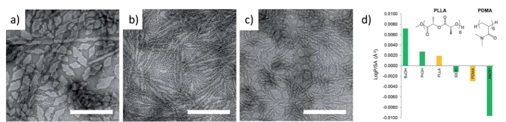
demonstrate that the hydrophobicity of the polymer can be correlated to the optimal hydrophobicity of the solvent in order to promote unimer solubility and allow access to well-dened constructs. Interestingly, the calculated logPoct values revealed that ethanol more closely resembled the hydrophobicity of PDMA compared to n-propanol, n-butanol, and methanol. This is in contrast to predictions made using the Hildebrand system, where the solubility parameters of PDMA (25.4) and the alcohols used (ethanol (26.5),n-propanol (24.6), andn-butanol (23.2))52predict
thatn-propanol would be the optimum solvent for PDMA. The results from the logPoctanalysis of our polymers were initially tested by investigating the self-assembly of PDMA600-b -PLLA48 (block ratio 12.5 : 1) in a range of alcoholic solvents. Assembly was performed in ethanol,n-propanol andn-butanol at 65C for 18 h followed by slow cooling to room temperature (analogous to the conditions used in our previous CDSA
studies).35 Consistent with logP
oct analysis, TEM imaging revealed that more well-dened 2D platelets and faceted lamellae were obtained from ethanol, whereas elongated or ill-dened structures were observed inn-propanol andn-butanol (Fig. 1). This conrmed ethanol as the optimum solvent for use in further investigations of well-dened 2D nanostructures and highlights the potential utility of logPoct as an indicator of solubility parameters for self-assembly.
Optimizing the conditions for self-assembly
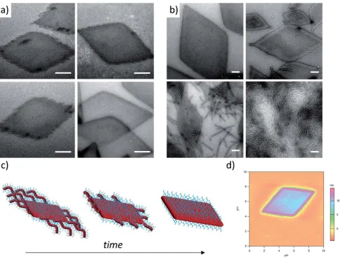
In order to enhance 2D particle formation, we theorised that increasing unimer solubility would reduce the dispersity of the assemblies. Our initial investigations into alternative solvents using logPoctanalysis demonstrated a poorer solubility withn -propanol andn-butanol, as shown previously, and methanol was found to fully solubilise the unimers (thus no structures were formed). Hence, we investigated the effect of elevated tempera-ture and prolonged heating in ethanol to increase the solubility of the unimers prior to assembly. Indeed, at longer heating times, kinetic studies at 90 C revealed increasingly well-dened dia-mond platelets (Fig. 2a) for the largest corona–core ratio (20 : 1) of up to 10mm in length andca.15 nm thick (Fig. 2d and S4†), where 8 h was determined to be the optimum time required to achieve consistently reproducible smooth diamond-shaped platelets. These structures are similar to those observed for PLLA single crystals where “lozenge” shaped crystals are re-ported.53,54 The concentration dependent assembly of the
dia-mond platelets showed no discernible change in morphology, particle dispersity or size at concentrations up to 25 mg mL1. Notably, all of the observed diamonds were consistently larger than those formed at 65 C due to the elevated dissolution temperatures reducing the number of crystalline nuclei, thus producing a smaller number of larger structures. Indeed, extending the heating time further resulted in more platelet structures, even for the smaller corona–core ratios (Fig. S5†).
Exploring the effect of polymer composition on self-assembly
[image:5.595.42.291.63.157.2]To further expand the scope of our investigation and determine how the solubility of the coronal block in ethanol affects the self-assembly process, a range of PDMA : PLLA block ratios were
Table 1 Characterization of block copolymers PDMAn-b-PLLAm
Mna
(kg mol1) ĐMa m:nb
Hydrophobic wt%c
PDMA1000-b-PLLA48 122.2 1.10 20 : 1 6.9 PDMA600-b-PLLA48 74.1 1.06 12.5 : 1 11.0 PDMA250-b-PLLA48 41.5 1.05 5 : 1 22.8 PDMA150-b-PLLA48 28.2 1.05 3 : 1 33.0 PDMA250-b-PLLA25 36.3 1.17 10 : 1 13.9 PDMA130-b-PLLA25 25.0 1.10 5 : 1 23.7
aApparent values based on SEC measurements.bRatio of degrees of
polymerization calculated from 1H NMR integration. cWeight
percentages calculated from1H NMR integration. Note that all PLLA wt% values lie within the previously identied region to undergo CDSA processes which yield cylindrical micelles.40
Fig. 1 TEM micrographs of corona–core ratio 12.5 : 1 PDMA600-b-PLLA48self-assembled in (a) ethanol, (b)n-propanol and (c)n-butanol at
65C for 18 h and cooled to room temperature. All samples were stained with uranyl acetate. Scale bar¼1mm. (d) Structure of hexameric models based on polylactide (PLLA) and poly(N,N-dimethylacrylamide) (PDMA) and logPocthydrophobicity calculations compared to methanol (MeOH),
ethanol (EtOH), propanol (PrOH), and butanol (BuOH). A similar trend was noted with oligomeric models composed of octamers. PLLA incorporated a MeO initiator with an OH endgroup. PDMA was hydrogen terminated (see Fig. S3†).
This journal is © The Royal Society of Chemistry 2017 Chem. Sci., 2017,8, 4223–4230 |4225
Edge Article Chemical Science
Open Access Article. Published on 13 April 2017. Downloaded on 19/10/2017 09:47:22.
This article is licensed under a
Creative Commons Attribution 3.0 Unported Licence.
[image:5.595.48.550.550.671.2]synthesized (Table 1). Decreasing the PDMA block length to give 12.5 : 1 and 5 : 1 block ratios (using a PLLA48 core block) resulted in mixed phases of structures primarily diamond in shape, with clear dispersity in size, and evidence of elongated ends and cylindrical micelles (Fig. 2b). In comparison, purely cylindrical structures were obtained from the lowest PDMA block length (3 : 1), which suggests that under these conditions, the crossover composition31has been reached in this system.
Similar observations (Fig. S6†) were made during the assembly of a second PLLA block which had a lower DP (PDMA250-b -PLLA25) but was more similar to the PLLA block lengths previ-ously reported by our groups (where no evidence of 2D struc-tures was observed).40These observations are in stark contrast
to the widely reported PFS system, where elongated structures are formed when the corona-forming block is much larger than the core-forming block24 to accommodate the large volume
occupied by the corona chains.55
Characterization of the assemblies
To further investigate the dimensions of specic 2D diamond-shaped nanostructures in solution, small angle X-ray scat-tering (SAXS) analysis was performed.56 It has been
demon-strated that at intermediateqvalues, the scattering intensityI(q) is proportional toqDwithDbeing the fractal exponent of the scattering objects, where dispersed plate-like objects have aD value of 2 while aggregates or folded structures have typicalD values between 3 and 4.57Upon examination, the 20 : 1 corona–
core ratio platelets (Fig. 2a) exhibit a slope of2 for interme-diateqvalues (Fig. S7a†), which conrms the presence of one-dimensional objects as observed by TEM. The early stage of a plateau is observable at lowqvalues,58with a repeat distance
which correlates closely with the platelet sizes observed by TEM analysis. The slope observed at lowqvalues is close to3, which suggests that some plates may have stacked together during analysis. The Guinier plot (Fig. S7b†) forat particles allows the determination of the thicknesssfrom the slopeRs2of the linear Fig. 2 (a) TEM micrographs of corona–core ratio 20 : 1 PDMA1000-b-PLLA48self-assembled in ethanol at 90C for 2 h (top left), 4 h (top right),
6 h (bottom left) and 8 h (bottom right) before cooling to room temperature. (b) TEM micrographs of a series of PDMAm-b-PLLA48block
copolymers of corona–core ratios of 20 : 1 (m¼1000, top left), 12.5 : 1 (m¼600, top right), 5 : 1 (m¼250, bottom left), and 3 : 1 (m¼150, bottom right). Samples were self-assembled in ethanol at 90C for 8 h and cooled to room temperature. All samples were stained with uranyl acetate. Scale bar¼1mm. (c) Schematic of diamond platelet formation kinetics. (d) AFM of diamond platelet assembled from 20 : 1 corona–core ratio diblock copolymer.
Chemical Science Edge Article
Open Access Article. Published on 13 April 2017. Downloaded on 19/10/2017 09:47:22.
This article is licensed under a
Creative Commons Attribution 3.0 Unported Licence.
[image:6.595.57.542.47.413.2]region with the following equation:s2¼12Rs2withRsbeing
the one-dimensional radius of gyration taken from the center of the platelet perpendicular to the face.59A thickness just below
2 nm is found for the 20 : 1 corona–core ratio PDMA1000-b -PLLA48platelets. It is expected this thickness mainly relates to the crystalline block as the scattering length density contrast between the solvent and the two blocks is much higher for the crystallized polylactide block than for the amorphous and solvated DMA block.
Selected area electron diffraction (SAED) was also performed on the diamond platelets, formed from the 20 : 1 corona–core ratio diblock copolymer, which conrmed the crystalline nature of the diamonds (Fig. S8†) in addition to wide-angle X-ray scattering (WAXS) analysis (Fig. S9†). The SAED patterns are consistent with the orthorhombic unit cell previously reported for PLLA (with reciprocal lattice parameters;a*¼0.935 nm1,
b* ¼ 1.626 nm1, g*¼ 90)44 and, along with the diamond
shape of the platelets, indicate the {110} growth plane. Furthermore, taking into consideration the ber repeat distance and molecular weights, we propose that the chain-folding occurs at lamellar surfaces of single crystals. The highly crystalline nature of the assembly also suggests the PLLA component crystallizes in a structure essentially identical to that expected for PLLA on its own.54Although various conned
crystal micelles in selective solvents for the amorphous block have been investigated extensively, the formation of well-dened block copolymer crystals is rarely reported.24,26,60–64
Understanding the assembly process
Unlike other systems that utilize CDSA, which rely on solvent quality for the core-forming block,21 the formation of these
bers and 2D nanostructures appears to be governed by the
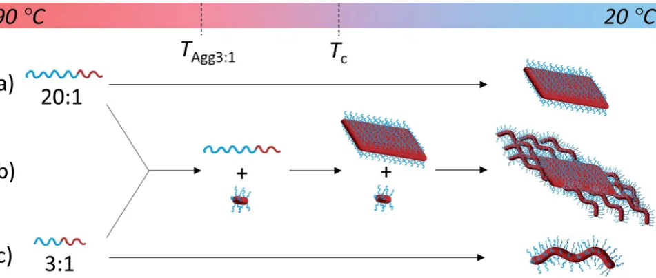
Scheme 2 Solvation-driven shape selectivity mechanism (using an arbitrary scale to represent sequential processes on cooling from 90C to 20C) for PDMAm-b-PLLA48block copolymers of block ratios (a) 20 : 1 (platelet-forming,m¼1000); (b) a mixture of 20 : 1 and 3 : 1 and (c) 3 : 1
(cylinder-forming,m¼150), whereTAgg3:1represents aggregation of the 3 : 1 block ratio cylinder-forming block copolymer.
Fig. 3 TEM micrographs of PDMAm-b-PLLA48blends of block ratios 20 : 1 (m¼1000) and 3 : 1 (m¼150), at blending ratios of (a) 25 : 75, (b)
50 : 50 and (c) 75 : 25, self-assembled in ethanol at 90C for 8 h and cooled to room temperature. Samples were stained with uranyl acetate. Scale bar¼1mm.
This journal is © The Royal Society of Chemistry 2017 Chem. Sci., 2017,8, 4223–4230 |4227
Edge Article Chemical Science
Open Access Article. Published on 13 April 2017. Downloaded on 19/10/2017 09:47:22.
This article is licensed under a
Creative Commons Attribution 3.0 Unported Licence.
[image:7.595.61.537.49.251.2]interplay between the crystallization of the PLLA core and the solubility of the corona block. On cooling, block copolymers that form unimers above the crystallization temperature of the PLLA block favor crystallization, thus reducing crystal defects and ultimately crystallizing similarly to PLLA homopolymers to form 2D diamond plates (Scheme 2a). In contrast, block copolymers that are less soluble above the crystallization temperature of the PLLA block form aggregates that undergo a crystallization event with epitaxial growth through a unimer exchange process akin to the well-established CDSA principle (Scheme 2c). To provide further evidence for this mechanism, we increased the solubility of the PDMA corona block of a cylinder-forming block copolymer (3 : 1 block ratio) by adding a single acid group to the chain end. Assembly under the same conditions resulted in a change in morphology from purebers towards a diamond platelet phase (Fig. S10†).
Exploring the versatility of this approach
To exploit this concept in creating complex nanostructures, and inspired by recent work in block copolymer blending,65–68we
explored the resultant assembly of mixtures of the two block copolymer compositions (Scheme 2b, Fig. 3). We postulated that blending different ratios of platelet-forming block copol-ymer (20 : 1 block ratio, PDMA1000-b-PLLA48) and cylinder-forming block copolymer (3 : 1 block ratio, PDMA150-b-PLLA48) and self-assembling in ethanol for 8 h at 90C followed by cooling to room temperature, would leadrst to assembly of the platelet-forming block copolymers which, in turn, would act as a seed for the aggregated cylinder-forming unimers to undergo epitaxial growth. Satisfyingly, the resultant assemblies did indeed exhibit a diamond center withbers attached parallel to the long axis of the diamond. While a small number of cylinders are observed in solution (presumably from unavoidable self-nucleation events), bers on the same side of the diamond grow unidirectionally and are quite uniform in length (Fig. S11†).
Conclusions
We have demonstrated a simple, single component solution-phase methodology that can be used as an alternative to the commonly applied surface growth approach for block copol-ymer single crystal preparation, greatly simplifying access to and the design of well-dened 2D organic nanomaterials. It is proposed that these advances will enable this eld to fully investigate the potential for these unique and interesting materials towards mimicking the success of their inorganic analogues.
We have further simplied synthetic access to hierarchical nanostructures by demonstrating how logPoctanalysis can be used to predict optimal solvents for CDSA processes to avoid laborious screening methods in solvent selection, a process that could, with further study, yield signicant insights into devel-oping methods to predict solvent systems to direct CDSA. Within this, the importance of solubility in obtaining novel structures has been highlighted, where two factors that
inuence the solubility of the copolymer were considered; the quality of the solvent for the corona block and the ratio of block lengths. In contrast to previous reports of platelet nano-particles,11,69diamond-shaped platelets were formed with good
solvent quality for the corona block and large corona–core ratios, while more elongated and less dened structures were formed with poorer solvent quality and smaller block ratios. As the polymer becomes less soluble (corona chain length decreases or the solvent quality becomes worse), there is not adequate time for the PLLA chains to adopt a preferred crystal conformation thus resulting in less dened or elongated structures. We propose that the ability of the corona block to solubilize, and thus stabilize, the block copolymer in solution allows the PLLA block to crystallize to a greater extent to yield diamonds which have the appearance of defect-free plates.
Given the high interest in 2D inorganic materials, the ability to readily access and control the assembly of polymers into 2D organic platelets through a simple assembly process provides a platform to develop a range of new materials. Moreover, given their well-dened size, morphology and high stability, applica-tions within nanocomposites, thermosets and platforms for nanoparticle delivery vehicles will be of high interest.
Acknowledgements
The authors would like to thank the University of Warwick, Materials GRP, EPSRC, The Royal Society and ERC for funding.
Notes and references
1 Y. Mai and A. Eisenberg, Chem. Soc. Rev., 2012,41, 5969– 5985.
2 N. J. Warren and S. P. Armes,J. Am. Chem. Soc., 2014,136, 10174–10185.
3 A. Blanazs, S. P. Armes and A. J. Ryan, Macromol. Rapid Commun., 2009,30, 267–277.
4 R. M. Van Horn, J. X. Zheng, H.-J. Sun, M.-S. Hsiao, W.-B. Zhang, X.-H. Dong, J. Xu, E. L. Thomas, B. Lotz and S. Z. Cheng,Macromolecules, 2010,43, 6113–6119.
5 P. Yang and Y. Han,Macromol. Rapid Commun., 2009,30, 1509–1514.
6 W. Huang, C. Luo, H. Wang and Y. Han,Polym. Int., 2010,59, 1064–1070.
7 E. D. Gomez, T. J. Rappl, V. Agarwal, A. Bose, M. Schmutz, C. M. Marques and N. P. Balsara,Macromolecules, 2005,38, 3567–3570.
8 R. M. Erb, R. Libanori, N. Rothfuchs and A. R. Studart, Science, 2012,335, 199–204.
9 L. J. Bonderer, A. R. Studart and L. J. Gauckler,Science, 2008,
319, 1069–1073.
10 D. Gournis and G. Floudas,Chem. Mater., 2004, 16, 1686– 1692.
11 C. Sinturel, M. Vayer, R. Erre and H. Amenitsch,Eur. Polym. J., 2009,45, 2505–2512.
12 B. Yu, X. Jiang and J. Yin,Chem. Commun., 2013,49, 603–605. 13 B. Li, B. Wang, R. C. Ferrier Jr and C. Y. Li,Macromolecules,
2009,42, 9394–9399.
Chemical Science Edge Article
Open Access Article. Published on 13 April 2017. Downloaded on 19/10/2017 09:47:22.
This article is licensed under a
Creative Commons Attribution 3.0 Unported Licence.
14 J. Wang, W. Zhu, B. Peng and Y. Chen,Polymer, 2013,54, 6760–6767.
15 B. Dong, T. Zhou, H. Zhang and C. Y. Li,ACS Nano, 2013,7, 5192–5198.
16 J. J. Crassous, P. Schurtenberger, M. Ballauff and A. M. Mihut,Polymer, 2015,62, A1–A13.
17 S. K. Patra, R. Ahmed, G. R. Whittell, D. J. Lunn, E. L. Dunphy, M. A. Winnik and I. Manners,J. Am. Chem. Soc., 2011,133, 8842–8845.
18 J. Schmelz, A. E. Schedl, C. Steinlein, I. Manners and H. Schmalz,J. Am. Chem. Soc., 2012,134, 14217–14225. 19 X. Wang, G. Guerin, H. Wang, Y. Wang, I. Manners and
M. A. Winnik,Science, 2007,317, 644–647.
20 A. Nazemi, C. E. Boott, D. J. Lunn, J. Gwyther,
D. W. Hayward, R. M. Richardson, M. A. Winnik and I. Manners,J. Am. Chem. Soc., 2016,138, 4484–4493. 21 S. F. Mohd Yusoff, M.-S. Hsiao, F. H. Schacher, M. A. Winnik
and I. Manners,Macromolecules, 2012,45, 3883–3891. 22 A. Presa Soto, J. B. Gilroy, M. A. Winnik and I. Manners,
Angew. Chem., Int. Ed., 2010,49, 8220–8223.
23 G. Molev, Y. Lu, K. S. Kim, I. C. Majdalani, G. Guerin, S. Petrov, G. Walker, I. Manners and M. A. Winnik, Macromolecules, 2014,47, 2604–2615.
24 L. Cao, I. Manners and M. A. Winnik,Macromolecules, 2002,
35, 8258–8260.
25 Y.-J. Kim, C.-H. Cho, K. Paek, M. Jo, M.-k. Park, N.-E. Lee, Y.-j. Kim, B. J. Kim and E. Lee, J. Am. Chem. Soc., 2014,
136, 2767–2774.
26 M. Su, H. Huang, X. Ma, Q. Wang and Z. Su,Macromol. Rapid Commun., 2013,34, 1067–1071.
27 W. Zhu, B. Peng, J. Wang, K. Zhang, L. Liu and Y. Chen, Macromol. Biosci., 2014,14, 1764–1770.
28 H. Qi, T. Zhou, S. Mei, X. Chen and C. Y. Li,ACS Macro Lett., 2016,5, 651–655.
29 B. Yu, X. Jiang and J. Yin,Macromolecules, 2014,47, 4761– 4768.
30 H. Qiu, Y. Gao, C. E. Boott, O. E. Gould, R. L. Harniman, M. J. Miles, S. E. Webb, M. A. Winnik and I. Manners, Science, 2016,352, 697–701.
31 Z. M. Hudson, C. E. Boott, M. E. Robinson, P. A. Rupar, M. A. Winnik and I. Manners,Nat. Chem., 2014,6, 893–898. 32 G. Cambridge, G. Guerin, I. Manners and M. A. Winnik,
Macromol. Rapid Commun., 2010,31, 934–938.
33 G. Rizis, T. G. van de Ven and A. Eisenberg,Angew. Chem., Int. Ed., 2014,53, 9000–9003.
34 G. Rizis, T. G. van de Ven and A. Eisenberg,ACS Nano, 2015,
9, 3627–3640.
35 N. Petzetakis, A. P. Dove and R. K. O'Reilly,Chem. Sci., 2011,
2, 955–960.
36 N. Petzetakis, D. Walker, A. P. Dove and R. K. O'Reilly,So Matter, 2012,8, 7408–7414.
37 L. Sun, A. Pitto-Barry, N. Kirby, T. L. Schiller, A. M. Sanchez, M. A. Dyson, J. Sloan, N. R. Wilson, R. K. O'Reilly and A. P. Dove,Nat. Commun., 2014,5, 5746–5754.
38 J. M. Becker, R. J. Pounder and A. P. Dove,Macromol. Rapid Commun., 2010,31, 1923–1937.
39 A. Pitto-Barry, N. Kirby, A. P. Dove and R. K. O'Reilly,Polym. Chem., 2014,5, 1427–1436.
40 L. Sun, N. Petzetakis, A. Pitto-Barry, T. L. Schiller, N. Kirby, D. J. Keddie, B. J. Boyd, R. K. O'Reilly and A. P. Dove, Macromolecules, 2013,46, 9074–9082.
41 M. Elsabahy and K. L. Wooley, Chem. Soc. Rev., 2012,41, 2545–2561.
42 Y. Geng, P. Dalhaimer, S. Cai, R. Tsai, M. Tewari, T. Minko and D. E. Discher,Nat. Nanotechnol., 2007,2, 249–255. 43 N. Daum, C. Tscheka, A. Neumeyer and M. Schneider,Wiley
Interdiscip. Rev.: Nanomed. Nanobiotechnol., 2012,4, 52–65. 44 S. E. Gratton, P. A. Ropp, P. D. Pohlhaus, J. C. Lu,
V. J. Madden, M. E. Napier and J. M. DeSimone,Proc. Natl. Acad. Sci. U. S. A., 2008,105, 11613–11618.
45 J. A. Champion and S. Mitragotri,Proc. Natl. Acad. Sci. U. S. A., 2006,103, 4930–4934.
46 S. Wilhelm, A. J. Tavares, Q. Dai, S. Ohta, J. Audet, H. F. Dvorak and W. C. Chan, Nat. Rev. Mater., 2016, 1, 16014.
47 E. Hinde, K. Thammasiraphop, H. T. Duong, J. Yeow, B. Karagoz, C. Boyer, J. J. Gooding and K. Gaus, Nat. Nanotechnol., 2016,12, 81–89.
48 D. Dakshinamoorthy, A. K. Weinstock, K. Damodaran, D. F. Iwig and R. T. Mathers,ChemSusChem, 2014,7, 2923– 2929.
49 A. J. Magenau, J. A. Richards, M. A. Pasquinelli, D. A. Savin and R. T. Mathers,Macromolecules, 2015,48, 7230–7236. 50 J. Waggel and R. T. Mathers,RSC Adv., 2016,6, 62884–62889.
51 E. Yildirim, D. Dakshinamoorthy, M. J. Peretic,
M. A. Pasquinelli and R. T. Mathers,Macromolecules, 2016,
49, 7868–7876.
52 J. E. Mark, Physical Properties of Polymers Handbook, Springer, 2007.
53 Y.-W. Chiang, Y.-Y. Hu, J.-N. Li, S.-H. Huang and S.-W. Kuo, Macromolecules, 2015,48, 8526–8533.
54 T. Iwata and Y. Doi,Macromolecules, 1998,31, 2461–2467. 55 G. Cambridge, M. J. Gonzalez-Alvarez, G. Guerin, I. Manners
and M. A. Winnik,Macromolecules, 2015,48, 707–716. 56 M. J. Hollamby,Phys. Chem. Chem. Phys., 2013,15, 10566–
10579.
57 E. M. Milner, N. T. Skipper, C. A. Howard, M. S. Shaffer, D. J. Buckley, K. A. Rahnejat, P. L. Cullen, R. K. Heenan, P. Lindner and R. Schweins, J. Am. Chem. Soc., 2012,134, 8302–8305.
58 C. Perry, P. H´ebraud, V. Gernigon, C. Brochon, A. Lapp, P. Lindner and G. Schlatter,SoMatter, 2011,7, 3502–3512. 59 E. K. Lin and A. P. Gast, Macromolecules, 1996,29, 4432–
4441.
60 A. P. Gast, P. K. Vinson and K. A. Cogan-Farinas, Macromolecules, 1993,26, 1774–1776.
61 M.-S. Hsiao, J. X. Zheng, S. Leng, R. M. Van Horn, R. P. Quirk, E. L. Thomas, H.-L. Chen, B. S. Hsiao, L. Rong, B. Lotz and S. Z. Cheng,Macromolecules, 2008,41, 8114–8123.
62 M.-S. Hsiao, W. Y. Chen, J. X. Zheng, R. M. Van Horn, R. P. Quirk, D. A. Ivanov, E. L. Thomas, B. Lotz and S. Z. Cheng,Macromolecules, 2008,41, 4794–4801.
This journal is © The Royal Society of Chemistry 2017 Chem. Sci., 2017,8, 4223–4230 | 4229
Edge Article Chemical Science
Open Access Article. Published on 13 April 2017. Downloaded on 19/10/2017 09:47:22.
This article is licensed under a
Creative Commons Attribution 3.0 Unported Licence.
63 M.-S. Hsiao, J. X. Zheng, R. M. Van Horn, R. P. Quirk, E. L. Thomas, H.-L. Chen, B. Lotz and S. Z. Cheng, Macromolecules, 2009,42, 8343–8352.
64 J. X. Zheng, H. Xiong, W. Y. Chen, K. Lee, R. M. Van Horn, R. P. Quirk, B. Lotz, E. L. Thomas, A.-C. Shi and S. Z. D. Cheng,Macromolecules, 2006,39, 641–650.
65 D. B. Wright, J. P. Patterson, A. Pitto-Barry, A. Lu, N. Kirby, N. C. Gianneschi, C. Chassenieux, O. Colombani and R. K. O'Reilly,Macromolecules, 2015,48, 6516–6522.
66 D. B. Wright, J. P. Patterson, N. C. Gianneschi, C. Chassenieux, O. Colombani and R. K. O'Reilly, Polym. Chem., 2016,7, 1577–1583.
67 J. Zhu, S. Zhang, K. Zhang, X. Wang, J. W. Mays, K. L. Wooley and D. J. Pochan,Nat. Commun., 2013,4, 2294.
68 Y. Chen, K. Zhang, X. Wang, F. Zhang, J. Zhu, J. W. Mays, K. L. Wooley and D. J. Pochan, Macromolecules, 2015,48, 5621–5631.
69 T. Vilgis and A. Halperin,Macromolecules, 1991, 24, 2090– 2095.
Chemical Science Edge Article
Open Access Article. Published on 13 April 2017. Downloaded on 19/10/2017 09:47:22.
This article is licensed under a
Creative Commons Attribution 3.0 Unported Licence.