ISSN Online: 2158-2750 ISSN Print: 2158-2742
DOI: 10.4236/ajps.2018.96085 May 17, 2018 1124 American Journal of Plant Sciences
Detection of Sugar-Regulated Gene Expression
and Signaling in Suspension-Cultured Rice Cells
Shin-Lon Ho
Department of Agronomy, National Chiayi University, Chiayi, Taiwan
Abstract
To better understand the mechanism of sugar signaling in rice cell, the sus-pension-cultured rice cells were transferred from sucrose-containing (+S) to sucrose-free (−S) of MS culture medium, we found that ribosomal RNAs (rRNAs) were degraded progressively. This suggests that carbon, nitrogen, and phosphate were recycled in this process and the reduction in cellular rRNAs might lead to decreased translation to save energy in response to sugar starvation. Differential screening revealed that two groups of genes, sug-ar-starvation-repressed (SSR) and sugar-starvation-activated (SSA) genes, were regulated by sugar in an opposing manner. Northern-blot analysis showed that two major hybridization signals of 0.8 and 1.9 kb were induced strongly under sugar starvation. The two populations of genes corresponded with homologs of α-amylases (1.9 kb) and the glycine-rich proteins (GRPs) gene family (0.8 kb), and all were SSA genes. Expression of GRP genes was strongly induced in sugar-starved cells, which suggests that GRPs may help to protect cells against nutritional stress. Treatment of +S and –S cells with the protein kinase (PK) inhibitor staurosporine (St) and the serine/theronine phosphoprotein phosphatases 1 (PP1) and 2A (PP2A) inhibitor okadaic acid (OA) revealed that PP1 and PP2A (PPs) might be involved in increasing SSR gene expression in +S cells, and that activation of the majority of the SSA genes in −S cells might be due to PKs activity. These results suggested that PKs and PPs might be involved in the sugar regulation of SSR and SSA gene expression. An in-gel PK activity assay demonstrated that the activity of two classes of PKs (50 and 66 kDa) may be induced rapidly after transfer of +S cells to −S medium. Following transfer of −S cells to +S medium, a novel class of 38 kDa PK was induced rapidly and showed high activity. The 38 kDa PK might play a role in sugar sensing, and the 50 and 66 kDa PKs might play roles in signal sensing under sugar starvation in rice cells. These results pro-vide valuable information on three classes of protein kinases that might play key roles in sugar sensing and signaling in rice.
How to cite this paper: Ho, S.-L. (2018) Detection of Sugar-Regulated Gene Expres-sion and Signaling in SuspenExpres-sion-Cultured Rice Cells. American Journal of Plant Sciences, 9, 1124-1142.
https://doi.org/10.4236/ajps.2018.96085
Received: April 4, 2018 Accepted: May 14, 2018 Published: May 17, 2018
Copyright © 2018 by author and Scientific Research Publishing Inc. This work is licensed under the Creative Commons Attribution International License (CC BY 4.0).
http://creativecommons.org/licenses/by/4.0/
DOI: 10.4236/ajps.2018.96085 1125 American Journal of Plant Sciences
Keywords
Suspension-Cultured Rice Cells, Glycine-Rich Proteins, Sugar-Starvation Repressed, Sugar-Starvation Activated, Protein Kinases, Phosphoprotein Phosphatases
1. Introduction
Carbohydrates are important nutrients in metabolism and important structural constituents in plants. Sugars, which are the basic units of carbohydrates, can serve as an energy source or a synthetic building block. Sugar availability may determine the direction of cellular metabolism and developmental responses [1] [2]. Many studies indicate that the expression of a variety of genes is regulated by sugar. In rice suspension-cultured cells, two sets of genes, namely growth-related and stress-related genes, are up- and down-regulated, respectively, by sugars [3]. In general, sugars favor the expression of genes in connection with the biosyn-thesis, respiration, and storage of reserves, and conversely repress the expression of genes associated with photosynthesis and reserve mobilization [4] [5]. For example, sugars positively regulate the expression of genes that encode storage proteins, such as patatin in potato and sporamin in sweet potato [6] [7]. Ac-cumulation of the mRNAs that encode proteins associated with starch biosyn-thesis, such as ADP glucose pyrophosphorylase, is also dependent on sugars [8]. In contrast, a variety of genes involved in key metabolic processes are ne-gatively regulated by sugars, such as α-amylase in rice [9] [10], and the ribulose 1,5-bisphosphate carboxylase/oxygenase small subunit (rbcS) and chlorophyll a/b binding protein (Cab) in maize protoplasts and suspension-cultured Chenopo-dium cells [11] [12]. A GeneChip analysis detected transcriptional up-regulation of 343 genes in sugar-starved suspension-cultured Arabidopsis cells [13]. These genes are involved in the recycling of cellular components and nutrient sca-venging, in a variety of defense and stress-response pathways, with specific pro-tein kinases and transcription factors regulating these processes.
DOI: 10.4236/ajps.2018.96085 1126 American Journal of Plant Sciences Many studies indicate that hexokinase acts as the primary sugar sensor [1] [20] [21] [22] [23] [24]. In Arabidopsis, antisense suppression and overexpres-sion of AtHXK1 resulted in insensitivity and hypersensitivity to exogenous glu-cose, respectively [20]. Other sugar-sensing pathways in plants have been pro-posed, such as hexose-dependent but hexokinase-independent, and su-crose-dependent pathways [21] [25] [26] [27] [28] [29]. Moreover, a gene (SnRK1) that encodes the yeast SNF1 homolog was cloned from potato and shown to be required for activation of sucrose synthase gene expression [30]. The expression of antisense SnRK1 in wheat embryos represses the activity of an α-amylase gene promoter, which suggests that SnRK1 is required for activation of glucose-repressing genes [31]. Rice SnRK1A functions upstream of the inte-raction between the transcription factor MYBS1 and a sugar response complex in the αAmy3 promoter to relieve glucose repression [32]. These results suggest that SnRK1 plays a role in sugar signal reception. An additional molecule in-volved in sugar signaling is trehalose 6-phosphate (T6P), which is generated from glucose 6-phosphate and UDP-glucose by trehalose 6-phosphate synthase (TPS) [33]. Overexpression of AtTPS1 in Arabidopsis results in insensitivity to glucose during seed germination [34], and increased quantities of T6P represses SnRK1 activity [35], which suggests that T6P and SnRK1 play contrasting roles in sugar responses.
In this study, we quantified the effect of sugar on cellular transcripts, and ob-served a high proportion of GRP homologs induced in suspension-cultured rice cells under sugar starvation. We showed that the protein kinase (PK) inhibitor staurosporine (St) and protein phosphatase (PP) inhibitor okadaic acid (OA) differentially affected the expression of genes subject to sugar-mediated regula-tion. Notably, addition or withdrawal of sugar from the culture medium resulted in rapid and transient activation of the specific activity of 38, 50, and 66 kDa PKs, which indicates that PKs may perform important roles in sugar sensing in rice.
2. Materials and Methods
2.1. Plant Material
The rice cultivar Oryza sativa L. cv Tainung 67 was used. Immature seeds were dehulled, sterilized with 2.4% NaOCl for 1 h, washed thoroughly with sterile water, and placed on N6D agar medium for callus induction. After 1 month, the callus derived from scutella was transferred to liquid Murashige and Skoog (MS) medium [36] supplemented with 3% sucrose and 10 µM 2,4-Dichlorophenoxyacetic
acid (2, 4-D, a synthetic plant hormone auxin, which is necessary for rice cell di-vision) to establish a suspension cell culture. Cells were cultured on a reciprocal shaker at 120 rpm and incubated at 26˚C in the dark.
2.2. Differential Screening of cDNA Library
supple-DOI: 10.4236/ajps.2018.96085 1127 American Journal of Plant Sciences mented with sucrose (+S) and cultured for 72 h, then the cells were transferred to sucrose-free (−S) medium for 4 h to isolate the candidate genes in response to sugar signaling in earlier phase. The cells were harvested and total RNA was purified. Poly (A)+ RNA was purified from the total RNA using an oligo (dT)-cellulose spin column. The Poly (A)+ RNA was used to construct a cDNA library with the λ gt22A vector and the SuperScript™ II reverse transcriptase cDNA library construction system (Invitrogen, Carlsbad, CA, USA). The pool of 32P-labeled single-stranded cDNA probes were prepared from Poly (A)+ RNA
derived from 24 h of +S or –S cells, respectively, using an oligo (dT) primer and SuperScriptTM II reverse transcriptase. Duplicated filters prepared from high-density platings of the cDNA library were differentially screened with the differential cDNA probes. The phage plaques that showed hybridization signals strongly associated with the cDNA probes from 24 h −S cells and weakly asso-ciated with the cDNA probes from 24 h +S cells were isolated. The cDNAs in-serted in the λ gt22A vector were cleaved with NotI and SalI and subcloned into the NotI–SalI sites of pBluescript vectors before being subjected to sequencing analysis.
2.3. Plasmids
The inserted DNA fragments of Act, ADH2, G3PD, HSP86, and SSP2 were di-gested from the pBluescript vector with SalI and NotI. The DNA fragments of salT, OsGRP1, OsGRP2, OsGRP3, OsGRP4, and OsGRP5 were digested with EcoRI. The αAmy3 and αAmy8 gene-specific DNA fragments were prepared as described by Sheu et al. [15]. The inserted DNAs were individually isolated, la-beled with 32P, and used as probes. A DNA fragment containing the 28S, 18S, and 5.8S rDNAs was excised from the pRY18 plasmid using BamHI, labeled with α-32P, and used as a probe to evaluate the quantity of the rRNA.
2.4. Northern-Blot Analysis
Total RNA was isolated from suspension-cultured cells using the TRIzol® Rea-gent (Invitrogen). The RNA gel-blot was analyzed as described by Ho et al. [3]. Ten micrograms of total RNA was electrophoresed in 1% agarose gel containing 10 mM sodium phosphate buffer (pH 6.5), transferred to a nylon membrane, and hybridized with random-primer-labeled [α-32P]dCTP cDNA probes at 42˚C. The membranes were exposed to X-ray film at −80˚C.
2.5. In-Gel Protein Kinase Activity Assay
Rice suspension-cultured cells were harvested and ground into powder. Proteins were purified with precooled extraction buffer (50 mM Hepes·KOH [pH 7.6], 1 mM EDTA, 1 mM EGTA, 10 mM NaF, 1 mM Na3VO4, 1 mM Na3MoO4, 20 mM β-glycerophosphate, 20% glycerol, 2 mM DTT, and 2 mM PMSF), supplemented with 2 µg/mL of three different protease inhibitors, namely leupeptin, E64, and
DOI: 10.4236/ajps.2018.96085 1128 American Journal of Plant Sciences the supernatants were collected and the protein concentration was quantified using the method of Bradford [37]. The in-gel PK activity assay was conducted in accordance with the methods of Mizoguch et al. [38] and Usami et al. [39] with minor revision. In brief, 50 µg proteins were separated by 10%
SDS-PAGE with or without 0.5 mg/mL histone III-S (Sigma). After electro-phoresis, the gel was rinsed in the wash solution (20% β-propanol and 50 mM Tris-HCl [pH 8.0] for 1 h to remove SDS, followed by soaking with buffer containing 50 mM Tris-HCl (pH 8.0) and 5 mM β-mercaptoethanol for re-moval of β-propanol residues. The gel was denatured at 25˚C for 2 h with de-naturation solution (6 M guanidine hydrochloride, 50 mM Tris-HCl [pH 8.0] and 5 mM β-mercaptoethanol), then renatured at 4˚C for at least 16 h by neutra-lization solution (50 mM Tris-HCl [pH 8.0], 5 mM β-mercaptoethanol, and 0.04% Tween 40). For kinase activity detection, the gel was soaked in kinase reaction buffer (40 mM Hepes·KOH [pH 7.6], 2 mM DTT, 15 mM MgCl2, 30 mM ATP, 100 μCi [γ-32P] ATP, and 0.1 mM EGTA) and incubated at 25˚C for 1 h, then rinsed in stop solution (5% TCA and 1% sodium pyrophosphate) for 30 min to terminate the reaction. The gel was washed several times with sterilized water to remove the free isotope, then the gel was vacuumed dried and exposed to X-ray film.
3. Results
3.1. Sugar Modulates Ribosomal RNA Turnover
DOI: 10.4236/ajps.2018.96085 1129 American Journal of Plant Sciences
Figure 1. Increased rRNA degradation in rice suspension-cultured cells under sucrose
starvation. After incubation for 72 h in sucrose-containing (+S) medium, cells were transferred to sucrose-free (−S) medium for 72 h, and then transferred to +S medium for an additional 24 h. Total RNA was purified from the cells and agarose gel electrophoresis was conducted. (a) After electrophoresis, the gel was stained with ethidium bromide and photographed; (b) The gel blot was hybridized with a 32P-labeled rDNA probe; (c) Levels of 28S and 18S rRNAs were quantified using a PhosphorImager (Molecular Dynamics, Sunnyvale, CA, USA). The positions of 28S and 18S rRNAs are as indicated. Arrows in-dicate the position of RNA degradation products.
[image:6.595.214.534.63.491.2]DOI: 10.4236/ajps.2018.96085 1130 American Journal of Plant Sciences progressively increased after cells were transferred from –S to +S medium. These results suggest that reduction in the amount of rRNAs might be due to increased degradation rather than decreased synthesis during sucrose starvation. Given that rRNAs are functionally correlated with protein synthesis, reduction in cel-lular rRNAs content probably leads to reduction in translation to conserve energy in response to sugar starvation.
3.2. Global Gene Expression in Sugar-Starved Cells
To evaluate global gene expression in rice cells under sugar starvation, total RNA was purified from suspension-cultured rice cells treated with or without sugar and subjected to northern-blot hybridization. The total poly (A+) RNA (mRNA) was prepared from cells cultured in +S or −S medium for 24 h. The pu-rified mRNA was used as the template to synthesize the pool of [α-32P]-labeled cDNA probes. Two major hybridization signals were distinctly induced in cells under sugar-starvation treatment (Figure 2, indicated by arrows). One induc-tion signal was similar in size to that of 18S rRNA (1.9 kb), which was induced abundantly when cells were starved of sucrose for 48 - 72 h and declined rapidly 1 - 4 h after cells were transferred from −S to +S medium. The other signal was about 0.8 kb in size. The mRNA populations gradually accumulated and attained the highest amounts at 12 - 72 h after sugar starvation (lanes 10 - 14) and rapidly decreased 1 - 24 h after transfer to culture medium supplemented with sucrose (lanes 15 - 19). These results suggested that the two distinct populations of 1.9 kb and 0.8 kb mRNAs might play important roles in the response to sugar star-vation in rice cells.
[image:7.595.215.535.489.629.2]To verify the two populations of corresponding genes induced under sugar starvation, differential screening of a cDNA library prepared from rice cells sug-ar-starved for 4 h was conducted. After screening, more than 100 cDNA
Figure 2. Northern-blot analysis of global gene expression in rice suspension cells
DOI: 10.4236/ajps.2018.96085 1131 American Journal of Plant Sciences clones that showed stronger signals (spots) after hybridization with either the pool of −S or +S cDNA probes were selected (Supplementary Figure S1) and cloned into plasmid vectors. Among these cDNA clones, five clones showed strongest hybridization signals with the +S cDNA probes and a weak signal with the –S cDNA probes (defined as the sugar-starvation-repressed [SSR] genes), and 12 clones showed stronger hybridization signals with the −S cDNA probes (defined as the sugar-starvation-activated [SSA] genes). These 17 cDNA clones were partially sequenced and analyzed. As described in our previous study [3], the five SSR cDNA clones were identified as actin (Act), alcohol dehydroge-nase 2 (ADH2), glyceraldehyde-3-phosphate dehydrogedehydroge-nase (G3PD), heat shock protein 86 (HSP86) and sucrose synthase P2 (SSP2), and the corres-ponding full-length cDNA clones were obtained from the Japan Rice Genome Research Program [40]. Among the 12 SSA cDNA clones, three clones were α-amylase homologous genes (two clones were αAmy3 [XM_015794560, the predicted mRNA length in GenBank was 1834 bp], one clone was αAmy8 [XM_015795212, the predicted mRNA length was 1687 bp]), and one clone was identical to sal T (S45168, 724 bp). Unexpectedly, the other eight clones were highly homologous to the glycine-rich proteins (GRPs) gene family of unknown function, of which three identical clones were designated OsGRP1 (X54449, pre-dicted mRNA length in GenBank was 834 bp), two clones were OsGRP2 (D21281, 852 bp), and one clone each as OsGRP3 (AF010579, 713 bp), OsGRP4 (U40708, 551 bp) and OsGRP5 (AJ002893, 780 bp). The predicted mRNA lengths of all the isolated SSA clones were consistent with the two major hybridization signals de-tected by −S cDNA probes at the positions of higher molecular weight (corres-ponding to the length of αAmy3 and αAmy8) (Figure 2, thin arrow) or lower molecular weight (this position similar to the length of GRPs and salT) (Figure 2, thick arrow). These results suggest that the expression of a high proportion of α-amylase and GRP homologs was induced abundantly during sugar starvation.
DOI: 10.4236/ajps.2018.96085 1132 American Journal of Plant Sciences
Figure 3. Protein kinase and protein phosphatase inhibitors alter expression of
su-crose-starvation-repressed genes. Rice suspension cells were cultured in sucrose-containing (+S) medium for 72 h and transferred to +S or sucrose-free (−S) medium lacking an inhi-bitor, or treated with the protein kinase inhibitor staurosporine (2 μM; St), or the phos-phoprotein phosphatase 1 (PP1) and 2A (PP2A) inhibitor okadaic acid (100 nM; OA), and incubated for an additional 2 or 12 h. All media contained 0.2% dimethyl sulforide (DMSO), which was used to dissolve St and OA. Total RNA was purified and subjected to RNA gel blot analysis. Sucrose-starvation-repressed genes were used as probes. Act, actin; ADH2, alcohol hydrogenase 2; G3PD, glyceraldehydes-3-phosphate dehydrogenase; HSP86, heat shock protein 86; SSP2, sucrose synthase P-2; αAmy, α-amylase.
Figure 4. Protein kinase and protein phosphatase inhibitors alter expression of
[image:9.595.304.443.69.255.2] [image:9.595.303.440.399.639.2]DOI: 10.4236/ajps.2018.96085 1133 American Journal of Plant Sciences findings indicate that the expression of all SSA clones was sensitive to sugar content in the culture medium and was activated in −S cells, whereas activation was diminished in +S cells, and the expression patterns are all converse in those SSR genes. Therefore, the SSR and SSA genes were considered to be an ideal sample to study the regulatory mechanisms of sugar regulation pathways.
3.3. Protein Kinase and Protein Phosphatase Effects on the Sugar
Regulation of Gene Expression
In the present study, we observed that two groups of genes (SSR and SSA genes) were regulated by sugar in opposing manners. To further verify whether the ef-fects of sugar were mediated by protein phosphorylation or dephosphorylation, suspension-cultured rice cells treated with PK or PP inhibitors were subjected to northern-blot analysis. In +S cells, accumulation of mRNAs of all five SSR genes was not affected by St (Figure 3, cf. lanes 2 and 6), but was repressed by OA (Figure 3, cf. lanes 2 and 10). In −S cells, the mRNA level for all of the SSR genes was repressed by St (Figure 3, cf. lanes 4 and 8), but was not altered by OA (Figure 3, cf. lanes 4 and 12). On the basis of these results, the activity of PPs may positively regulate SSR gene expression in +S cells, whereas the activity of PKs somewhat activated expression of SSR genes in −S cells.
In +S cells, St slightly activated the expression of SSA genes, but not that of αAmy3 and αAmy8 (Figure 4, cf. lanes 2 and 6). In contrast, OA dramatically increased the expression of salT, OsGRP1, OsGRP2, and OsGRP3, and slightly activated OsGRP4 and OsGRP5. No effect of OA on αAmy3 and αAmy8 was observed (Figure 4, cf. lanes 2 and 10). In –S cells, St enhanced expression of salT but suppressed that of the other seven SSA genes (Figure 4, cf. lanes 4 and 8). Okadaic acid enhanced the expression of αAmy3, salT, OsGRP2, and OsGRP3, but suppressed αAmy8 expression. Okadaic acid had no effect on ex-pression of OsGRP1, OsGRP4, and OsGRP5 (Figure 4, cf. lanes 4 and 12). On the basis of these results, in +S cells the activities of both PKs and PPs may sup-press the exsup-pression of SSA genes, but not that of αAmy3 and αAmy8. In –S cells, the activity of some PKs may negatively regulate the expression of salT and positively regulate expression of other SSA genes. Some PPs suppressed the ex-pression of αAmy3, salT, OsGRP2, and OsGRP3, but activated the exex-pression of αAmy8, and no effect on the expression of OsGRP1, OsGRP4, and OsGRP5 was observed. These results suggest that PKs, PP1, and PP2A might be involved in the regulation of SSR and SSA gene expression by sugars.
3.4. Detection of Protein Kinase Activity Involved in Sugar
Sensing
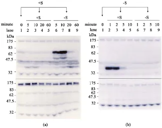
DOI: 10.4236/ajps.2018.96085 1134 American Journal of Plant Sciences cultured in +S medium were then transferred to either +S or –S medium, and cultured for an additional 5, 10, 20, and 60 min (Figure 5(a)). Similarly, the cells cultured in −S medium were then transferred to either +S or –S medium, and cultured for an additional 1, 2, 5, and 10 min (Figure 5(b)). The cells were har-vested at the specified time points and total proteins were purified for the in-gel PK activity assay by supplementation with (Figure 5(a) and Figure 5(b), upper panel) or without (Figure 5(a) and Figure 5(b), lower panel) Histone III-S in the gel as the PK substrate. We therefore could distinguish the signals of PKs ac-tivity derived from autophosphorylation of PKs (without Histone III-S in gel) (Figure 5, lower panel) or phosphorylation of the substrate by the specific PK activity (with Histone III-S in gel) (Figure 5, upper panel). After the +S cells were transferred to +S medium, no obvious difference in the specific signal of PKs activity was detected, nor in the presence or absence of Histone III-S in the gel (Figure 5(a)). However, after cells were transferred from +S to –S medium, two specific signals of PKs activity, one stronger signal located in the position of about 66 kDa and a weaker signal at about 50 kDa, were induced rapidly within 5 - 10 min, and thereafter declined quickly and were only weakly detected at 20 min (Figure 5(a), lanes 6 - 9). Moreover, four constitutive PKs activity detected
Figure 5. Activation of protein kinases in response to alteration in sucrose level. Rice
[image:11.595.212.535.352.606.2]DOI: 10.4236/ajps.2018.96085 1135 American Journal of Plant Sciences at the positions of about 30, 45, 66, and 170 kDa, irrespective of +S and –S cells and with or without Histone III-S in the gel (Figure 5), revealed that at least four classes of autophosphorylation of PKs were exhibited in rice cells. Similar to the experimental results shown in Figure 5(a), after –S cells were transferred to +S and –S medium followed by incubation for an additional 5, 10, 20, and 60 min, only a weaker signal was detected specifically at the position of about 38 kDa at 5 min after the transfer of –S cells to +S medium. This result indicates that activity of the 38 kDa PK may be induced rapidly within 5 min. We therefore shortened the incubation time points to 1, 2, 5, and 10 min after –S cells were transferred to +S and –S medium. As shown in Figure 5(b), after –S cells were transferred to +S medium, we observed that a strong signal of PKs activity was induced rapidly at the position of 38 kDa, and that no inducible signal of PKs activity was ob-served after –S cells were transferred to –S medium. Altogether, the present re-sults demonstrate that activity of two classes of PKs, of 50 and 66 kDa, were in-duced after +S rice cells were transferred to –S medium, and activity of a 38 kDa class of PK was induced after –S cells were transferred to +S medium. These re-sults indicate that the 38 kDa class of PK might play a role in sugar sensing, and that the 50 and 66 kDa classes of PKs might play roles in signal transduction of sugar starvation in rice cells.
4. Discussion
DOI: 10.4236/ajps.2018.96085 1136 American Journal of Plant Sciences and growth, autophagy, reduced protein synthesis, and strong activation of α-amylase and proteolytic enzymes [29]. Similar to the aforementioned studies, the present results show that extension of the duration of sugar starvation of rice cells was accompanied by global degradation of rRNAs and mRNAs (Figure 1), which might be intended to supplement the nitrogen and carbon sources in the cell. Moreover, two populations of mRNAs, namely those of α-amylases and GRPs, were induced and expressed abundantly during 12 - 72 h of sugar starva-tion, which suggests that α-amylases and GRPs might play important roles in the response to sugar starvation (Figure 2). The α-amylases hydrolyze starch into glucose to provide a carbon source [5]. Although GRPs form a group of struc-tural protein components in the cell walls of many higher plants [46] [47], the biological functions of GRPs in response to biotic and abiotic stresses in plant cells is still largely unknown. We observed that under the energy-limited condi-tion of sugar starvacondi-tion, a populacondi-tion of GRPs is still generated, which suggests that GRPs might play novel roles in adaptation or response to nutritional stress in plant cells. In addition, the synchronized induction and repression of GRP homologs in −S and +S cells, respectively (Figure 3 and Figure 4), might imply that a common signaling pathway is operational in sugar sensing. Given that GRP transcript levels are synergistically regulated and sensitive to sugar supply, GRPs be a useful indicator in investigations of the sugar signaling pathway.
respec-DOI: 10.4236/ajps.2018.96085 1137 American Journal of Plant Sciences tively. Interestingly, although αAmy3 and αAmy8 are homologous genes, the expression of αAmy3 and αAmy8 may be coordinately up-regulated by PKs, whereas PPs differentially down- and up-regulate the expression of αAmy3 and αAmy8, respectively. These results demonstrate that sugar signaling pathways function via a complex crosstalk between phosphorylation and dephosphoryla-tion to control expression of a variety of genes.
In signaling transduction pathways, the signaling molecules usually stimulate a rapid, transient response, and the effects can be rapidly reversed [51]. Consis-tent with this observation, the present results demonstrated that activity of two classes of PKs, of 50 and 66 kDa, was induced rapidly 5 - 10 min after transfer of +S rice cells to –S medium, and the induced signals were almost non-detectable 20 min after transfer (Figure 5(a)). Moreover, when –S cells were transferred to +S medium for 1 - 2 min, a novel class of 38 kDa PK was hypersensitive to sugar and high activity was induced immediately, but then declined quickly and al-most vanished at 5 min after transfer (Figure 5(b)). These results suggest that the 38 kDa PK class might play a role in sugar sensing, and the 50 and 66 kDa PK classes might play roles in signal sensing in response to sugar starvation in rice cells. To the best of our knowledge, no known class of PK is induced rapidly and is highly expressed in response to sugar and sugar-starvation signaling in plant cells. These results provide valuable information that indicates that 38, 50 and 66 kDa protein kinases might play key roles as sensors in sugar sensing and signaling.
5. Conclusion
Our results provided novel information that a population of glycine-rich pro-teins might play important roles in response to sugar starvation in rice cells. And a class of 38 kDa protein kinases could act as a sugar sensor, and the class of 50 and 66 kDa protein kinases might function as a sensor in response to sugar star-vation in rice cells.
Acknowledgements
This work was supported by grants from the Ministry of Science and Technology of the Republic of China (Grant Nos. MOST 105-2313-B-415-009- and MOST 106-2313-B-415-006-).
References
[1] Granot, D., David-Schwartz, R. and Kelly, G. (2013) Hexose Kinases and Their Role in Sugar-Sensing and Plant Development. Frontiers in Plant Science, 4, 44.
https://doi.org/10.3389/fpls.2013.00044
[2] Lastdrager, J., Hanson, J. and Smeekens, S. (2014) Sugar Signals and the Control of Plant Growth and Development. Journal of Experimental Botany,65, 799-807.
https://doi.org/10.1093/jxb/ert474
DOI: 10.4236/ajps.2018.96085 1138 American Journal of Plant Sciences
Signal Transduction Network and Multiple Control Mechanisms. Plant Physiology, 125, 877-890. https://doi.org/10.1104/pp.125.2.877
[4] Koch, K.E. (1996) Carbohydrate-Modulated Gene Expression in Plants. Annual Re-view of Plant Physiology and Plant Molecular Biology, 47, 509-540.
https://doi.org/10.1146/annurev.arplant.47.1.509
[5] Yu, S.M. (1999) Cellular and Genetic Responses of Plants to Sugar Starvation. Plant Physiology, 121, 687-693. https://doi.org/10.1104/pp.121.3.687
[6] Jefferson, R., Goldsbrough, A. and Bevan, M. (1990) Transcriptional Regulation of a Patatin-1 Gene in Potato. Plant Molecular Biology,14, 995-1006.
https://doi.org/10.1007/BF00019396
[7] Hattori, T., Nakagawa, S. and Nakamura, K. (1990) High Level Expression of Tu-berous Root Storage Protein Genes of Sweet Potato in Stems of Plantlets Grown in Vitro on Sucrose Medium. Plant Molecular Biology,14, 595-604.
https://doi.org/10.1007/BF00027505
[8] Müller-Röber, B.T., Kobmann, J., Hannah, L.C., Willmitzer, L. and Sonnewald, U. (1990) One of Two Different ADP-Glucose Pyrophosphorylase Genes from Potato Responds Strongly to Elevated Levels of Sucrose. Molecular Genetics and Genom-ics,224, 136-146.
[9] Yu, S.M., Kuo, Y.H., Sheu, G., Sheu, Y.J. and Liu, L.F. (1991) Metabolic Derepres-sion of α-Amylase Gene Expression in Suspension-Cultured Cells of Rice. Journal of Biological Chemistry, 266, 21131-21137.
[10] Yu, S.M., Lee, Y.C., Fang, S.C., Chan, M.T., Hwa, S.F. and Liu, L.F. (1996) Sugar Act as Signal Molecules and Osmotica to Regulate the Expression of α-Amylase Genes and Metabolic Activities in Germinating Cereal Grains. Plant Molecular Biology, 30, 1277-1289. https://doi.org/10.1007/BF00019558
[11] Krapp, A., Hofmann, B., Schäfer, C. and Stitt, M. (1993) Regulation of the Expres-sion of rbcS and Other Photosynthetic Genes by Carbohydrates: A Mechanism for the “Sink Regulation” of Photosynthesis? The Plant Journal, 3, 817-828.
https://doi.org/10.1111/j.1365-313X.1993.00817.x
[12] Sheen, J. (1990) Metabolic Repression of Transcription in Higher Plants. The Plant Cell, 2, 1027-1038. https://doi.org/10.1105/tpc.2.10.1027
[13] Contento, A.L., Ki, S.J. and Bassham, D.C. (2004) Transcriptome Profiling of the Response of Arabidopsis Suspension Culture Cells to Suc Starvation. Plant Physiol-ogy,135, 2330-2347. https://doi.org/10.1104/pp.104.044362
[14] Sheu, J.J., Jan, S.P., Lee, H.T. and Yu, S.M. (1994) Control of Transcription and mRNA Turnover as Mechanisms of Metabolic Repression of α-Amylase Gene Ex-pression. The Plant Journal,5, 655-664.
https://doi.org/10.1111/j.1365-313X.1994.00655.x
[15] Sheu, J.J., Yu, T.S., Tong, W.F. and Yu, S.M. (1996) Carbohydrate Starvation Sti-mulates Differential Expression of Rice α-Amylase Genes That Is Modulated through Complicated Transcriptional and Posttranscriptional Processes. Journal of Biological Chemistry, 271, 26998-27004. https://doi.org/10.1074/jbc.271.43.26998
[16] Chan, M.T. and Yu, S.M. (1998) The 3’-Untranslated Region of a Rice α-Amylase Gene Functions as a Sugar-Dependent mRNA Stability Determinant. Proceedings of the National Academy of Sciences of the United States of America, 95, 6543-6547. https://doi.org/10.1073/pnas.95.11.6543
DOI: 10.4236/ajps.2018.96085 1139 American Journal of Plant Sciences https://doi.org/10.1023/A:1005956104636
[18] Lu, C.A., Lim, E.K. and Yu, S.M. (1998) Sugar Response Sequence in the Promoter of a Rice α-Amylase Gene Serves as a Transcriptional Enhancer. Journal of Biologi-cal Chemistry,273, 10120-10131. https://doi.org/10.1074/jbc.273.17.10120
[19] Lu, C.A., Ho, T.H.D., Ho, S.L. and Yu, S.M. (2002) Three Novel MYB Proteins with One DNA Binding Repeat Mediate Sugar and Hormone Regulation of α-Amylase Gene Expression. The Plant Cell,14, 1963-1980. https://doi.org/10.1105/tpc.001735
[20] Jang, J.C. and Sheen, J. (1997) Sugar Sensing in Higher Plants. Trends in Plant Science, 2, 208-213. https://doi.org/10.1016/S1360-1385(97)89545-3
[21] Sheen, J., Zhou, L. and Jang, J.C. (1999) Sugars as Signaling Molecules. Current Opinion in Plant Biology,2, 410-418.
https://doi.org/10.1016/S1369-5266(99)00014-X
[22] Rolland, F., Moore, B. and Sheen, J. (2002) Sugar Sensing and Signaling in Plants.
The Plant Cell, 14,S185-S205. https://doi.org/10.1105/tpc.010455
[23] Moore, B., Zhou, L., Rolland, F., Hall, Q., Cheng, W.H., Liu, Y.X., et al. (2003) Role of the Arabidopsis Glucose Sensor HXK1 in Nutrient, Light, and Hormonal Signal-ing. Science,300, 332-336.
[24] Rolland, F., Baena-Gonzalez, E. and Sheen, J. (2006) Sugar Sensing and Signaling in Plants: Conserved and Novel Mechanisms. Annual Review of Plant Biology, 57, 675-709.
[25] Smeekens, S. and Rook, F. (1997) Sugar Sensing and Sugar-Mediated Signal Trans-duction in Plants. Plant Physiology,115, 7-13. https://doi.org/10.1104/pp.115.1.7
[26] Halford, N.G., Purcell, P.C. and Grahame, H.D. (1999) Is Hexokinase Really a Sugar Sensor in Plants? Trends in Plant Science, 4, 117-120.
https://doi.org/10.1016/S1360-1385(99)01377-1
[27] Smeekens, S. (2000) Sugar-Induced Signal Transduction in Plants. Annual Review of Plant Physiology and Plant Molecular Biology, 51, 49-81.
https://doi.org/10.1146/annurev.arplant.51.1.49
[28] Hanson, J. and Smeekens, S. (2009) Sugar Perception and Signaling—An Update.
Current Opinion in Plant Biology,12, 562-567.
https://doi.org/10.1016/j.pbi.2009.07.014
[29] Smeekens, S. and Hellmann, H.A. (2014) Sugar Sensing and Signaling in Plants.
Frontier in Plant Sciences,5, 113.
[30] Purcell, P.C., Smith, A.M. and Halhord, N.G. (1998) Antisense Expression of a Su-crose Non-Fermenting-1-Related Protein Kinase Sequence in Potato Results in De-creased Expression of Sucrose Synthase in Tubers and Loss of Sucrose-Inducibility of Sucrose Synthase Transcripts in Leaves. ThePlant Journal,14, 195-202.
https://doi.org/10.1046/j.1365-313X.1998.00108.x
[31] Laurie, S., Mckibbin, R.S. and Halford, N.G. (2003) Antisense SNF1-Related (SnRK1) Protein Kinase Gene Represses Transient Activity of an α-Amylase (α-Amy2) Gene Promoter in Cultured Wheat Embryos. Journal of Experimental Botany,54, 739-747. https://doi.org/10.1093/jxb/erg085
[32] Lu, C.A., Lin, C.C., Lee, K.W., Chen, J.L., Huang, L.F., Ho, S.L., Liu, H.J., Hsing, Y.I. and Yu, S.M. (2007) The SnRK1A Protein Kinase Plays a Key Role in Sugar Signal-ing durSignal-ing Germination and SeedlSignal-ing Growth of Rice. The Plant Cell, 19, 2484-2499. https://doi.org/10.1105/tpc.105.037887
DOI: 10.4236/ajps.2018.96085 1140 American Journal of Plant Sciences https://doi.org/10.1146/annurev.arplant.59.032607.092945
[34] Avonce, N., Leyman, B., Mascorro-Gallardo, J.O., Van Dijck, P., Thevelein, J.M. and Iturriaga, G. (2004) The Arabidopsis Trehalose-6-P Synthase AtTPS1 Gene Is a Regulator of Glucose, Abscisic Acid, and Stress Signaling. Plant Physiology, 136, 3649-3659. https://doi.org/10.1104/pp.104.052084
[35] Zhang, Y., Primavesi, L.F., Jhurreea, D., Andralojc, P.J., Mitchell, R.A., Powers, S.J., Schluepmann, H., Delatte, T., Wingler, A. and Paul, M.J. (2009) Inhibition of SNF1-Related Protein Kinase1 Activity and Regulation of Metabolic Pathways by Trehalose-6-Phosphate. Plant Physiology,149, 1860-1871.
https://doi.org/10.1104/pp.108.133934
[36] Murashige, T. and Skoog, F. (1962) A Revised Medium for Rapid Growth and Bio-assays with Tobacco Tissue Cultures. Physiologia Plantarum,15, 473-497.
https://doi.org/10.1111/j.1399-3054.1962.tb08052.x
[37] Braford, M.M. (1976) A Rapid and Sensitive Method for the Quantification of Mi-crogram Quantities of Protein Utilizing the Principle of Protein Dye Binding. Ana-lytical Biochemistry,112, 195-203.
[38] Mizoguchi, T., Gotoh, Y., Nishida, E., Yamaguchi-Shinozaki, K., Hayashida, N., Iwasaki, T., Kamada, H. and Shinozaki, K. (1994) Characterization of Two cDNAs that Encode MAP Kinase Homologues in Arabidopsis thaliana and Analysis of the Possible Role of Auxin in Activating Such Kinase Activities in Cultured Cells. The Plant Journal, 5, 111-122. https://doi.org/10.1046/j.1365-313X.1994.5010111.x
[39] Usami, S., Banno, H., Ito, Y., Nishihama, R. and Machida, Y. (1995) Cutting Acti-vates a 46-Kilodalton Protein Kinase in Plants. Proceedings of the National Acade-my of Sciences of the United States of America,92, 8660-8664.
https://doi.org/10.1073/pnas.92.19.8660
[40] Uchimiya, H., Kidou, S.I., Shimazaki, T., Aotsuka, S., Takamatsu, S., Nishi, R., Ha-shimoto, H., Matsubayashi, Y., Kidou, N., Umeda, M. and Kato, A. (1992) Random Sequencing of cDNA Libraries Reveals a Variety of Expressed Genes in Cultured Cells of Rice (Oryza sativa L.). The Plant Journal, 2, 1005-1009.
https://doi.org/10.1111/j.1365-313X.1992.01005.x
[41] Condit, C.M. and Meagher, R.B. (1987) Expression of a Gene Encoding a Gly-cine-Rich Protein in Petunia. Molecular Cell Biology, 7, 4273-4279.
https://doi.org/10.1128/MCB.7.12.4273
[42] Gómez, J., Sanchez-Martinez, D., Stiefel, R., Rigau, J., Puigdomènech, P. and Pagès, M. (1998) A Gene Induced by the Plant Hormone Abscisic Acid in Response to Water Stress Encodes a Glycine-Rich Protein. Nature, 334, 262-264.
https://doi.org/10.1038/334262a0
[43] De Oliveira, D.E., Seurinck, J., Inzé, D., van Montagu, M. and Botterman, J. (1990) Differential Expression of Five Arabidopsis Genes Encoding Glycine-Rich Proteins.
The Plant Cell,2, 427-436. https://doi.org/10.1105/tpc.2.5.427
[44] Fang, R.X., Pang, Z., Gao, D.M., Mang, K.Q. and Chua, N.H. (1991) cDNA Se-quence of a Virus-Inducible, Glycine-Rich Protein Gene from Rice. Plant Molecular Biology,17, 1255-1257. https://doi.org/10.1007/BF00028742
[45] Cheng, S.H., Keller, B. and Condit, C.M. (1996) Common Occurrence of Homolo-gues of Petunia Glycine-Rich Protein-1 among Plants. Plant Molecular Biology,31, 163-168. https://doi.org/10.1007/BF00020616
[46] Showalter, A.M. (1993) Structure and Function of Plant Cell Wall Proteins. The Plant Cell, 5, 9-23. https://doi.org/10.1105/tpc.5.1.9
DOI: 10.4236/ajps.2018.96085 1141 American Journal of Plant Sciences
Components of Plant Cell Walls. Cellular and Molecular Life Sciences, 58, 1430-1441. https://doi.org/10.1007/PL00000786
[48] Pego, J.V., Weisbeek, P.T. and Smeekens, S.C.M. (1999) Mannose Inhibits Arabi-dopsis Germination via a Hexokinase-Mediated Step. Plant Physiology, 119, 1017-1024. https://doi.org/10.1104/pp.119.3.1017
[49] Sheen, J. (1993) Protein Phosphatase Activity Is Required for Light-Inducible Gene Expression in Maize. TheEMBO Journal, 12, 3497-3505.
[50] Ehness, R., Ecker, M., Godt, D.E. and Roitsch, T. (1997) Glucose and Stress Inde-pendently Regulate Source and Sink Metabolism and Defense Mechanisms via Sig-nal Transduction Pathways Involving Protein Phosphorylation. The Plant Cell, 9, 1825-1841. https://doi.org/10.1105/tpc.9.10.1825
DOI: 10.4236/ajps.2018.96085 1142 American Journal of Plant Sciences
Supplementary
Figure S1. Differential screening to identify sugar-regulated genes. The cDNA library was