http://dx.doi.org/10.4236/ojo.2012.22013 Published Online June 2012 (http://www.SciRP.org/journal/ojo)
Use of Synteny Conversion in Identification of Candidate
Genes for Somitogenesis in Humans
Philip F. Giampietro1, Cathy L. Raggio2, Robert D. Blank1
1
University of Wisconsin, Madison, USA; 2Hospital for Special Surgery, New York, USA. Email: pfgiampietro@pediatrics.wisc.edu
Received March 2nd, 2012; revised April 5th, 2012; accepted May 13th, 2012
ABSTRACT
Understanding the genetic component of scoliosis in humans has relied on the assumption that spine development is conserved across species. Since evolutionary conserved genes tend to lie within synteny blocks (HSBs) and genes which are not conserved lie within evolutionary breakpoint regions (EBRs), HSB analysis may be used to determine if spine development is conserved across species. We hypothesized that vertebral patterning genes are conserved in amni-otes and their location is within stable or “syntenic” regions of chromosomes. Seventy seven patterning genes involved in Fgf, Wnt and Notch signaling pathways were analyzed to determine their location within HSBs or EBRs in the ge- nomes of several amniotic species. The human genome was divided into 1 Mbp intervals and a comparison was made to determine whether these genes were preferentially localized within HSBs or EBRs associated with rapid evolution. The results indicate that genes associated with somite development in humans are preferentially located away from the EBRs: 0.014 genes in EBRs on genome average vs. 0.030 on average in other parts of the genome (p-value = 0.01). The concentration of vertebral patterning genes in HSBs, provides evidence that developmental pathways involved in verte- bral morphogenesis are likely conserved across amniotes, consistent with their known function. These data support prior observations indicating that gene networks associated with major developmental processes such as neuronal, central nervous system, bone and blood vessel development, some mediated by Wnt and Notch signaling pathways, were less likely to be localized at EBRs.
Keywords: Synteny; Candidate Genes; Vertebral Patterning Genes; Congenital Scoliosis; Idiopathic Scoliosis
1. Introduction
Congenital and idiopathic scoliosis constitute two major categories of spinal curvature. Idiopathic scoliosis, as defined by the Scoliosis Research Society refers to a lat- eral curvature of 10˚ or greater on plane radiographs, is not associated with any underlying cause [1]. Congenital scoliosis is a spinal curvature that results from develop- mental abnormalities in vertebral bodies, which are re- ferred to as congenital vertebral malformations (CVM). These abnormalities may further be divided into disor- ders of formation (wedge vertebrae, hemivertebrae) or disorders of segmentation (vertebral bar). Both congeni- tal and idiopathic scoliosis are clinically and etiologically heterogeneous. Although the genetic mechanisms respon- sible for both conditions are not well understood, there is an observed prevalence of 17.3% of congenital scoliosis in families with idiopathic scoliosis, suggesting similar underlying pathogenic mechanisms [2].
Mutations in genes associated with somitogenesis rep- resent ideal candidates for scoliosis. Mesodermally de- rived somites are paired structures that give rise to the
genes that are syntenic to mouse genes are associated with spine development [3-9].
There are inherent difficulties in the identification of genes contributing to scoliosis in humans. Most cases of congenital vertebral malformations (CVMs) represent sporadic occurrences within a single family, thus making traditional linkage approaches difficult to utilize. The large number of potential candidate genes to choose from, compounded by a clinical heterogeneity of CVM phenol- types, makes this a difficult area to provide genetic di- agnosis and counseling for families.
Multiple factors may contribute to the development of idiopathic scoliosis including muscle imbalance and chan- ges in the connective tissue matrix. Linkage and associa- tion studies have identified a number of genetic regions associated with idiopathic scoliosis [10-13]. Polymor- phisms in CHD7, a chromeodomain helicase which is associated with CHARGE syndrome (Coloboma, Atresia Choanae, Retarded Growth, Ear Anomalies), have been associated with idiopathic scoliosis [11]. CHD7 is asso- ciated with embryonic axial development in mice, pro- viding additional evidence that congenital and idiopathic scoliosis may have a unifying pathogenetic mechanism [14].
Prior studies have demonstrated that considerable evo- lutionary activity exists at the evolutionary breakpoint re- gions (EBRs) which are located between homologous synteny blocks (HSBs) including reuse, increased gene density, segmental duplication accumulation and the emergence of centromeres and telomeres [15]. EBR is defined as an interval between two adjacent HSBs that is demarcated by the end-sequence coordinates of those
HSBs on each side. Because the process of spinal column development is similar among amniotes, we hypothesized that genes associated with scoliosis are conserved in am- niotes and their location is within the regions of con- served synteny of chromosomes in different mammals.
2. Methods
Ninety seven patterning genes including genes from the Wnt, Fgf, and Notch signaling pathways in addition to other patterning genes operative in mice somitogenesis and associated with scoliosis phenotypes, were initially identified for synteny block analyses [3]. The analysis was performed in order to determine whether these genes involved in somitogenesis and scoliosis are preferentially located in the regions of mammalian chromosomes that are stable in evolution, or whether they are located in the regions that correspond to positions of EBRs in the ge- nomes of several amniotic species (human, chimp, ma- caque, mouse, rat, dog, pig, cattle, opossum, chicken).
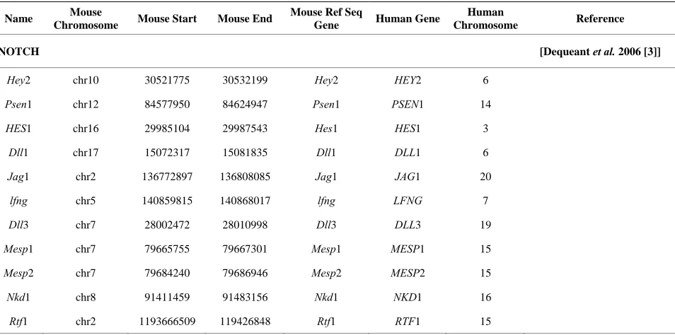
[image:2.595.61.538.497.734.2]Mouse gene coordinates corresponding to the human chromosome coordinates for the 77 genes from Table 1 (20 of the original 97 patterning genes did not have corre- sponding mouse coordinates) were obtained by using En- semble homology tables [16]. The human genome was divided into 2980, 1 Mbp intervals; the number of the genes from Table 1 was counted in each of those inter-vals. A determination was made as to which bins are overlapping with positions of the HSBs or EBRs. Stu- dent’s t-test analysis with unequal variances, as described previously, was performed to determine whether the so- mite patterning genes are preferentially located in the EBRs or HSBs [17,18].
Table 1. Mouse genes and coordinates studied with corresponding human syntenic gene region.
Name Mouse
Chromosome Mouse Start Mouse End
Mouse Ref Seq
Gene Human Gene
Human
Chromosome Reference
NOTCH [Dequeant et al. 2006 [3]]
Hey2 chr10 30521775 30532199 Hey2 HEY2 6
Psen1 chr12 84577950 84624947 Psen1 PSEN1 14
HES1 chr16 29985104 29987543 Hes1 HES1 3
Dll1 chr17 15072317 15081835 Dll1 DLL1 6
Jag1 chr2 136772897 136808085 Jag1 JAG1 20
lfng chr5 140859815 140868017 lfng LFNG 7
Dll3 chr7 28002472 28010998 Dll3 DLL3 19
Mesp1 chr7 79665755 79667301 Mesp1 MESP1 15
Mesp2 chr7 79684240 79686946 Mesp2 MESP2 15
Nkd1 chr8 91411459 91483156 Nkd1 NKD1 16
Continued
FGF [Dequeant et al. 2006[3]]
dusp6 chr10 98692919 98697172 Dusp6 DUSP6 12
FRS2 chr10 116474239 110912758 Frs2 FRS2 12
Grb2 chr11 115460135 115524687 Grb2 GRB 2 17
SOS1 chr17 80305779 80388265 Sos1 SOS1 2
FGF8 chr19 45790109 45796226 Fgf8 FGF8 10
Bcl2l11 chr2 127817479 127853988 Bcl2l11 BCL2L11 2
Efna1 chr3 89357663 89365568 Efna1 EFNA1 1
Erk chr4 135919615 136108064 Ephb3 EPHB2 1
Hspg2 chr4 136740845 136842706 Hspg2 HSPG2 1
Shh chr5 28787602 28797888 shh SHH 7
Shp-2 chr5 121391150 121452014 Ptpn11 PTPN11 12
Gab1 chr8 836601080 83776225 Gab1 GAB1 4
WNT [Dequeant et al. 2006 [3]]
Wnt3a chr11 103590240 103634047 Wnt3 WNT3 17
Axin2 chr11 108736439 108766873 Axin2 AXIN2 17
Fzd7 chr1 59426970 59431505 Fzd7 FZD7 2
Fzd5 chr1 64668689 64672026 Fzd5 FZD5 2
Cdc73 chr1 145368379 145464902 Cdc73 CDC73 1
Phlda1 chr10 110910396 110912758 Phlda1 PHLDA1 12
Dvl2 chr11 69816790 69828496 Dvl2 DVL2 17
HDAC chr12 34663701 35022647 Hdac9 HDAC9 7
Dact1 chr12 72228589 72237499 Dact1 DACT1 14
Tnfrsf19 chr14 59918146 60000579 Tnfrsf19 TNFRSF19 13
Fzd3 chr14 64155136 64216673 Fzd3 FZD3 8
Sprouty2 chr14 104778114 104782418 Spry2 SPRY2 13
Fzd6 chr15 38836426 38868268 Fzd6 FZD6 8
Has2 chr15 56495712 56524587 Has2 HAS2 8
c-myc chr15 61815052 61820027 Myc MYC 8
Ppp2r1a chr17 20650008 20670613 Ppp2r1a PPP2R1A 19
Fzd8 chr18 921918 9214975 Fzd8 FZD8 10
APC chr18 34345794 34443382 Apc APC 5
Smad4 chr18 73764378 73829149 Smad4 SMAD4 18
LRP5/6 chr19 3584836 3686546 Lrp5 LRP5 11
Dkk1 chr19 30611873 30615516 Dkk1 DKK1 10
FrzB chr2 80212809 80248464 Frzb FRZB 2
Tcf15 chr2 151835002 151840538 Tcf15 TCF15 20
Fzd1 chr5 4759879 4764041 Fzd1 FZD1 7
CtBP1 chr5 33564581 33591839 Ctbp1 CTBP1 4
Fzd9 chr5 135533565 135535857 Fzd9 FZD9 7
Fzd4 chr7 89279586 89285277 Fzd4 FZD4 11
Smarca5 chr8 83595689 83635205 Smarca5 SMARCA5 4
Continued
HOX
Related
Hoxc8 chr15 10281573 102821404 Hoxc8 HOXC8 12 [Yueh et al. 1998 [26]]
Hoxc-4 chr15 102862429 102864631 Hoxc4 HOXC4 12 [Apiou et al. 1996 [27]]
Hoxd11 chr2 74480397 74487855 Hoxd11 HOXD11 2 [Kessel and Gruss 1990 [28]]
Hoxd10 chr2 74492730 74495942 Hoxd10 HOXD10 2 [Kessel and Gruss 1990 [28]]
Hoxd3 chr2 74512768 74549113 Hoxd3 HOXD3 2 [Kessel and Gruss 1990 [28]]
Hoxa7 chr6 52144074 52151437 Hoxa7 HOXA7 7 [Kessel and Gruss 1990 [28]]
Hoxb7 chr11 96100653 96106426 Hoxb7 HOXB7 17 [Kessel and Gruss 1990 [28]]
Leo1 chr7 28101775 28108283 Paf1 PAF1 19 [Apiou et al.1996 [27]]
PAX
Pax9 chr12 57613651 57629242 Pax9 PAX9 14 [Peters et al.1999 [29]]
Pax1 chr2 147053366 147083649 Pax1 PAX1 20 [Peters et al.1999[29]]
Pax7 chr4 139009138 139105044 Pax7 PAX7 1 [Basch et al. 2006; Relaix et al.
2005 [30,31]]
TGFβ
Nodal chr10 60813329 60820695 Nodal NODAL 10 [Brennan et al. 2001 [32]]
Tgfbr2 chr9 115932995 116023987 Tbfbr2 TGFBR2 3 [Baffi et al. 2006 [33]]
Other
Rab23 chr1 33664428 33687110 Rab23 RAB23 6 [Eggenschwiler et al. 2001 [34]]
Ihh chr1 74878522 74884858 Ihh IHH 2 [Vortkamp et al. 1996 [35]]
Plxdc1 chr11 97739328 97802534 Plxdc1 PLXDC1 17 [Kanda et al. 2007 [36]]
TWIST1 chr12 34542918 34545078 Twist1 TWIST1 7 [Bialek et al. 2004 [37]]
Gli3 chr13 15254867 15517860 Gli3 GLI3 7 [Aruga et al. 1999 [38]]
FlnB chr14 6608366 6743464 Flnb FLNB 3 [Krakow et al. 2004 [39]]
Slc35a3 chr3 116662802 116704284 Slc35a3 SLC35A3 1 [Thomsen et al. 2006 [40]]
Mxd4 chr5 34492821 34504537 Mxd4 MXD4 4 [Yokoyama et al. 2009 [41]]
PDGFRA chr5 75434033 75479895 Pdgfra PDGFRA 4 [Soriano 1997 [42]]
Tbx6 chr7 126572631 126576696 Tbx6 TBX6 16 [Chapman and others 2003 [43]]
Acd chr8 108584989 108590214 Acd ACD 16 [Keegan et al. 2005 [44]]
Mid1 chrX 165029304 165334903 MID1 MID1 X [Quaderi et al.1997 [45]]
3. Results
Vertebral patterning and scoliosis associated genes in Ta- ble 1 were found to be preferentially located away from the EBRs, with approximately twice as many genes on average occurring in other parts of the genome as com- pared to the breakpoint intervals (p-value = 0.011). While this does not appear to be a large difference, if all genes in the genome are counted, the EBRs on average contain ~2 times more genes than the rest of the genome. In general, breakpoint intervals are significantly enriched for genes, and the results of this analysis indicate that they are not enriched for vertebral patterning genes. Examination of large blocks (>3 Mb) of homologous synteny (approxi- mately 7 of these occur in amniote genomes, which are
>16.3 Mbp in human coordinates) indicated 0.04 genes from Table 1 localized in these blocks on genome aver- age, while 0.03 genes localized to the rest of the genome [19]. This result is not statistically significant, probably because of the small number of genes in this comparison.
4. Discussion
ciated with somitogenesis in amniotic species and pro- vides additional genetic evidence for similarities in spine development in amniotes.
A prior analysis of mouse scoliotic phenotypes using the Mouse Genome Database (MGD), followed by use of the Online Mendelian Inheritance in Man (OMIM), yielded 45 genes with possible scoliosis phenotypes. Twenty eight genes were translated to the human genome coordinates using mouse and human synteny maps [8]. These in-cluded WNT3A and DLL3 genes, also members of the co-hort of genes in Table 1. During this analysis it was not possible to determine whether each vertebral patterning gene was located within EBRs or away from breakpoint intervals. The localization of patterning genes associated with human vertebral development to regions away from synteny breakpoint intervals provides evidence for con- servation of the basic vertebral patterning scheme during amniote development.
These data are consistent with prior analyses per- formed by Larkin et al. [19]. Gene networks associated with major developmental processes such as neuronal development, central nervous system, bone and blood vessel development, some of which were mediated by Wnt and Notch signaling pathways, were significantly enriched in HSBs and, therefore, less likely to be local- ized at EBRs. Gene networks associated with responses to external stimuli such as inflammatory responses and muscle contraction were more likely to be localized to EBRs. Our study focused on 77 genes associated with somitogenesis including 11 NOTCH, 12 FGF, 29 WNT, 8 HOX, 3 PAX, 2 TGFβ pathway and 12 additional genes associated with scoliosis phenotypes and the results demonstrated these patterning genes were significantly overrepresented in the evolutionary conserved regions.
Using a series of bioinformatic approaches including neighbor-joining (NJ) and maximum parsimony (MP), contained within the PHYLIP (PHYLogeny inference package) software package [20], the evolution of Notch family proteins in species from worm to human was ana- lyzed in C. elegans, D. melanogaster, C. intestinalis, and
H. sapiens using Mapviewer, Geneview, and the BlastP and TBlastN algorithm [21]. The chromosomal distribu- tion of PBX (pre-B cell leukemia homeobox), LHX3(Lim homeobox 3), NRARP (Notch regulated ankyrin repeat protein), BRD (bromodomain) and CAMSAP1 (calmo- dulin regulated spectrin-associated protein 1) was found to follow the distribution of Notch, providing evidence for co-evolution with Notch signaling pathway genes by segmental duplication. The close proximity of these genes may reflect a functional relationship. For instance, in C. elegans, PBX appears to be responsible for tran- scriptional control of Notch signaling [22].
This study has several limitations. While genes in the FGF, WNT and Notch signaling pathways were analyzed,
genes in other pathways such as the BMP signaling pathway were not studied. Corresponding mouse coordi- nates were identified for 77 of the 97 patterning human genes originally identified. Due to the relatively small number of genes studied, it was not possible to determine whether the vertebral patterning genes were preferen- tially localized to large blocks of homologous synteny.
Besides playing a crucial role in somitogenesis, the Fgf, Wnt and Notch signaling pathways also are involved in the embryogenesis of other organs. Fgf’s have impor-tant roles in development of the limbs, skin, central nerv- ous system, ear, lungs, liver and have major involvement in the wound healing process [23]. Fibroblast growth factor receptor related disorders in humans include cra- niosynostosis syndromes such as Apert and Crouzon syndrome, and skeletal dsyplasias, of which achondro- plasia is the most common. The Wnt canonical pathway is active in neural tube development [24]. Notch signal- ing is involved in developmental pathways which affect the vasculature, heart, eye and liver [25]. Both Notch and Wnt pathways are involved in autonomous phenotypes including cellular development and proliferation. It is possible that the conservation of Fgf, Wnt and Notch signaling related genes in HSBs reflects conservation of non-somite signaling functions or conservation of multi- ple signaling functions. A previous study by Larkin et al. focused on association of HSBs with respect to gene net-works, while our study was aimed at localization of genes associated with a specific disease process, namely scoliosis with respect to HSBs [19].
In summary we provide further evidence that devel- opmental pathways associated with somitogenesis are conserved across amniotes which is consistent with their known function.
5. Acknowledgements
We appreciate the comments and advice provided by Drs. Harris Lewin and Denis Larkin.
REFERENCES
[1] Scoliosis Research Society, “Glossary of Scoliosis Terms,” Spine, Vol. 1, No. 1, 1976, pp. 57-58.
doi:10.1097/00007632-197603000-00008
[2] S. B. Purkiss, B. Driscoll, W. G. Cole and B. Alman, “Idiopathic Scoliosis in Families of Children with Con-genital Scoliosis,” Clinical Orthopaedics and Related Research, Vol. 401, 2002, pp. 27-31.
doi:10.1097/00003086-200208000-00005
[3] M. Dequeant, E. Glynn, K. Gaundenz, M. Wahl, J. Chen, A. Mushegian and O. Pourquie, “A Complex Oscillating Network of Signaling Genes Underlies the Mouse Seg-mentation Clock,” Science, Vol. 314, No. 5805, 2006, pp. 1595-1598. doi:10.1126/science.1133141
McPherson, L. Ivacic, K. Rasmussen, F. Jacobsen, T. Faciszewski, J. K. Burmester, R. M. Pauli, O. Boachie- Adjei, I. Glurich and P. F. Giampietro, “A Missense T (Brachyury) Mutation Contributes to Vertebral Malfor- mations,” Journal of Bone and Mineral Research, Vol. 23, No. 10 2008, pp. 1576-1583. doi:10.1359/jbmr.080503
[5] N. Ghebranious, J. Burmester, I. Glurich, E. McPherson, L. Ivacic, J. Kislow, K. Rasmussen, V. Kumar, C. Raggio, R. Blank, F. S. Jacobsen, T. Faciszewski, J. Womack and P. F. Giampietro, “Evaluation of SLC35A3 as a Candidate Gene for Human Vertebral Malformations,” American Journal of Medical Genetics, Vol. 140A, No. 12, 2006, pp. 1346-1348. doi:10.1002/ajmg.a.31307
[6] N. Ghebranious, C. L. Raggio, R. D. Blank, E. McPher-son, J. K. Burmester, L. Ivacic, K. Rasmussen, J. Kislow, I. Glurich, F. S. Jacobsen, T. Faciszewski, R. M. Pauli, O. Boachie-Adjei and P. F. Giampietro, “Lack of Evidence of WNT3A as a Candidate Gene for Congenital Vertebral Malformations,” Scoliosis, Vol. 2, 2007, p. 13.
doi:10.1186/1748-7161-2-13
[7] P. Giampietro, C. Raggio, C. Reynolds, N. Ghebranious, J. Burmester, I. Glurich, K. Rasmussen, E. McPherson, R. Pauli, S. K. Shukla, S. Merchant, F. S. Jacobsen, T. Faciszewski and R. D. Bland, “DLL3 as a Candidate for Vertebral Malformations,” American Journal of Medical Genetics, Vol. 140A, No. 22, 2006, pp. 2447-2453. doi:10.1002/ajmg.a.31509
[8] P. F. Giampietro, C. L. Raggio and R. D. Blank, “Synteny- Defined Candidate Genes for Idiopathic and Congenital Scoliosis,” American Journal of Medical Genetics, Vol. 83, No. 3, 1999, pp. 164-177.
doi:10.1002/(SICI)1096-8628(19990319)83:3<164::AID-AJMG5>3.0.CO;2-D
[9] P. F. Giampietro, C. L. Raggio, C. E. Reynolds, S. K. Shukla, E. McPherson, N. Ghebranious, F. S. Jacobsen, V. Kumar, T. Faciszewski, R. M. Pauli, K. Rasmussen, J. K. Burmester, C. Zaleski, S. Merchant, D. David, J. L. We-ber, I. Glurich and R. D. Blank, “An Analysis of PAX1 in the Development of Vertebral Malformations,” Clinical Genetics, Vol. 68, No. 5, 2005, pp. 448-453.
doi:10.1111/j.1399-0004.2005.00520.x
[10] K. J. Alden, B. Marosy, N. Nzegwu, C. M. Justice, A. F. Wilson and N. H.Miller, “Idiopathic Scoliosis: Identifica-tion of Candidate Regions on Chromosome 19, p13,” Spine, Vol. 31, No. 16, 2006, pp. 1815-1819.
doi:10.1097/01.brs.0000227264.23603.dc
[11] X. Gao, D. Gordon, D. Zhang, R. Browne, C. Helms, J. Gillum, S. Weber, S. Devroy, S. Swaney, M. Dobbs, J. Morcuende, V. Sheffield, M. Lovett, A. Bowcock, J. Her-ring and C. Wise, “CHD7 Gene Polymorphisms Are As-sociated with Susceptibility to Idiopathic Scoliosis,” American Journal of Human Genetics, Vol. 80, No. 5, 2007, pp. 957-965. doi:10.1086/513571
[12] N. H. Miller, C. M. Justice, B. Marosy, K. F. Doheny, E. Pugh, J. Zhang, H. C. Dietz 3rd and A. F. Wilson, “Iden-tification of Candidate Regions for Familial Idiopathic Scoliosis,” Spine, Vol. 30, No. 10, 2005, pp. 1181-1187. doi:10.1097/01.brs.0000162282.46160.0a
[13] N. H. Miller, B. Marosy, C. M. Justice, S. M. Novak, E. Y. Tang, P. Boyce, J. Pettengil, K. F. Doheny, E. W.
Pugh and A. F. Wilson, “Linkage Analysis of Genetic Loci for Kyphoscoliosis on Chromosomes 5, p13,” 13q13.3, and 13q32,” American Journal of Medical Ge-netics A, Vol. 140, No. 10, 2006, pp. 1059-1068. doi:10.1002/ajmg.a.31211
[14] V. Subramanian, B. I. Meyer and P. Gruss, “Disruption of the Murine Homeobox Gene Cdx1 Affects Axial Skeletal Identities by Altering the Mesodermal Expression Do-mains of Hox Genes,” Cell, Vol. 83, No. 4, 1995, pp. 641-653. doi:10.1016/0092-8674(95)90104-3
[15] W. J. Murphy, D. M. Larkin, A. E. van der Wind, G. Bourque, G. Tesler, L. Auvil, J. E. Beever, B. P. Chowd-hary, F. Galibert, L. Gatzke, C. Hitte, S. N. Meyers, D. Milan, E. A. Ostrander, G. Pape, H. G. Parker, T. Raud-sepp, M. B. Roqatcheva, L. B. Schook, L. C. Skow, M. Welge, J. E. Womack, S. J. O’brien, P. A. Pevzner and H. A. Lewin, “Dynamics of Mammalian Chromosome Evo-lution Inferred from Multispecies Comparative Maps,” Science, Vol. 309, No. 5734, 2005, pp. 613-617.
doi:10.1126/science.1111387
[16] ensemble.org
[17] E. Skovlund and G. U. Fenstad, “Should We Always Choose a Nonparametric Test When Comparing Two Apparently Nonnormal Distributions?” Journal of Clini- cal Epidemiology, Vol. 54, No. 1, 2001, pp. 86-92. doi:10.1016/S0895-4356(00)00264-X
[18] A. V. Smith, D. J. Thomas, H. M. Munro and G. R. Abe-casis, “Sequence Features in Regions of Weak and Strong Linkage Disequilibrium,” Genome Research, Vol. 15, No. 11, 2005, pp. 1519-1534. doi:10.1101/gr.4421405
[19] D. M. Larkin, G. Pape, R. Donthu, L. Auvil, M. Welge and H. A. Lewin, “Breakpoint Regions and Homologous Synteny Blocks in Chromosomes Have Different Evolu-tionary Histories,” Genome Research, Vol. 19, No. 5, 2009, pp. 770-777. doi:10.1101/gr.086546.108
[20] evolution.gs.org
[21] A. Theodosiou, S. Arhondakis, M. Baumann and S. Kos-sida, “Evolutionary Scenarios of Notch Proteins,” Mo-lecular Biology and Evolution, Vol. 26, No. 7, 2009, pp. 1631-1640. doi:10.1093/molbev/msp075
[22] K. Takacs-Vellai, T. Vellai, E. B. Chen, Y. Zhang, F. Guerry, M. J. Stern and F. Muller, “Transcriptional Con-trol of Notch Signaling by a HOX and a PBX/EXD Pro-tein during Vulval Development in C. elegans,” Devel-opmental Biology, Vol. 302, No. 2, 2007, pp. 661-669. doi:10.1016/j.ydbio.2006.09.049
[23] X. Coumoul and C. X. Deng, “Roles of FGF Receptors in Mammalian Development and Congenital Diseases,” Birth Defects Research C: Embryo Today, Vol. 69, No. 4, 2003, pp. 286-304. doi:10.1002/bdrc.10025
[24] R. Alvarez-Medina, J. Cayuso, T. Okubo, S. Takada and E. Marti, “Wnt Canonical Pathway Restricts Graded Shh/Gli Patterning Activity through the Regulation of Gli 3 Expression,” Development, Vol. 135, 2008, pp. 237- 247. doi:10.1242/dev.012054
[25] T. Gridley, “Notch Signaling and Inherited Human Dis-ease Syndromes,” Human Molecular Genetics, Vol. 12, No. 1, 2003, pp. R9-R13. doi:10.1093/hmg/ddg052
Regulation of Cartilage Differentiation by the Homeobox Gene Hoxc-8,” Proceedings of the National Academy of Sciences of the United States of America, Vol. 95, No. 17, 1998, pp. 9956-9961. doi:10.1073/pnas.95.17.9956
[27] F. Apiou, D. Flagiello, C. Cillo, B. Malfoy, M. F. Poupon and B. Dutrillaux, “Fine Mapping of Human HOX Gene Clusters,” Cytogenetics and Cell Genetics, Vol. 73, No. 1-2, 1996, pp. 114-115. doi:10.1159/000134320
[28] M. Kessel and P. Gruss, “Murine Developmental Control Genes,” Science, Vol. 249, No. 4967, 1990, pp. 374-379. doi:10.1126/science.1974085
[29] H. Peters, B. Wilm, N. Sakai, K. Imai, R. Mass and R. Balling, “Pax1 and Pax9 Synergistically Regulate Verte-bral Column Development,” Development, Vol. 126, 1999, pp. 5399-5408.
[30] M. L. Basch, M. Bronner-Fraser and M. I. Garcia-Castro, “Specification of the Neural Crest Occurs during Gas-trulation and Requires Pax7,” Nature, Vol. 441, No. 7090, 2006, pp. 218-222. doi:10.1038/nature04684
[31] F. Relaix, D. Rocancourt, A. Mansouri and M. Bucking- ham, “A Pax3/Pax7-Dependent Population of Skeletal Muscle Progenitor Cells,” Nature, Vol. 435, No. 7044, 2005, pp. 948-953. doi:10.1038/nature03594
[32] J. Brennan, C. C. Lu, D P. Norris, T. A. Rodriguez, R. S. Beddington and E. J. Robertson, “Nodal Signaling in the Epiblast Patterns the Early Mouse Embryo,” Nature, Vol. 411, No. 6840, 2001, pp. 965-969. doi:10.1038/35082103
[33] M. O. Baffi, M. A. Moran and R. Serra, “Tgfbr2 Regu-lates the Maintenance of Boundaries in the Axial Skele-ton,” Developmental Biology, Vol. 296, No. 2, 2006, pp. 363-374. doi:10.1016/j.ydbio.2006.06.002
[34] J. T. Eggenschwiler, E. Espinoza and K. V. Anderson, “Rab23 Is an Essential Negative Regulator of the Mouse Sonic Hedgehog Signalling Pathway,” Nature, Vol. 412, No. 6843, 2001, pp. 194-198. doi:10.1038/35084089
[35] A. Vortkamp, K. Lee, B. Lanske, G. V. Segre, H. M. Kronenberg and C. J. Tabin, “Regulation of Rate of Car-tilage Differentiation by Indian Hedgehog and PTH-Re- lated Protein [See Comments],” Science, Vol. 273, No. 5275, 1996, pp. 613-622.
doi:10.1126/science.273.5275.613
[36] T. Kanda, Y. Yoshida, Y. Izu, A. Nifuji, Y. Ezura, K. Nakashima and M. Noda, “PlexinD1 Deficiency Induces Defects in Axial Skeletal Morphogenesis,” Journal of Cellular Biochemistry, Vol. 101, No. 6, 2007, pp. 1329- 1337. doi:10.1002/jcb.21306
[37] P. Bialek, B. Kern, X. Yang, M. Schrock, D. Sosic, N. Hong, H. Wu, K. Yu, D. M. Ornitz, E. N. Olson, M. J. Justice and G. Karsenty, “A Twist Code Determines the Onset of Osteoblast Differentiation,” Developmental Cell, Vol. 6, No. 3, 2004, pp. 423-435.
doi:10.1016/S1534-5807(04)00058-9
[38] J. Aruga, K. Mizugishi, H. Koseki, K. Imai, R. Balling, T.
Noda and K. Mikoshiba, “Zic1 Regulates the Patterning of Vertebral Arches in Cooperation with Gli3,” Mecha-nisms of Development, Vol. 89, No. 1-2, 1999, pp. 141- 150. doi:10.1016/S0925-4773(99)00220-8
[39] D. Krakow, S. P. Robertson, L. M. King, T. Morgan, E. T. Sebald, C. Bertolotto, S. Wachsmann-Hogiu, D. Acuna, S. S. Shapiro, T. Takafuta, S. Aftimos, C. A. Kim, H. Firth, C. E. Steiner, V. Cormier-Daire, A. Superti Furga, L. Bonafe, J. M. Graham, A. Grix, C. A. Bacino, J. Allanson, M. G. Bialer, R. S. Lachman, D. L. Rimoin and D. H. Cohn, “Mutations in the Gene Encoding Filamin B Dis-rupt Vertebral Segmentation, Joint Formation and Skele- togenesis,” Nature Genetics, Vol. 36, No. 4, 2004, pp. 405-410. doi:10.1038/ng1319
[40] B. Thomsen, P. Horn, F. Panitz, E. Bendixen, A. H. Pe-tersen, L. E. Holm, V. H. Nielsen, J. S. Agerholm, J. Arnbjerg and C. Bendixen, “A Missense Mutation in the Bovine SLC35A3 Gene, Encoding a UDP-N-Acetylgluco- samine Transporter, Causes Complex Vertebral Malfor- mation,” Genome Research, Vol. 16, No. 1, 2006, pp. 97- 105. doi:10.1101/gr.3690506
[41] S. Yokoyama, Y. Ito, H. Ueno-Kudoh, H. Shimizu, K. Uchibe, S. Albini, K. Mitsuoka, S. Miyaki, M. Kiso, A. Nagai, T. Kikata, T. Osada, N. Fukuda, S. Yamashita, D. Harada, V. Mezzano, M. Kasai, P. L. Puri, Y. Hayashi-zaki, H. Okado, M. Hashimoto and H. Ashara, “A Sys-tems Approach Reveals That the Myogenesis Genome Network Is Regulated by the Transcriptional Repressor RP58,” Developmental Cell, Vol. 17, No. 6, 2009, pp. 836-848. doi:10.1016/j.devcel.2009.10.011
[42] P. Soriano, “The PDGF Alpha Receptor Is Required for Neural Crest Cell Development and for Normal Pattern-ing of the Somites,” Development, Vol. 124, No. 14, 1997, pp. 2691-2700.
[43] D. Chapman, A. Cooper-Morgan, Z. Harrelson and V. Papaioannou, “Critical Role for Tbx6 in Mesoderm Specification in the Mouse Embryo,” Mechanisms of De-velopment, Vol. 120, No. 7, 2003, pp. 837-847. doi:10.1016/S0925-4773(03)00066-2
[44] C. Keegan, J. Hutz, T. Else, M. Adamska, S. Shah, A. Kent, J. Howes, W. Beamer and G. Hammer, “Urogenital and Caudal Dysgenesis in Adrenocortical Dysplasia (acd) Is Caused by a Splicing Mutation in a Novel Telomeric Regulator,” Human Molecular Genetics, Vol. 14, 2005, pp. 113-123. doi:10.1093/hmg/ddi011