Supported by the National Key Basic Research Program of China (Project No. 2011CB100805).
Comparative proteomics analysis of plasma proteins
during the transition period in dairy cows
with or without subclinical mastitis after calving
Y.X. Yang
1,2, J.Q Wang
1, D.P. Bu
1, S.S.
Li
1, T.J. Yuan
1, L.Y. Zhou
1, J.H. Yang
1,
P. Sun
11State Key Laboratory of Animal Nutrition, Institute of Animal Science, Chinese Academy of Agricultural Sciences, Beijing, P.R.China
2Institute of Animal Science and Veterinary Medicine, Anhui Academy of Agricultural Sciences, Hefei, P.R.China
ABSTRACT: The transition period is the most critical time of the cow’s lactation cycle that is associated with
the onset of mastitis. In this study, changes of plasma proteins in cows (n = 12) with or without subclinical mastitis after calving were determined by two-dimensional electrophoresis (2-DE), which detected 18 spots with variations in protein spots abundance. These spots were identified by liquid chromatography coupled with tandem mass spectrometry. The changes in protein profile from day 21 before calving to day 1 after calving were similar in cows with or without subclinical mastitis. Abundance of α1 acid glycoprotein (AGP) and hap-toglobin was dramatically increased at parturition, while transthyretin was down-regulated at parturition, and apolipoprotein E and immunoglobulin gamma 1 were up-regulated at postpartum compared with prepartum in periparturient dairy cows. In cows infected with subclinical mastitis, AGP, haptoglobin, and serum amyloid A were dramatically increased and continued to be elevated in plasma from day 1 to day 21 after calving compared with cows free of mastitis. Changes of protein in plasma at parturition may serve as an immune system response to parturition and lactation process at the protein level and suggest that these altered proteins would not serve as a potential marker for predicting if the periparturient dairy cows are susceptible to subclinical mastitis.
Keywords: dairy cow; plasma proteome; periparturient; mass spectrometry; mastitis
The transition period is traditionally considered to last for approximately 6 weeks − 3 weeks be-fore and 3 weeks after calving (Drackley, 1999). During this period the dairy cow’s immune system including neutrophil and lymphocyte function is suppressed (Kehrli et al., 1989), which is associated with increases in the incidence of infectious diseas-es, especially mastitis (Valde et al., 2004; Pyörälä, 2008). Thus, the transition period is the most criti-cal time of the lactation cycle and can determine to a large measure how profitable the entire lactation will be. For this reason, many researchers have at-tempted to reveal complex physiological adaptive
mechanisms using transcriptomic and proteomic methodologies (Madsen et al., 2002; Loor et al., 2005; Cairoli et al., 2006), the latter of which is a powerful tool for comparative analysis of complex protein mixtures based on high-resolution two-dimensional electrophoresis (2-DE) coupled with mass spectrometry. This proteomic approach can provide useful information on temporal and dy-namic processes occurring in the proteome.
alteration of plasma proteins can serve as potential diagnostic markers for abnormal physiological or pathological conditions.
To investigate the host immune system response to parturition and lactation, neutrophil functionality was first investigated in cows using amine-reactive isobaric tagging reagents. Over 40 proteins in neu-trophils were differentially expressed at parturition as compared to prepartum, including a bovine my-eloid antimicrobial peptide of 28 residues with an-timicrobial activity and several proteins involved in arachidonic acid metabolism and transport (Lippolis et al., 2006). Additionally, several serum proteins were shown to change during the last phase of pregnancy and early postpartum, which included a decrease of α2-HS-glycoprotein, an increase of α1-antichymotrypsin, and a dramatic variation in orosomucoid and haptoglobin (Cairoli et al., 2006). These results indicated that parturition and lactation induced changes in protein synthesis of the neu-trophil and liver. However, few data are currently available on the plasma proteome during the pe-riparturient period in dairy cows with or without subclinical mastitis after calving.
The objective of this study was to identify tempo-ral changes of proteins during the transition of cows using 2-DE and liquid chromatography coupled with tandem mass spectrometry. The findings of this study may provide additional insight into the regulation of plasma proteins during this period and potentially discover markers that can be used to indentify cows that are susceptible to mastitis.
MATERIAL AND METHODS
Sample collection and preparation
The procedures used for caring for the dairy cows in this experiment were reviewed and approved by the Animal Care Advisory Committee of the Chinese Academy of Agricultural Sciences. Blood samples (9 ml) were collected from the tail vein of sixty Chinese Holstein primiparous cows from a farm near Beijing, using BD vacationer tubes con-taining ethylenediaminetetraacetic acid-anticoag-ulant 20.4 ± 1.6 days before the expected date of calving, and 1 and 21 days postpartum. The cows were housed in free-stalls, fed a total mixed ra-tion, and milked three times daily. Twelve healthy cows considered free of mastitis (somatic cell count less than 500 000 cells/ml) as measured by
Fossomatic 5000 (Foss Electric, Hilerød, Denmark) on day 14 and 21 after calving and 12 cows with subclinical mastitis (somatic cell counts > 500 000 cells/ml) (Liu et al., 2010; Mollenhorst et al., 2010) were selected for proteomic analysis. Cows with any other diseases or disorders according to vet-erinarian records, such as retained placenta and metritis, were excluded from analysis in agreement with this record. Blood samples were centrifuged at 4°C and 3000 g for 15 min, plasma was collected and then stored at −80°Cuntil analysis.
Enrichment of minor abundance proteins
After thawing, the plasma samples from twelve healthy cows were pooled as three samples, and from twelve cows with subclinical mastitis the plas-ma samples were also pooled as three samples col-lected at the same time during the transition period. These pooled samples were then enriched using a ProteoMiner kit (Bio-Rad Laboratories, Hercules, USA) per manufacturer’s protocols. A 1 ml plasma sample was mixed with the beads after centrifuging and the storage solution was decanted off. The col-umn containing the beads was rotated end-to-end at room temperature for 2 h and centrifuged at 1000 g
to remove any remaining storage solution. Then the beads were rinsed with a buffer solution and then re-suspended in the elution buffer and mixed by gent- lely vortexing. Following centrifugation at 1000 g
at room temperature, the supernatant containing the released proteins was collected. Fresh elution buffer was added to the bead and the above process was repeated twice to ensure maximum protein re-covery. The released proteins were pooled and the resulting enriched protein samples were analyzed for protein content using the bicinchoninic acid method (Kao et al., 2008).
Two-dimensional gel electrophoresis and software analysis
for 10 min with gentle mixing. Plasma samples en-riched with ProteoMiner were diluted with rehy-dration solution to 350 μl. The resulting solution then was rehydrated on 17 cm, pH 4−7 IPG strips (Bio-Rad Laboratories, Hercules, USA). After 12 h of passive rehydration, isoelectric focusing (IEF) was performed at 80 kVh using a Protean IEF Cell (Bio-Rad Laboratories, Hercules, USA) at 20°C as follows: 30 min at 250 V, 1 h at 1000 V, then in-creased from 1000 V to 10 000 V over a period of 5 h, and finally 10 000 V for 8 h. Subsequently, IPG strips were equilibrated with an equilibration solution (6 mol/l urea, 2% SDS, 20% glycerol, 50 mmol/l Tris-HCl, 0.01% w/v bromophenol blue) containing 2% DTT for 12 min with shaking and followed by equilibration solution containing 2.5% w/v iodoacetamide for an additional 12 min with shaking. Strips were transferred onto a 12% poly-acrylamide gel for second dimension separation using an Ettan DALTsix Electrophoresis Unit (GE Healthcare, Waukesha, USA). The gels were run at 1 W for the first 30 min and then at 46 W until the bromophenol blue reached the bottom of the gels. Then the gels were stained with Coomassie Brilliant Blue G-250 as described by Candiano et al. (2004). Gel images were analyzed using the ImageMaster 2-D Platinum software, Version 6.0 (GE Healthcare, Waukesha, USA). For comparisons of relative abun-dance of proteins among gels, protein spots were automatically detected and manually confirmed. Three biological replicate gels were superimposed and a master gel was obtained. In each master gel protein spot position, shape and optical density were averaged, and individual spot intensity volume was normalized with total intensity volume. Protein spots were considered to be differentially expressed when there was at least a two-fold increase or de-crease in spot stains density, and values were then analyzed for statistical significance (Gonçalves et al., 2010).
In-gel digestion and extraction of peptides
Protein spots that showed differential expression were excised manually using pipette tips. Each spot was placed into a 1.5 ml microtube and washed twice with MilliQ water for 15 min. After washing, gel pieces were incubated in 50% acetonitrile at room temperature for 15 min, and then shrunk in acetonitrile. The protein spots were digested in a buffer containing 50 mmol/l of ammonium
bicar-bonate and 10 ng/ml of sequence-grade trypsin by incubating at 37°C overnight. Then, peptides were extracted twice with 50% acetonitrile and 0.2% for-mic acid, with each extraction being followed by vortexing and sonication (10 min each).
Protein identification
Peptide mass was determined by ion trap mass spectrometry (LCQ Deca XP, Thermo Finnigan, San Chose, USA) equipped with a Surveyor high perfor-mance liquid chromatography system. A BioBasic-18 column (50 ± 0.18 mm) (Thermo Electron, San Jose, USA) was equilibrated with mobile phase A consist-ing of MilliQ water and 0.1% formic acid. A 20 μl aliquot was injected onto the BioBasic-18 column and the proteins were eluted using mobile phase B consisting of 0.1% formic acid in acetonitrile at a flow rate of 120 μl/min with a linear gradient over 120 min. Using the full scan mode m/z 400−2000, tandem mass spectrometry was performed in a data-dependent mode. After acquisition of a full scan, three tandem mass spectrometry scans were acquired for the next three most intense ions using dynamic exclusion. Peptides were identified using the SEQUEST software (Bioworks 2.0, Thermo Finnigan, San Chose, USA), which uses the mass spectrometry and tandem mass spectrometry spec-tra of peptide ions to search the NCBInr database. The following search parameters were used with trypsin as the cleavage enzyme, one missed cleav-age, carbamidomethylation of cysteine residues as a fixed modification, and methionine oxidation selected as a variable modification. The protein identification criteria were based on Delta CN ≥ 0.1 and Xcorr (+ 1 ≥ 1.9, + 2 ≥ 2.2, + 3 ≥ 3.75).
Western blot analysis
The recombinant protein of haptoglobin was ex-pressed in Escherichia coli BL21 strain after IPTG inducement and purified. Polyclonal antibodies against haptoglobin were produced to immunize rabbits and then the antiserum was collected and purified with protein A affinity resin for use.
membrane was blocked with tris-buffered saline tween-20 (TBST) containing 1% ovalbumin for 1 h at room temperature and then incubated with antibody for 1 h at 37°C. The membrane was then washed with TBST and visualized with horserad-ish peroxidase-coupled secondary antibody using diaminobenzidine as substrate. The relative quantity of the protein was defined as the gray scale of the gels. The resulting values were tested for significance using the statistic software SPSS, Version 16.0 (SPSS Inc., Chicago, USA), statistically significant of group and day relative to calving for relative quantity of the protein were performed by two-way analysis of variance (General Linear Model) followed by Tukey’s test; two groups were the transition cows with or without subclinical mastitis after calving, and the three levels of the repeated measurement of time were on day 21 before calving, and on days 1 and 21 after calving. P values lower than 0.05 were con-sidered as statistically significant.
RESULTS
Identification of changes in plasma proteins
Each pooled sample analysis was repeated three times in order to evaluate gel reproducibility and improve the reliability of the qualitative and quan-titative changes in protein expression measured by
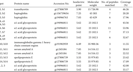
means of electrophoresis. The protein profiles either before or after calving were similar in periparturient dairy cows and representative gels were chosen to il-lustrate the protein profiles generated in the current study (Figure 1). After the software analysis, fourteen of the protein spots increased at parturition when compared with day 21 before calving in healthy cows and eighteen of the protein spots increased on days 1 and 21 after calving in cows with subclinical mastitis (Figure 1). Protein profile from day 21 before calving to day 1 after calving was similar in cows with or without subclinical mastitis, and protein profile on day 21 after calving exhibited a significant difference. Figure 1 presents relative abundance of protein spots from transition dairy cows with or without subclini-cal mastitis after subclini-calving. These protein spots were selected for follow-up identification. Proteins were identified in plasma samples from dairy cows with or without subclinical mastitis by the sequencing of at least 2 peptides as shown in Table 1.
[image:4.595.69.533.511.761.2]The α1 acid glycoprotein (AGP) and haptoglo-bin were abruptly increased at parturition and then decreased on day 21, while transthyretin was down-regulated at parturition and then increased on day 21 after calving in healthy dairy cows. After plasma enrichment with ProteoMiner, serum amy-loid A was increased before calving and down-reg-ulated on day 21 after calving, while apolipoprotein E was up-regulated after calving in healthy cows. AGP and haptoglobin were abruptly increased,
Table 1. Changes of plasma proteins in the transition cows identfied by ion trap mass spectrometry
Spot Protein name Accession No. Isoelectric point Molecular weight No. of peptides matched Coverage (%)
a1, b1 transthyretin gi:27806789 5.90 15 726.98 4 46.26
a2, b2 haptoglobin gi:156739654 7.25 43 383 7 20.20
a3, b3 haptoglobin gi:94966763 7.83 45 629 6 17.96
a4, a5
b4, b5 α1 acid glycoprotein gi:94966811 5.62 23 182.5 9 43.56 a6, b6 α1 acid glycoprotein gi:94966811 5.62 23 182.5 11 53.96 a7, b7 α1 acid glycoprotein gi:94966811 5.62 23 182.5 7 37.13 a8, a9
b8, b9 α1 acid glycoprotein gi:94966811 5.62 23 182.5 6 32.67 a10, b10 immunoglobulin gamma 1 heavy chain constant region gi:91982959 6.49 35 900.36 3 11.55
a11, b11 serum amyloid A gi:245184 7.85 14 516.12 2 30.63
a12, b12 serum amyloid A gi:245184 7.85 14 516.12 3 22.52
a13, b13 apolipoprotein E gi:27806739 5.55 35 979.85 5 23.42 a14, b14 apolipoprotein E gi:27806739 5.55 35 979.85 4 17.09 1, 2, 3 α1 acid glycoprotein gi:94966811 5.62 23 182.5 7 42.08
Figure 1. Analysis of expression protein patterns of plasma in transition dairy cows with or without subclinical mastitis
while transthyretin was down-regulated on days 1 and 21 after calving compared with day 21 before calving in cows with subclinical mastitis. Serum am-yloid A in cows with subclinical mastitis was similar to that in healthy cows before calving, but sharply increased on day 21 after calving. In addition, there were four spots on day 21 after calving identified as AGP that were up-regulated in cows with subclinical mastitis when compared with healthy cows.
Western blot analysis
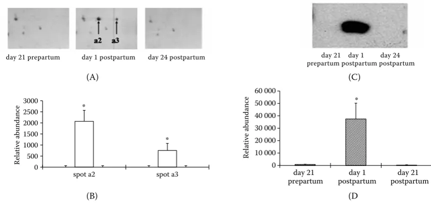
[image:6.595.77.508.80.283.2]To confirm the proteomic data, expression of haptoglobin in plasma samples from transition dairy cows with or without subclinical mastitis after calving was further confirmed by Western blot analysis (Figures 2 and 3). The proteomic data showed an abrupt increase in level of haptoglobin at parturition in healthy dairy cows, and continued Figure 2. Changes of haptoglobin in plasma of healthy dairy cows during the transition period
(A) representative images of haptoglobin spots in the 2-DE map and (B) densitometric analysis; (C) Western blot analysis of haptoglobin and (D) densitometric analysis
*P < 0.05 (n = 3)
Figure 3. Changes of haptoglobin in plasma of dairy cows during the transition period associated with a new intramam-mary infection after calving
(A) representative images of haptoglobin spots in the 2-DE map and (B) densitometric analysis; (C) Western blot analysis of haptoglobin and (D) densitometric analysis
*P < 0.05 (n = 3)
0 500 1000 1500 2000 2500 3000
spot b2 spot b3
Re
la
tive
a
bun
da
nc
e
(A) (C)
0 10 000 20 000 30 000 40 000 50 000 60 000
day 21
prepartum postpartum day 1 postpartumday 21
Re
la
tive
a
bun
da
nce
(B) (D)
day 21 prepartum day 1 postpartum day 24 postpartum day 21 day 1 day 24 prepartum postpartum postpartum
* *
* *
*
*
0 500 1000 1500 2000 2500 3000
spot a2 spot a3
Re
la
tive
a
bun
da
nc
e
0 10 000 20 000 30 000 40 000 50 000 60 000
Re
la
tive
a
bu
nd
ance
day 21
prepartum postpartum day 1 postpartumday 21
(A) (C)
(B) (D
day 21 prepartum day 1 postpartum day 24 postpartum day 21 day 1 day 24 prepartum postpartum postpartum
*
*
[image:6.595.74.520.485.684.2]to be up-regulated in plasma 21 day postpartum in cows with subclinical mastitis. Results from Western blot analysis were consistent with data from 2-DE followed by mass spectrometry.
One protein with multiple spots
There were multiple spots with varying masses and isoelectric points present on the gels of plasma col-lected after calving that revealed a marked increase in abundance of AGP. Two spots (spots a2 and a3, or spots b2 and b3) were detected on the gels with different isoelectric points but similar molecular weight, which were identified as haptoglobin, and two other spots (spots a11 and a12, or spots b11 and b12) were identified as serum amyloid A. Multiple spots may be due to the presence of genetic variants or post-translational modifications. These spots, identified by ion trap mass spectrometry as identi-cal proteins, had similar peptide sequences (Table 1).
DISCUSSION
Results of the current study demonstrate that protein profiles change in transition dairy cows with or without subclinical mastitis after calv-ing. The changes of AGP, haptoglobin, and serum amyloid A indicate that the current methods were successful in identifying differentially expressed plasma proteins in periparturient dairy cows.
Western blot analyses of haptoglobin in plasma in transition dairy cows with or without subclini-cal mastitis were found to be highly correlated and agree with the 2-DE in combination with mass spectrometry data.
Enhanced detection of minor serum proteins can be achieved by selective removal of major proteins using specific immunoaffinity columns and/or pro-tein fractionations based on charge, size or hydro-phobicity (Fountoulakis et al., 2004; Echan et al., 2005). Recently, hexapeptides library was shown to enable detection of many new proteins and to im-prove detection of minor proteins as compared to the depletion technique (Sihlbom et al., 2008). This new method could lead to determining both ma-jor proteins that are reduced and less predominant proteins that are concentrated, thus enabling detec-tion of all proteins within a single sample (Boschetti and Righetti, 2008; Hartwig et al., 2009). Recently, Marco-Ramell and Bassols (2010) have analyzed the
efficiency and reproducibility of the ProteoMiner with bovine and porcine serum samples enriched with low-abundance proteins.
Haptoglobin, AGP, and serum amyloid A are pri-marily synthesized and secreted by hepatocytes. Indeed, parturition is an inflammatory process (Norman et al., 2007). Haptoglobin, which acts as an acute phase protein, was shown by ELISA detection to be higher at week 1 postpartum, and then decreased in cows without a new subclini-cal intramammary infection (Piccinini et al., 2004; Rezamand et al., 2007). Using 2-DE and mass spec-trometry to analyze serum in periparturient dairy cows, haptoglobin and orosomucoid were abruptly increased at parturition as compared with the last phase of pregnancy (Cairoli et al., 2006). In ad-dition, haptoglobin has been shown to increase during inflammation (Humblet et al., 2006). This is associated with several non-specific immune parameter changes, such as decreased respiratory burst and decreased nitric oxide concentration after calving (Piccinini et al., 2004). In cows infected with mastitis, the concentrations of haptoglobin, AGP, and serum amyloid A in serum were significantly increased as compared to those of healthy cows (Eckersall et al., 2001; Lehtolainen et al., 2004). Our results were consistent with previous obser-vations that haptoglobin in plasma was increased at parturition in cows with and without mastitis but haptoglobin continued to be elevated 21 days postpartum in cows with subclinical mastitis.
AGP belongs to the subfamily of immunocalins, a group of binding proteins that also have immuno-modulatory functions. Glycosylation is one of the most important ways to modulate both protein func-tion and lifespan. Glycosylafunc-tion microheterogeneity of AGP can be modified during disease (Ceciliani and Pocacqua, 2007). The AGP can also be used to adjust inflammation-induced hyporetinolemia in vitamin A-sufficient animals (Gieng and Rosales, 2006).
cows and up-regulated in cows with mastitis. This phenomenon is probably caused by the inflamma-tory process that was occurring during this period (Norman et al., 2007), which altered the plasma proteins at parturition. These changes may serve as an acute and temporal regulatory mechanism in response to parturition- and lactation-induced im-munosuppression. Thus, cows are more susceptible to an intramammary infection after calving.
Our results show that apolipoprotein E is up-reg-ulated in plasma after calving in early lactation cows with and without mastitis. It was reported that apo-lipoprotein E increased lipid uptake by macrophages in lipoprotein lipase deficient cows during pregnancy (Steinberg et al., 1996). Other researchers have shown that apolipoprotein E would stimulate the phospho-lipid transfer protein-mediation of surface fragments of triglyceride-rich lipoproteins as mediated by high-density lipoproteins (HDL) and promote HDL remod-elling (Dallinga-Thie et al., 2007), and also interfere with lipolysis by interacting with the lipoprotein li-pase activator, apolipoprotein C2 (Chalas et al., 2002).
CONCLUSION
In the current research with cows during the tran-sition period, the plasma proteome was separated by 2-DE and several proteins that were associated with immune function were identified by LC-MS/MS. Expression of serum amyloid A, AGP, and hapto-globin, which serve as positive acute phase reactant proteins in response to parturition and lactation stress, was found to be up-regulated at parturition. Proteins that were altered at parturition were acute and temporal regulatory mechanisms in response to stressors of parturition and lactation, and so cannot serve as potential markers for periparturient cows susceptible to subclinical mastitis. These findings may provide valuable insight on the changes occur-ring in plasma protein in periparturient dairy cows with or without subclinical mastitis after calving.
REFERENCES
Boschetti E., Righetti P.G. (2008): Hexapeptide combi-natorial ligand libraries: the march for the detection of the low-abundance proteome continues. Biotech-niques, 44, 663−665.
Cairoli F., Battocchio M., Veronesi M.C., Brambilla D., Conserva F., Eberini I., Wait R., Gianazza E. (2006):
Serum protein pattern during cow pregnancy: acute-phase proteins increase in the peripartum period. Elec-trophoresis, 27, 1617−1625.
Candiano G., Bruschi M., Musante L., Santucci L., Ghig-geri G.M., Carnemolla B., Orecchia P., Zardi L., Righetti P.G. (2004): Blue silver: a very sensitive colloidal Coomassie G-250 staining for proteome analysis. Elec-trophoresis, 25, 1327−1333.
Ceciliani F., Pocacqua V. (2007): The acute phase protein alpha1-acid glycoprotein: a model for altered glycosyla-tion during diseases. Current Protein and Peptide Sci-ence, 8, 91−108.
Chalas J., Audibert F., Francoual J., Le Bihan B., Frydman R., Lindenbaum A. (2002): Concentrations of apolipo-proteins E, C2, and C3 and lipid profile in preeclamp-sia. Hypertension in Pregnancy, 21, 199−204.
Dallinga-Thie G.M., Dullaart R.P., van Tol A. (2007): Con-certed actions of cholesteryl ester transfer protein and phospholipid transfer protein in type 2 diabetes: effects of apolipoproteins. Current Opinion in Lipidology, 18, 251−257.
Drackley J.K. (1999): ADSA Foundation Scholar Award. Biology of dairy cows during the transition period: the final frontier? Journal of Dairy Science, 82, 2259−2273. Echan L.A., Tang H.Y., Ali-Khan N., Lee K., Speicher D.W. (2005): Depletion of multiple high-abundance proteins improves protein profiling capacities of human serum and plasma. Proteomics, 5, 3292−3303.
Eckersall P.D., Young F.J., McComb C., Hogarth C.J., Safi S., Weber A., McDonald T., Nolan A.M., Fitzpatrick J.L. (2001): Acute phase proteins in serum and milk from dairy cows with clinical mastitis. The Veterinary Record, 148, 35−41.
Fountoulakis M., Juranville J.F., Jiang L., Avila D., Roder D., Jakob P., Berndt P., Evers S., Langen H. (2004): De-pletion of the high-abundance plasma proteins. Amino Acids, 27, 249−259.
Gieng S.H., Rosales F.J. (2006): Plasma alpha1-acid gly-coprotein can be used to adjust inflammation-induced hyporetinolemia in vitamin A-sufficient, but not vita-min A-deficient or -supplemented rats. Journal of Nu-trition, 136, 1904−1909.
Gonçalves L.D.R., Soares M.R., Nogueira F., Garcia C., Camisasca D.R., Domont G., Feitosa A.C.R., Pereira D.A., Zingali R.B., Alves G. (2010): Comparative pro-teomic analysis of whole saliva from chronic periodon-titis patients. Journal of Proteomics, 73, 1334−1341. Hartwig S., Czibere A., Kotzka J., Passlack W., Haas R.,
Humblet M.F., Guyot H., Boudry B., Mbayahi F., Hanzen C., Rollin F., Godeau J.M. (2006): Relationship between haptoglobin, serum amyloid A, and clinical status in a survey of dairy herds during a 6-month period. Vet-erinary Clinical Pathology, 35, 188−193.
Jafari A., Emmanuel D.G., Christopherson R.J., Thomp-son J.R., Murdoch G.K., Woodward J., Field C.J., Ame- taj B.N. (2006): Parenteral administration of glutamine modulates acute phase response in postparturient dairy cows. Journal of Dairy Science, 89, 4660−4668. Jensen L.E., Whitehead A.S. (1998): Regulation of serum
amyloid A protein expression during the acute-phase response. The Biochemical Journal, 334, 489−503. Kao S.H., Wong H.K., Chiang C.Y., Chen H.M. (2008):
Evaluating the compatibility of three colorimetric pro-tein assays for two-dimensional electrophoresis ex-periments. Proteomics, 8, 2178−2184.
Kehrli M.E., Nonnecke Jr. B.J., Roth J.A. (1989): Altera-tions in bovine neutrophil function during the peri-parturient period. American Journal of Veterinary Research, 50, 207−214.
Lehtolainen T., Rontved C., Pyorala S. (2004): Serum amyloid A and TNF alpha in serum and milk during experimental endotoxin mastitis. Veterinary Research, 35, 651−659.
Lippolis J.D., Peterson-Burch B.D., Reinhardt T.A. (2006): Differential expression analysis of proteins from neu-trophils in the periparturient period and neuneu-trophils from dexamethasone-treated dairy cows. Veterinary Immunology and Immunopathology, 111, 149−164. Liu G., Zhang C., Wang J., Bu D., Zhou L., Zhao S., Li S.
(2010): Canonical correlation of milk immunoglobu-lins, lactoferrin concentration and Dairy Herd Im-provement data of Chinese Holstein cows. Livestock Science, 128, 197−200.
Loor J.J., Dann H.M., Everts R.E., Oliveira R., Green C.A., Guretzky N.A., Rodriguez-Zas S.L., Lewin H.A., Drack-ley J.K. (2005): Temporal gene expression profiling of liver from periparturient dairy cows reveals complex adaptive mechanisms in hepatic function. Physiologi-cal Genomics, 23, 217−226.
Madsen S.A., Weber P.S., Burton J.L. (2002): Altered ex-pression of cellular genes in neutrophils of periparturient dairy cows. Veterinary Immunology and Immunopathol-ogy, 86, 159−175.
Marco-Ramell A., Bassols A. (2010): Enrichment of low-abundance proteins from bovine and porcine serum samples for proteomic studies. Research in Veterinary Science, 89, 340−343.
Meglia G.E., Johannisson A., Agenas S., Holtenius K., Waller K.P. (2005): Effects of feeding intensity during the dry period on leukocyte and lymphocyte sub-popu- lations, neutrophil function and health in periparturi-ent dairy cows. The Veterinary Journal, 169, 376−384. Mollenhorst H., Van der Tol P., Hogeveen H. (2010): So-matic cell count assessment at the quarter or cow milk-ing level. Journal of Dairy Science, 93, 3358−3364. Norman J.E., Bollapragada S., Yuan M., Nelson S.M. (2007):
Inflammatory pathways in the mechanism of parturition. BMC Pregnancy and Childbirth, 7 (Suppl. 1), S7. Piccinini R., Binda E., Belotti M., Casirani G., Zecconi
A. (2004): The evaluation of non-specific immune sta-tus of heifers in field conditions during the peripartu-rient period. Veterinary Research, 35, 539−550. Pyörälä S. (2008): Mastitis in post-partum dairy cows.
Re-production in Domestic Animals, 43 (Suppl. 2), 252−259. Rezamand P., Hoagland T.A., Moyes K.M., Silbart L.K.,
Andrew S.M. (2007): Energy status, lipid-soluble vita-mins, and acute phase proteins in periparturient Hol-stein and Jersey dairy cows with or without subclinical mastitis. Journal of Dairy Science, 90, 5097−5107. Sihlbom C., Kanmert I., Bahr H., Davidsson P. (2008):
Evaluation of the combination of bead technology with SELDI-TOF-MS and 2-D DIGE for detection of plasma proteins. Journal of Proteome Research, 7, 4191−4198. Steel L.F., Shumpert D., Trotter M., Seeholzer S.H., Evans
A.A., London W.T., Dwek R., Block T.M. (2003): A strategy for the comparative analysis of serum pro-teomes for the discovery of biomarkers for hepatocel-lular carcinoma. Proteomics, 3, 601−609.
Steinberg F.M., Tsai E.C., Brunzell J.D., Chait A. (1996): ApoE enhances lipid uptake by macrophages in lipo-protein lipase deficiency during pregnancy. Journal of Lipid Research, 37, 972−984.
Valde J.P., Lawson L.G., Lindberg A., Agger J.F., Saloniemi H., Osteras O. (2004): Cumulative risk of bovine mas-titis treatments in Denmark, Finland, Norway and Sweden. Acta Veterinaria Scandinavica, 45, 201−210.
Received: 2011−09−22 Accepted after corrections: 2012−06−01
Corresponding Author
Prof. Jiaqi Wang, Chinese Academy of Agricultural Sciences, Institute of Animal Science, State Key Laboratory of Animal Nutrition, No.2 Yuan Ming Yuan West Road, Haidian District, 100193 Beijing, P.R.China