Received 4 December 2016 Accepted 19 December 2016
Edited by A. Van der Lee, Universite´ de Montpellier II, France
Keywords:crystal structure; powder diffraction; density functional theory; citrate; rubidium.
CCDC references:1523504; 1523503
Supporting information:this article has supporting information at journals.iucr.org/e
Crystal structure of dirubidium hydrogen citrate
from laboratory X-ray powder diffraction data and
DFT comparison
Alagappa Rammohanaand James A. Kadukb*
a
Atlantic International University, Honolulu, HI, USA, andbIllinois Institute of Technology, Chicago, IL, USA. *Correspondence e-mail: [email protected]
The crystal structure of dirubidium hydrogen citrate, 2Rb+HC6H5O7 2
, has been solved and refined using laboratory X-ray powder diffraction data, and optimized using density functional techniques. The un-ionized carboxylic acid group forms helical chains of very strong hydrogen bonds (O O2.42 A˚ ) along the b axis. The hydroxy group participates in a chain of intra- and intermolecular hydrogen bonds along thecaxis. These hydrogen bonds result in corrugated hydrogen-bonded layers in the bc plane. The Rb+cations are
six-coordinate, and share edges and corners to form layers in the ab plane. The interlayer contacts are composed of the hydrophobic methylene groups.
1. Chemical context
In the course of a systematic study of the crystal structures of Group 1 (alkali metal) citrate salts to understand the confor-mational flexibility, ionization, coordination tendencies, and hydrogen bonding of the anion, we have determined several new crystal structures. Most of the new structures were solved using powder diffraction data (laboratory and/or synchro-tron), but single crystals were used where available. The general trends and conclusions about the 16 new compounds and 12 previously characterized structures are being reported separately (Rammohan & Kaduk, 2017). Six of the new structures, i.e. NaKHC6H5O7, NaK2C6H5O7, Na3C6H5O7,
NaH2C6H5O7, Na2HC6H5O7, and K3C6H5O7, have been
published recently (Rammohan & Kaduk, 2016a,b,c,d,e; Rammohan et al., 2016), and two additional structures, i.e. KH2C6H5O7 and KH2C6H5O7(H2O)2, have been
commu-nicated (Kaduk & Stern, 2016a,b) to the Cambridge Structural Database (Groomet al., 2016).
2. Structural commentary
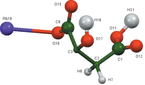
The asymmetric unit of the title compound is shown in Fig. 1. The r.m.s. deviation of the non-H atoms in the Rietveld refined and DFT-optimized structures is 0.052 A˚ (Fig. 2), and the maximum deviation is 0.083 A˚ , at atom C1. The good agreement between the two structures is strong evidence that the experimental structure is correct (van de Streek & Neumann, 2014). This discussion uses the DFT-optimized structure. Most of the bond lengths, bond angles, and torsion angles fall within the normal ranges indicated by a Mercury Mogul Geometry Check(Macraeet al., 2008). The C1—C2— C3 angle of 111.1 is flagged as unusual [Z-score = 2.7;
average = 114.3 (11)]. The Z-score is the result of the low
standard uncertainty on the average; the absolute difference of 3.2 is well within the expected range of such angles. The
citrate anion occurs in the trans,trans-conformation, which is one of the two low-energy conformations of an isolated citrate. The central carboxylate group and the hydroxy group lie on the mirror plane. The central carboxylate O15 atom and the terminal carboxylic acid O11 atom chelate to Rb19, and the central carboxylate O16 atom and the terminal carboxylic acid O11 atom chelate to another Rb19. The Mulliken overlap populations and atomic charges indicate that the metal– oxygen bonding is ionic.
The Bravais–Friedel–Donnay–Harker (Bravais, 1866; Friedel, 1907; Donnay & Harker, 1937) morphology suggests that we might expect a platy morphology for dirubidium hydrogen citrate, with {020} as the principal faces. A 4th order spherical harmonic texture model was included in the
refine-ment. The texture index was 1.078, indicating that preferred orientation was significant for this rotated flat plate specimen.
3. Supramolecular features
The Rb cation is six-coordinate (bond-valence sum = 0.96). The coordination polyhedra share corners and edges to form layers in theabplane (Fig. 3). The un-ionized terminal carb-oxylic acid forms a very strong symmetric hydrogen bond (Table 1). The Mulliken overlap population in the hydrogen-acceptor bond is 0.161 e. By the correlation in Rammohan & Kaduk (2017), this hydrogen bond accounts for 21.9 kcal mol 1of crystal energy. The hydroxy group participates in two hydrogen bonds to ionized central carboxylate groups; one is intramolecular with graph-set motif S(5), and the other is intermolecular. These hydrogen bonds contribute 9.3 and 8.6 kcal mol 1to the crystal energy.
4. Database survey
Details of the comprehensive literature search for citrate structures are presented in Rammohan & Kaduk (2017). A reduced cell search of the cell of dirubidium hydrogen citrate in the Cambridge Structural Database (Groom et al., 2016) (increasing the default tolerance from 1.5 to 2.0%) yielded 12 hits, but limiting the chemistry to C, H, Rb, and O only resulted in no hits. The powder pattern is now contained in the the Powder Diffraction File (ICDD, 2016) as entry 00-063-1541.
research communications
Acta Cryst.(2017). E73, 92–95 Rammohan and Kaduk 2Rb+HC
[image:2.610.314.564.69.175.2] [image:2.610.44.295.103.153.2]6H5O72
93
Figure 1The asymmetric unit of the title compound, showing the atom numbering. The atoms are represented by 50% probability spheroids.
Figure 2
Comparison of the refined and optimized structures of dirubidium hydrogen citrate. The refined structure is in red and the DFT-optimized structure is in blue.
Table 1
Hydrogen-bond geometry (A˚ , ) for the DFT-optimized structure of dirubidium hydrogen citrate.
D—H A D—H H A D A D—H A
O11—H21 O11 1.209 1.209 2.418 180.0
O17—H18 O15 0.979 1.992 2.611 119.0
O17—H18 O16 0.979 1.992 3.216 148.6
Figure 3
The crystal structure of dirubidium hydrogen citrate, viewed down thea
[image:2.610.46.299.567.716.2] [image:2.610.306.569.624.714.2]5. Synthesis and crystallization
H3C6H5O7(H2O) (2.0768 g, 10.0 mmol) was dissolved in 10 ml
deionized water. Rb2CO3 (10.0 mmol, 2.3170 g, Sigma–
Aldrich) was added to the citric acid solution slowly with stirring. The resulting clear colorless solution was evaporated to dryness in an oven at 333 K.
6. Refinement
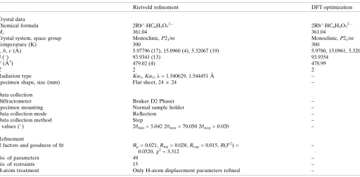
Crystal data, data collection and structure refinement details are summarized in Table 2. Entering 22 peaks (after manually applying a constant 2shift to approximate specimen displa-cement effects) into ITO/CRYSFIRE (Visser, 1969; Shirley, 2002) yielded a primitive monoclinic cell havinga= 5.978,b= 15.096, c = 5.320 A˚ , = 93.93, V = 478.33 A˚3
, and Z = 2. Processing the pattern in DASH3.2 (David et al., 2006) suggested that the most probable space group wasP21, but no
acceptable solution was found. A peak list was created from the results of a Le Bail fit using the REFLIST option inGSAS, and imported intoEndeavour1.7b(Putzet al., 1999). Using a citrate, two Rb atoms, and the O atom of a water molecule as fragments yielded a successful structure solution. In the initial refinements, the water molecule moved very close to one of the Rb atoms, and so was removed from the refinement.
Pseudo-Voigt profile coefficients were as parameterized in Thompsonet al.(1987) with profile coefficients for Simpson’s rule integration of the pseudo-Voigt function according to Howard (1982). The asymmetry correction of Finger et al. (1994) was applied, and microstrain broadening by Stephens
[image:3.610.49.561.90.342.2](1999). The structure was refined by the Rietveld (Fig. 4) method usingGSAS/EXPGUI(Larson & Von Dreele, 2004; Toby, 2001). All C—C and C—O bond lengths were restrained, as were all bond angles. The H atoms were included at fixed positions, which were recalculated during the course of the refinement using Materials Studio (Dassault Table 2
Experimental details.
Rietveld refinement DFT optimization
Crystal data
Chemical formula 2Rb+HC6H5O72 2Rb+HC6H5O72
Mr 361.04 361.04
Crystal system, space group Monoclinic,P21/m Monoclinic,P21/m
Temperature (K) 300 300
a,b,c(A˚ ) 5.97796 (17), 15.0960 (4), 5.32067 (19) 5.9780, 15.0961, 5.3207
(
) 93.9341 (13) 93.9354
V(A˚3) 479.02 (4) 478.99
Z 2 2
Radiation type K1,K2,= 1.540629, 1.544451 A˚ –
Specimen shape, size (mm) Flat sheet, 2424 –
Data collection
Diffractometer Bruker D2 Phaser –
Specimen mounting Normal sample holder –
Data collection mode Reflection –
Data collection method Step –
values (
) 2min= 5.042 2max= 70.050 2step= 0.020 –
Refinement
Rfactors and goodness of fit Rp= 0.021,Rwp= 0.028,Rexp= 0.015,R(F2) = 0.0520,2
= 3.312
–
No. of parameters 49 –
No. of restraints 15 –
H-atom treatment Only H-atom displacement parameters refined –
[image:3.610.314.565.479.668.2]Computer programs:DIFFRAC.Measurement(Bruker, 2009),Endeavour(Putzet al., 1999),GSAS(Larson & Von Dreele, 2004),DIAMOND(Crystal Impact, 2015) andpublCIF (Westrip, 2010).
Figure 4
Systemes, 2014). TheUisoof the atoms in the central and outer
portions of the citrate were constrained to be equal, and the Uisoof the H atoms were constrained to be 1.3 times those of
the atoms to which they are attached.
The structure was solved and initially refined in the space groupP21. The ADDSYM module ofPLATON(Spek, 2009)
suggested the presence of an additional centre of symmetry, and that the space group wasP21/m. Refinement in this space
group yielded slightly better residuals (Rwp = 0.0277 and
reduced2= 3.3236, compared toRwp= 0.0282 and 2
= 3.454 forP21), and we believe thatP21/mis the correct space group.
Stoichiometry requires one carboxylic acid proton per citrate. The space group P21 is consistent with ordered
asymmetric hydrogen bonds, while P21/m is consistent with
both disordered asymmetric hydrogen bonds or symmetric hydrogen bonds. Crystallographically, it would be difficult to distinguish these two possibilities, especially using X-ray powder diffraction data. DFT calculations on the asymmetric (P21) and symmetric (P21/m) hydrogen-bond models indicate
that the symmetric model is 0.2 kcal mol 1 lower in energy. This difference is within the expected error range of such calculations. Since the crystallography strongly indicates the higher symmetry, we believe that the P21/m model with
symmetric hydrogen bonds is the best model for this structure.
7. DFT calculations
After the Rietveld refinement, a density functional geometry optimization (fixed experimental unit cell) was carried out usingCRYSTAL14(Dovesiet al., 2014). The basis sets for the C, H, and O atoms were those of Peintingeret al.(2012), and the basis set for Rb was that of Schoenes et al.(2008). The calculation was run on eight 2.1 GHz Xeon cores (each with 6 Gb RAM) of a 304-core Dell Linux cluster at IIT, used 8 k-points and the B3LYP functional, and took about 5 h. TheUiso
from the Rietveld refinement were assigned to the optimized fractional coordinates.
Acknowledgements
We thank Andrey Rogachev for the use of computing resources at IIT.
References
Bravais, A. (1866). In Etudes Cristallographiques. Paris: Gauthier Villars.
Bruker (2009).DIFFRAC.Measurement. Bruker AXS Inc., Madison, Wisconsin, USA.
Crystal Impact (2015). DIAMOND. Crystal Impact GbR, Bonn, Germany. http://www.crystalimpact.com/diamond.
Dassault Systemes (2014). Materials Studio. BIOVIA, San Diego, California, USA.
David, W. I. F., Shankland, K., van de Streek, J., Pidcock, E., Motherwell, W. D. S. & Cole, J. C. (2006).J. Appl. Cryst.39, 910– 915.
Donnay, J. D. H. & Harker, D. (1937).Am. Mineral.22, 446–467. Dovesi, R., Orlando, R., Erba, A., Zicovich-Wilson, C. M., Civalleri,
B., Casassa, S., Maschio, L., Ferrabone, M., De La Pierre, M., D’Arco, P., Noel, Y., Causa, M., Rerat, M. & Kirtman, B. (2014).Int. J. Quantum Chem.114, 1287–1317.
Finger, L. W., Cox, D. E. & Jephcoat, A. P. (1994).J. Appl. Cryst.27, 892–900.
Friedel, G. (1907).Bull. Soc. Fr. Mineral.30, 326–455.
Groom, C. R., Bruno, I. J., Lightfoot, M. P. & Ward, S. C. (2016).Acta Cryst.B72, 171–179.
Howard, C. J. (1982).J. Appl. Cryst.15, 615–620.
ICDD (2016).PDF-4+ 2015 and PDF-4 Organics 2016 (Databases), edited by S. Kabekkodu. International Centre for Diffraction Data, Newtown Square PA, USA.
Kaduk, J. A. & Stern, C. (2016a). CSD Communication 1446457-1446458. CCDC, Cambridge, England.
Kaduk, J. A. & Stern, C. (2016b). CSD Communication 1446460-1446461. CCDC, Cambridge, England.
Larson, A. C. & Von Dreele, R. B. (2004).General Structure Analysis System (GSAS). Report LAUR 86-784 Los Alamos National Laboratory, New Mexico, USA.
Macrae, C. F., Bruno, I. J., Chisholm, J. A., Edgington, P. R., McCabe, P., Pidcock, E., Rodriguez-Monge, L., Taylor, R., van de Streek, J. & Wood, P. A. (2008).J. Appl. Cryst.41, 466–470.
Peintinger, M. F., Vilela Oliveira, D. & Bredow, T. (2012).J. Comput. Chem.34, 451–459.
Putz, H., Scho¨n, J. C. & Jansen, M. (1999).J. Appl. Cryst.32, 864–870. Rammohan, A. & Kaduk, J. A. (2017). Submitted toActa Cryst.B72
[hw5042].
Rammohan, A. & Kaduk, J. A. (2016a).Acta Cryst.E72, 170–173. Rammohan, A. & Kaduk, J. A. (2016b).Acta Cryst.E72, 403–406. Rammohan, A. & Kaduk, J. A. (2016c).Acta Cryst.E72, 793–796. Rammohan, A. & Kaduk, J. A. (2016d).Acta Cryst.E72, 854–857. Rammohan, A. & Kaduk, J. A. (2016e).Acta Cryst.E72, 1159–1162. Rammohan, A., Sarjeant, A. A. & Kaduk, J. A. (2016).Acta Cryst.
E72, 943–946.
Schoenes, J., Racu, A.-M., Doll, K., Bukowski, Z. & Karpinski, J. (2008).Phys. Rev. B,77, 134515.
Shirley, R. (2002).The Crysfire 2002 system for automatic powder indexing. Guildford, Surrey, England.
Spek, A. L. (2009).Acta Cryst.D65, 148–155. Stephens, P. W. (1999).J. Appl. Cryst.32, 281–289.
Streek, J. van de & Neumann, M. A. (2014).Acta Cryst.B70, 1020– 1032.
Thompson, P., Cox, D. E. & Hastings, J. B. (1987).J. Appl. Cryst.20, 79–83.
Toby, B. H. (2001).J. Appl. Cryst.34, 210–213. Visser, J. W. (1969).J. Appl. Cryst.2, 89–95. Westrip, S. P. (2010).J. Appl. Cryst.43, 920–925.
research communications
Acta Cryst.(2017). E73, 92–95 Rammohan and Kaduk 2Rb+HC
sup-1 Acta Cryst. (2017). E73, 92-95
supporting information
Acta Cryst. (2017). E73, 92-95 [https://doi.org/10.1107/S2056989016020168]
Crystal structure of dirubidium hydrogen citrate from laboratory X-ray powder
diffraction data and DFT comparison
Alagappa Rammohan and James A. Kaduk
Computing details
Data collection: DIFFRAC.Measurement (Bruker, 2009) for RAMM020C_publ. Program(s) used to solve structure:
Endeavour (Putz et al., 1999) for RAMM020C_publ. Program(s) used to refine structure: GSAS for RAMM020C_publ.
Molecular graphics: DIAMOND (Crystal Impact, 2015) for RAMM020C_publ. Software used to prepare material for
publication: publCIF (Westrip, 2010) for RAMM020C_publ.
(RAMM020C_publ) Dirubidium hydrogen citrate
Crystal data
2Rb+·HC6H5O72− Mr = 361.04 Monoclinic, P21/m Hall symbol: -P 2yb a = 5.97796 (17) Å b = 15.0960 (4) Å c = 5.32067 (19) Å β = 93.9341 (13)°
V = 479.02 (4) Å3 Z = 2
Dx = 2.503 Mg m−3
Kα1, Kα2 radiation, λ = 1.540629, 1.544451 Å T = 300 K
white
flat_sheet, 24 × 24 mm
Specimen preparation: Prepared at 333 K
Data collection
Bruker D2 Phaser diffractometer
Specimen mounting: Normal sample holder
Data collection mode: reflection Scan method: step
2θmin = 5.042°, 2θmax = 70.050°, 2θstep = 0.020°
Refinement
Least-squares matrix: full Rp = 0.021
supporting information
sup-2 Acta Cryst. (2017). E73, 92-95
Profile function: CW Profile function number 4 with 21 terms Pseudovoigt profile coefficients as parameterized in P. Thompson, D.E. Cox & J.B. Hastings (1987). J. Appl. Cryst.,20,79-83. Asymmetry correction of L.W. Finger, D.E. Cox & A. P. Jephcoat (1994). J. Appl.
Cryst.,27,892-900. Microstrain broadening by P.W. Stephens, (1999). J. Appl.
Cryst.,32,281-289. #1(GU) = 142.783 #2(GV) = 0.000 #3(GW) = 4.751 #4(GP) = 0.000 #5(LX) = 5.874 #6(ptec) = -0.71 #7(trns) = 1.83 #8(shft) = -25.7226 #9(sfec) = 0.00 #10(S/L) = 0.0441 #11(H/L) = 0.0005 #12(eta) = 0.0000 #13(S400 ) = 3.9E-02 #14(S040 ) = 8.9E-05 #15(S004 ) = 3.1E-01 #16(S220 ) = 9.7E-03 #17(S202 ) = -6.4E-02 #18(S022 ) = 5.7E-04 #19(S301 ) = -8.2E-03 #20(S103 ) = 2.5E-02 #21(S121 ) = -8.8E-03 Peak tails are ignored where the intensity is below 0.0100 times the peak Aniso. broadening axis 0.0 1.0 0.0
49 parameters 15 restraints
Only H-atom displacement parameters refined Weighting scheme based on measured s.u.'s (Δ/σ)max = 0.08
Background function: GSAS Background function number 1 with 4 terms. Shifted Chebyshev function of 1st kind 1: 3185.87 2: -310.317 3: -87.5319 4: 63.9418
Fractional atomic coordinates and isotropic or equivalent isotropic displacement parameters (Å2)
x y z Uiso*/Ueq
C1 0.2727 (12) 0.4151 (4) 0.5145 (14) 0.0556 (15)*
C2 0.3859 (11) 0.33308 (16) 0.4236 (14) 0.026 (4)*
C3 0.2821 (12) 0.25 0.5368 (15) 0.026 (4)*
C6 0.0268 (13) 0.25 0.463 (2) 0.0556 (15)*
H7 0.56395 0.33136 0.47541 0.018 (5)*
H8 0.35795 0.33169 0.21841 0.018 (5)*
O11 0.1036 (11) 0.4467 (5) 0.3841 (15) 0.0556 (15)*
O12 0.3502 (12) 0.4512 (5) 0.7178 (14) 0.0556 (15)*
O15 −0.0968 (16) 0.25 0.650 (2) 0.0556 (15)*
O16 −0.0415 (16) 0.25 0.232 (2) 0.0556 (15)*
O17 0.3416 (14) 0.25 0.7999 (15) 0.0556 (15)*
H18 0.17127 0.25 0.87091 0.068 (2)*
Rb19 −0.2264 (3) 0.10523 (10) −0.0313 (3) 0.0590 (9)*
H21 0.0 0.0 0.5 0.07*
Geometric parameters (Å, º)
C1—C2 1.5070 (17) O12—Rb19iv 2.907 (7)
C1—O11 1.278 (5) O15—C6 1.281 (5)
sup-3 Acta Cryst. (2017). E73, 92-95
C2—C1 1.5070 (17) O15—Rb19vi 2.903 (7)
C2—C3 1.5400 (17) O16—C6 1.267 (5)
C2—H7 1.081 (7) O16—Rb19 2.784 (7)
C2—H8 1.093 (7) O16—Rb19i 2.784 (7)
C3—C2 1.5400 (17) O17—C3 1.420 (5)
C3—C2i 1.5400 (17) O17—H18 1.110 (8)
C3—C6 1.5499 (18) O17—Rb19vii 3.454 (7)
C3—O17 1.420 (5) O17—Rb19iv 3.455 (7)
C6—C3 1.5499 (18) H18—O17 1.110 (8)
C6—O15 1.281 (5) Rb19—O11viii 3.159 (7)
C6—O16 1.267 (5) Rb19—O11i 2.965 (7)
H7—C2 1.081 (7) Rb19—O12ix 2.984 (6)
H8—C2 1.093 (8) Rb19—O12x 2.907 (7)
O11—C1 1.278 (5) Rb19—O15xi 2.903 (7)
O11—Rb19ii 3.159 (7) Rb19—O16 2.784 (7)
O11—Rb19i 2.965 (7) Rb19—O17xii 3.454 (7)
O11—H21iii 1.209 (6) H21—O11ix 1.209 (6)
O12—C1 1.270 (5) H21—O11i 1.209 (6)
O12—Rb19iii 2.984 (6)
C2—C1—O11 119.2 (4) C6—O15—Rb19v 130.21 (19)
C2—C1—O12 118.7 (4) C6—O15—Rb19xv 130.21 (19)
O11—C1—O12 122.1 (4) Rb19v—O15—Rb19xv 97.7 (3)
C1—C2—C3 110.0 (4) C6—O16—Rb19 125.3 (2)
C1—C2—H7 113.2 (4) C6—O16—Rb19xiii 125.3 (2)
C1—C2—H8 17.29 (4) Rb19—O16—Rb19xiii 103.5 (4)
C3—C2—H7 107.3 (5) C3—O17—H18 99.3 (7)
C3—C2—H8 109.7 (5) O11viii—Rb19—O11xiii 94.2 (2)
H7—C2—H8 109.5 (5) O11viii—Rb19—O12ix 79.54 (16)
C2—C3—C2xiii 109.1 (4) O11viii—Rb19—O12xvi 74.7 (2)
C2—C3—C6 108.5 (4) O11viii—Rb19—O15xi 98.11 (18)
C2—C3—O17 107.8 (4) O11viii—Rb19—O16 142.5 (2)
C2xiii—C3—C6 108.5 (4) O11xiii—Rb19—O12ix 63.59 (19)
C2xiii—C3—O17 107.8 (4) O11xiii—Rb19—O12xvi 141.13 (14)
C6—C3—O17 115.2 (7) O11xiii—Rb19—O15xi 116.3 (2)
C3—C6—O15 114.4 (9) O11xiii—Rb19—O16 67.2 (2)
C3—C6—O16 119.5 (9) O12ix—Rb19—O12xvi 77.7 (2)
O15—C6—O16 126.1 (9) O12ix—Rb19—O15xi 177.6 (2)
C1—O11—Rb19ii 113.5 (7) O12ix—Rb19—O16 115.8 (3)
C1—O11—Rb19xiii 140.0 (4) O12xvi—Rb19—O15xi 102.2 (2)
Rb19ii—O11—Rb19xiii 85.8 (2) O12xvi—Rb19—O16 139.9 (2)
C1—O12—Rb19iii 136.0 (4) O15xi—Rb19—O16 65.8 (2)
C1—O12—Rb19xiv 121.4 (4) O11ix—H21—O11xiii 180.0
Rb19iii—O12—Rb19xiv 102.3 (2)
supporting information
sup-4 Acta Cryst. (2017). E73, 92-95
(ramm020c_DFT)
Crystal data
C6H6O7Rb2 Mr = 361.04 Monoclinic, P21/m Hall symbol: -P 2yb a = 5.9780 Å b = 15.0961 Å
c = 5.3207 Å β = 93.9354° V = 478.99 Å3 Z = 2
T = 300 K
Data collection
h = → k = →
l = →
Fractional atomic coordinates and isotropic or equivalent isotropic displacement parameters (Å2)
x y z Uiso*/Ueq
C1 0.28187 0.41704 0.52143 0.05270*
C2 0.38508 0.33363 0.42243 0.01400*
C3 0.27797 0.25000 0.53116 0.01400*
C6 0.02276 0.25000 0.45797 0.05270*
H7 0.56395 0.33136 0.47541 0.01820*
H8 0.35795 0.33169 0.21841 0.01820*
O11 0.10613 0.44671 0.38734 0.05270*
O12 0.35379 0.45283 0.72015 0.05270*
O15 −0.10430 0.25000 0.63801 0.05270*
O16 −0.03680 0.25000 0.22772 0.05270*
O17 0.31806 0.25000 0.79999 0.05270*
H18 0.17127 0.25000 0.87091 0.06840*
Rb19 −0.22414 0.10319 −0.03836 0.05980*
H21 0.00000 0.00000 0.50000 0.07000*
Bond lengths (Å)
C1—C2 1.513 Rb19—O11ii 2.997
C1—O11 1.308 Rb19—O16 2.820
C1—O12 1.238 Rb19—O12viii 2.966
C2—C3 1.546 C3—C2ii 1.546
C2—H7 1.087 C3—C6 1.548
C2—H8 1.087 C3—O17 1.434
O11—Rb19i 3.115 C6—O15 1.263
O11—Rb19ii 2.997 C6—O16 1.252
O11—H21iii 1.209 O15—Rb19ix 2.926
O12—Rb19iv 2.879 O15—Rb19x 2.926
O12—Rb19iii 2.966 O16—Rb19ii 2.820
Rb19—O12v 2.879 O17—H18 0.979
Rb19—O15vi 2.926 H21—O11ii 1.209
Rb19—O11vii 3.115 H21—O11viii 1.209
sup-5 Acta Cryst. (2017). E73, 92-95
Hydrogen-bond geometry (Å, º)
D—H···A D—H H···A D···A D—H···A
O11—H21···O11 1.209 1.209 2.418 180.0
O17—H18···O15 0.979 1.992 2.611 119.0