2-[1-(2-Hydroxy-6-methoxyphenyl)ethyl-idene]-
N
-methylhydrazinecarbothio-amide acetonitrile monosolvate
Brian J. Anderson, Alexander M. Keeler, Kelly A. O’Rourke, Shannon T. Krauss and Jerry P. Jasinski*
Department of Chemistry, Keene State College, 229 Main Street, Keene, NH 03435–2001, USA
Correspondence e-mail: jjasinski@keene.edu
Received 20 November 2012; accepted 27 November 2012
Key indicators: single-crystal X-ray study;T= 173 K; mean(C–C) = 0.003 A˚;
Rfactor = 0.063;wRfactor = 0.170; data-to-parameter ratio = 16.0.
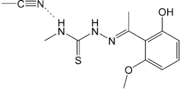
In the title compound, C11H15N3O2SC2H3N, the dihedral
angle between the benzene ring and the mean plane of the hydrazinecarbothioamide group is 75.1 (2). In the crystal, the
main molecule is linked to the solvent molecule by a weak N— H N hydrogen bond while O—H S hydrogen bonds link the molecules into columns along [100].
Related literature
For thiosemicarbazone structures and their biological activity, see: Lobanaet al.(2009). For thiosemicarbazone as ligands for hydrogenations or metal-catalysed reactions, see: Pelagattiet al. (1998); Xie et al. (2010). For a related structure, see: Andersonet al.(2012). For standard bond lengths, see: Allen
et al.(1987).
Experimental
Crystal data
C11H15N3O2SC2H3N
Mr= 294.38
Triclinic,P1
a= 7.6232 (10) A˚
b= 9.4004 (9) A˚
c= 11.8031 (12) A˚
= 80.121 (8)
= 71.555 (10)
= 74.732 (10)
V= 770.44 (16) A˚3
Z= 2
CuKradiation
= 1.93 mm1
T= 173 K
0.440.280.12 mm
Data collection
Agilent Xcalibur (Eos, Gemini CCD) diffractometer
Absorption correction: multi-scan (CrysAlis RED; Agilent, 2012)
Tmin= 0.575,Tmax= 1.000
4716 measured reflections 2968 independent reflections 2622 reflections withI> 2(I)
Rint= 0.020
Refinement
R[F2> 2(F2)] = 0.063
wR(F2) = 0.170
S= 1.06 2968 reflections
186 parameters
H-atom parameters constrained
max= 1.13 e A˚
3
min=0.26 e A˚
[image:1.610.82.261.525.617.2]3
Table 1
Hydrogen-bond geometry (A˚ ,).
D—H A D—H H A D A D—H A
O2—H2 S1i
0.84 2.34 3.1823 (17) 177 N1—H1 N1A 0.88 2.25 3.039 (3) 149
Symmetry code: (i)x;yþ2;zþ2.
Data collection: CrysAlis PRO(Agilent, 2012); cell refinement: CrysAlis PRO; data reduction: CrysAlis RED (Agilent, 2012); program(s) used to solve structure: SHELXS97 (Sheldrick, 2008); program(s) used to refine structure:SHELXL97(Sheldrick, 2008); molecular graphics:SHELXTL(Sheldrick, 2008); software used to prepare material for publication:SHELXTL.
JPJ acknowledges the NSF–MRI program (grant No. CHE1039027) for funds to purchase the X-ray diffractometer.
Supplementary data and figures for this paper are available from the IUCr electronic archives (Reference: RK2388).
References
Agilent (2012). CrysAlis PRO and CrysAlis RED Agilent Technologies, Yarnton, England.
Allen, F. H., Kennard, O., Watson, D. G., Brammer, L., Orpen, A. G. & Taylor, R. (1987).J. Chem. Soc. Perkin Trans. 2, pp. S1–19.
Anderson, B. J., Kennedy, C. J. & Jasinski, J. P. (2012).Acta Cryst.E68, o2982. Lobana, T. S., Sharma, R., Bawa, G. & Khanna, S. (2009).Coord. Chem. Rev.
253, 977–1055.
Pelagatti, P., Venturini, A., Carcelli, M., Costa, M., Bacchi, A., Pelizzi, G. & Pelizza, C. (1998).J. Chem. Soc. Dalton Trans.pp. 2715–2721.
Sheldrick, G. M. (2008).Acta Cryst.A64, 112–122.
Xie, G., Chellan, P., Mao, J., Chibale, K. & Smith, G. S. (2010).Adv. Synth. Catal.352, 1641–1647.
Acta Crystallographica Section E Structure Reports
Online
supporting information
Acta Cryst. (2013). E69, o11 [https://doi.org/10.1107/S1600536812048799]
2-[1-(2-Hydroxy-6-methoxyphenyl)ethylidene]-
N
-methylhydrazinecarbothio-amide acetonitrile monosolvate
Brian J. Anderson, Alexander M. Keeler, Kelly A. O
′
Rourke, Shannon T. Krauss and Jerry P.
Jasinski
S1. Comment
Thiosemicarbazones are an important class of ligands and their metal complexes and biological activity have been
investigated (Lobana, et al., 2009). More recently, thiosemicarbazones have been studied as ligands for metal catalyzed
reactions such as Mizoroki–Heck couplings (Xie et al., 2010) and hydrogenations (Pelagatti et al., 1998). A similar and
related structure has been reported (Anderson, et al., 2012). The crystal structure of a novel thiosemicarbazone molecule,
C13H18N4O2S, I, is reported here.
In I the dihedral angle between least squares planes of the benzene ring (C4–C9) and hydrazinecarbothioamide (N1/C2/S1/N2/N3) group is 75.1 (2)° (Fig. 1). Bond lengths are in normal ranges (Allen et al., 1987). Weak H—H···N
intramolecular interactions and O—H···S intermolecular interactions (Table 1) link the molecules into columns along [1 0
0] (Fig. 2).
S2. Experimental
A 50 mL round bottom flask was charged with 0.208 g (1.2 mmol) of 2′-hydroxy-6′-methoxyacetophenone and 0.126 g
(1.2 mmol) of 4-methyl-3-thiosemicarbazide followed by 20 mL of methanol, resulting in a clear yellow solution. The
solution was refluxed for 48 hours, and then the solvent was removed by rotary evaporation. The product was dissolved
into acetonitrile at 313 K and slowly allowed to cool to 273 K. Translucent crystals were observed after 48 hours. M.p.:
415–420 K.
S3. Refinement
All of the H atoms were placed in their calculated positions and then refined using the riding model with C—H lengths of
0.95Å (CH), 0.96Å (CH3), 0.88Å (NH) or 0.84Å (OH). The isotropic displacement parameters for these atoms were set to
Figure 1
The molecular structure of the title compound with the atom numbering scheme. Displacement ellipsoids are drawn at the
50% probability level. A weak H—H···N intramolecular interaction with the solvent acetonitrile molecule is shown with a
dashed line. H atoms are presented as small spheres of arbitrary radius.
Figure 2
Packing diagram viewed along the c axis. Weak H—H···N intramolecular interactions and O—H···S intermolecular
interactions which link the molecules into columns along [1 0 0] are shown by dashed lines.
2-[1-(2-Hydroxy-6-methoxyphenyl)ethylidene]-N- methylhydrazinecarbothioamide acetonitrile monosolvate
Crystal data
C11H15N3O2S·C2H3N Mr = 294.38
Triclinic, P1 Hall symbol: -P 1
[image:3.610.136.474.330.561.2]β = 71.555 (10)° γ = 74.732 (10)° V = 770.44 (16) Å3 Z = 2
F(000) = 312 Dx = 1.269 Mg m−3
Melting point = 415–420 K
Cu Kα radiation, λ = 1.54184 Å Cell parameters from 2112 reflections θ = 4.0–72.3°
µ = 1.93 mm−1 T = 173 K Chunk, colourless 0.44 × 0.28 × 0.12 mm
Data collection
Agilent Xcalibur (Eos, Gemini CCD) diffractometer
Radiation source: Enhance (Cu) X–ray Source Graphite monochromator
Detector resolution: 16.0416 pixels mm-1 ω scans
Absorption correction: multi-scan
(CrysAlis PRO and CrysAlis RED; Agilent, 2012)
Tmin = 0.575, Tmax = 1.000 4716 measured reflections 2968 independent reflections 2622 reflections with I > 2σ(I) Rint = 0.020
θmax = 72.4°, θmin = 4.0° h = −9→9
k = −7→11 l = −12→14
Refinement
Refinement on F2 Least-squares matrix: full R[F2 > 2σ(F2)] = 0.063 wR(F2) = 0.170 S = 1.06 2968 reflections 186 parameters 0 restraints
Primary atom site location: structure-invariant direct methods
Secondary atom site location: difference Fourier map
Hydrogen site location: inferred from neighbouring sites
H-atom parameters constrained w = 1/[σ2(F
o2) + (0.115P)2 + 0.1027P] where P = (Fo2 + 2Fc2)/3
(Δ/σ)max < 0.001 Δρmax = 1.13 e Å−3 Δρmin = −0.26 e Å−3
Special details
Geometry. All s.u.'s (except the s.u. in the dihedral angle between two l.s. planes) are estimated using the full covariance matrix. The cell s.u.'s are taken into account individually in the estimation of s.u.'s in distances, angles and torsion angles; correlations between s.u.'s in cell parameters are only used when they are defined by crystal symmetry. An approximate (isotropic) treatment of cell s.u.'s is used for estimating s.u.'s involving l.s. planes.
Refinement. Refinement of F2 against ALL reflections. The weighted R–factor wR and goodness of fit S are based on F2, conventional R–factors R are based on F, with F set to zero for negative F2. The threshold expression of F2 > σ(F2) is used only for calculating R–factors(gt) etc. and is not relevant to the choice of reflections for refinement. R–factors based on F2 are statistically about twice as large as those based on F, and R–factors based on ALL data will be even larger.
Fractional atomic coordinates and isotropic or equivalent isotropic displacement parameters (Å2)
x y z Uiso*/Ueq
S1 0.25112 (8) 1.21212 (6) 0.65436 (4) 0.0389 (2)
O1 0.6434 (2) 0.76106 (19) 0.83823 (13) 0.0433 (4)
O2 0.0253 (2) 0.7583 (2) 1.08053 (14) 0.0453 (4)
H2 −0.0446 0.7645 1.1514 0.068*
N1 0.2464 (3) 0.9666 (2) 0.57065 (15) 0.0378 (4)
H1 0.2499 0.8710 0.5813 0.045*
N2 0.2694 (3) 0.94030 (19) 0.76141 (15) 0.0362 (4)
H2A 0.2792 0.9762 0.8227 0.043*
C1 0.2309 (3) 1.0463 (3) 0.45605 (19) 0.0430 (5)
H1A 0.2248 0.9777 0.4042 0.064*
H1B 0.3419 1.0893 0.4176 0.064*
H1C 0.1155 1.1255 0.4691 0.064*
C2 0.2555 (3) 1.0302 (2) 0.66005 (18) 0.0330 (4)
C3 0.2951 (3) 0.7115 (2) 0.86217 (18) 0.0342 (5)
C4 0.3366 (3) 0.7630 (2) 0.96197 (17) 0.0334 (5)
C5 0.1988 (3) 0.7793 (2) 1.07313 (18) 0.0360 (5)
C6 0.2433 (3) 0.8147 (2) 1.16953 (19) 0.0403 (5)
H6 0.1500 0.8267 1.2449 0.048*
C7 0.4234 (3) 0.8320 (2) 1.15433 (19) 0.0404 (5)
H7 0.4531 0.8546 1.2206 0.049*
C8 0.5626 (3) 0.8174 (2) 1.0454 (2) 0.0378 (5)
H8 0.6857 0.8310 1.0363 0.045*
C9 0.5175 (3) 0.7824 (2) 0.94952 (18) 0.0348 (5)
C10 0.2904 (4) 0.5518 (2) 0.8712 (2) 0.0459 (6)
H10A 0.2653 0.5322 0.7996 0.069*
H10B 0.1898 0.5289 0.9426 0.069*
H10C 0.4129 0.4896 0.8774 0.069*
C11 0.8359 (3) 0.7636 (3) 0.8237 (2) 0.0465 (6)
H11A 0.9138 0.7373 0.7433 0.070*
H11B 0.8831 0.6921 0.8841 0.070*
H11C 0.8428 0.8632 0.8337 0.070*
N1A 0.2277 (5) 0.6691 (3) 0.5126 (3) 0.0736 (8)
C1A 0.2497 (4) 0.5798 (3) 0.4541 (2) 0.0530 (6)
C2A 0.2725 (7) 0.4659 (4) 0.3788 (3) 0.0844 (12)
H2AA 0.2316 0.5115 0.3076 0.127*
H2AB 0.1952 0.3951 0.4238 0.127*
H2AC 0.4062 0.4141 0.3541 0.127*
Atomic displacement parameters (Å2)
U11 U22 U33 U12 U13 U23
S1 0.0540 (4) 0.0364 (3) 0.0265 (3) −0.0125 (2) −0.0111 (2) −0.0006 (2)
O1 0.0434 (8) 0.0578 (10) 0.0286 (8) −0.0144 (7) −0.0074 (6) −0.0046 (7)
O2 0.0443 (9) 0.0651 (11) 0.0280 (8) −0.0151 (7) −0.0071 (6) −0.0096 (7)
N1 0.0521 (11) 0.0381 (9) 0.0228 (8) −0.0071 (8) −0.0123 (7) −0.0041 (7)
N2 0.0521 (10) 0.0347 (9) 0.0246 (8) −0.0098 (7) −0.0147 (7) −0.0029 (7)
N3 0.0423 (9) 0.0350 (9) 0.0264 (8) −0.0076 (7) −0.0101 (7) −0.0049 (7)
C1 0.0565 (13) 0.0500 (13) 0.0232 (10) −0.0094 (10) −0.0137 (9) −0.0049 (9)
C2 0.0343 (10) 0.0386 (10) 0.0235 (9) −0.0062 (8) −0.0066 (7) −0.0026 (8)
C3 0.0382 (10) 0.0375 (11) 0.0265 (10) −0.0088 (8) −0.0083 (8) −0.0035 (8)
C4 0.0463 (11) 0.0300 (10) 0.0238 (9) −0.0075 (8) −0.0123 (8) 0.0004 (7)
C5 0.0443 (11) 0.0373 (11) 0.0259 (9) −0.0072 (8) −0.0118 (8) −0.0010 (8)
C6 0.0496 (12) 0.0445 (12) 0.0248 (10) −0.0054 (9) −0.0104 (9) −0.0064 (8)
C7 0.0549 (13) 0.0399 (11) 0.0302 (10) −0.0077 (9) −0.0183 (9) −0.0058 (8)
C8 0.0456 (11) 0.0358 (11) 0.0351 (11) −0.0093 (9) −0.0166 (9) −0.0011 (8)
C10 0.0625 (14) 0.0360 (12) 0.0407 (12) −0.0123 (10) −0.0160 (11) −0.0031 (9) C11 0.0419 (12) 0.0558 (14) 0.0383 (12) −0.0108 (10) −0.0086 (10) −0.0008 (10) N1A 0.098 (2) 0.0602 (15) 0.0683 (17) −0.0126 (14) −0.0275 (15) −0.0228 (14) C1A 0.0675 (16) 0.0453 (13) 0.0412 (13) −0.0115 (11) −0.0082 (11) −0.0068 (11) C2A 0.152 (4) 0.0515 (16) 0.0383 (14) −0.0300 (19) −0.0020 (18) −0.0111 (12)
Geometric parameters (Å, º)
S1—C2 1.692 (2) C5—C6 1.398 (3)
O1—C9 1.371 (3) C6—C7 1.377 (3)
O1—C11 1.429 (3) C6—H6 0.9500
O2—C5 1.361 (3) C7—C8 1.385 (3)
O2—H2 0.8400 C7—H7 0.9500
N1—C2 1.327 (3) C8—C9 1.393 (3)
N1—C1 1.453 (3) C8—H8 0.9500
N1—H1 0.8800 C10—H10A 0.9800
N2—C2 1.357 (3) C10—H10B 0.9800
N2—N3 1.375 (2) C10—H10C 0.9800
N2—H2A 0.8800 C11—H11A 0.9800
N3—C3 1.278 (3) C11—H11B 0.9800
C1—H1A 0.9800 C11—H11C 0.9800
C1—H1B 0.9800 N1A—C1A 1.123 (3)
C1—H1C 0.9800 C1A—C2A 1.448 (4)
C3—C4 1.495 (3) C2A—H2AA 0.9800
C3—C10 1.496 (3) C2A—H2AB 0.9800
C4—C9 1.397 (3) C2A—H2AC 0.9800
C4—C5 1.400 (3)
C9—O1—C11 117.25 (18) C5—C6—H6 120.3
C5—O2—H2 109.5 C6—C7—C8 122.0 (2)
C2—N1—C1 123.52 (19) C6—C7—H7 119.0
C2—N1—H1 118.2 C8—C7—H7 119.0
C1—N1—H1 118.2 C7—C8—C9 118.3 (2)
C2—N2—N3 119.82 (17) C7—C8—H8 120.9
C2—N2—H2A 120.1 C9—C8—H8 120.9
N3—N2—H2A 120.1 O1—C9—C8 124.1 (2)
C3—N3—N2 117.10 (17) O1—C9—C4 114.70 (18)
N1—C1—H1A 109.5 C8—C9—C4 121.23 (19)
N1—C1—H1B 109.5 C3—C10—H10A 109.5
H1A—C1—H1B 109.5 C3—C10—H10B 109.5
N1—C1—H1C 109.5 H10A—C10—H10B 109.5
H1A—C1—H1C 109.5 C3—C10—H10C 109.5
H1B—C1—H1C 109.5 H10A—C10—H10C 109.5
N1—C2—N2 116.28 (19) H10B—C10—H10C 109.5
N1—C2—S1 124.32 (16) O1—C11—H11A 109.5
N2—C2—S1 119.40 (16) O1—C11—H11B 109.5
N3—C3—C4 124.78 (19) H11A—C11—H11B 109.5
C4—C3—C10 117.71 (18) H11A—C11—H11C 109.5
C9—C4—C5 119.05 (19) H11B—C11—H11C 109.5
C9—C4—C3 120.69 (18) N1A—C1A—C2A 178.4 (4)
C5—C4—C3 119.99 (19) C1A—C2A—H2AA 109.5
O2—C5—C6 123.43 (19) C1A—C2A—H2AB 109.5
O2—C5—C4 116.64 (18) H2AA—C2A—H2AB 109.5
C6—C5—C4 119.9 (2) C1A—C2A—H2AC 109.5
C7—C6—C5 119.5 (2) H2AA—C2A—H2AC 109.5
C7—C6—H6 120.3 H2AB—C2A—H2AC 109.5
C2—N2—N3—C3 175.26 (18) C3—C4—C5—C6 174.31 (19)
C1—N1—C2—N2 179.94 (19) O2—C5—C6—C7 179.1 (2)
C1—N1—C2—S1 −0.2 (3) C4—C5—C6—C7 −0.5 (3)
N3—N2—C2—N1 −1.7 (3) C5—C6—C7—C8 0.9 (3)
N3—N2—C2—S1 178.38 (14) C6—C7—C8—C9 −0.8 (3)
N2—N3—C3—C4 −3.1 (3) C11—O1—C9—C8 5.1 (3)
N2—N3—C3—C10 178.83 (18) C11—O1—C9—C4 −173.45 (18)
N3—C3—C4—C9 −77.2 (3) C7—C8—C9—O1 −178.17 (19)
C10—C3—C4—C9 100.9 (2) C7—C8—C9—C4 0.3 (3)
N3—C3—C4—C5 108.7 (2) C5—C4—C9—O1 178.65 (18)
C10—C3—C4—C5 −73.2 (3) C3—C4—C9—O1 4.5 (3)
C9—C4—C5—O2 −179.60 (18) C5—C4—C9—C8 0.0 (3)
C3—C4—C5—O2 −5.4 (3) C3—C4—C9—C8 −174.16 (19)
C9—C4—C5—C6 0.1 (3)
Hydrogen-bond geometry (Å, º)
D—H···A D—H H···A D···A D—H···A
O2—H2···S1i 0.84 2.34 3.1823 (17) 177
N1—H1···N1A 0.88 2.25 3.039 (3) 149