1444
doi:10.1107/S2056989015020538 Acta Cryst.(2015).E71, 1444–1446research communications
Received 26 September 2015 Accepted 29 October 2015
Edited by G. Smith, Queensland University of Technology, Australia
†
Keywords:crystal structure; organic–inorganic hybrid; piperidinium salts; hydrogen-bonding
CCDC reference:1434209
Supporting information:this article has supporting information at journals.iucr.org/e
Crystal structure of tris(piperidinium)
hydrogen sulfate sulfate
Tamara J. Lukianova,* Vasyl Kinzhybalo and Adam Pietraszko
Institute of Low Temperature and Structure Research, Polish Academy of Sciences, Okolna str. 2, PO Box 1410, 50-950 Wroclaw, Poland. *Correspondence e-mail: t.lukianova@int.pan.wroc.pl
In the title molecular salt, 3C5H12N +
HSO4
SO4
2
, each cation adopts a chair conformation. In the crystal, the hydrogen sulfate ion is connected to the sulfate ion by a strong O—H O hydrogen bond. The packing also features a number of N—H O hydrogen bonds, which lead to a three-dimensional network structure. The hydrogen sulfate anion accepts four hydrogen bonds from two cations, whereas the sulfate ion, as an acceptor, binds to five separate piperidinium cations, forming seven hydrogen bonds.
1. Chemical context
Hydrogen bonding is a powerful and versatile tool commonly used in crystal engineering to design, combine and organize individual organic molecules in solids, thus creating new materials with tunable physical properties. Simple organic– inorganic salts seem to be good candidates for this purpose because of the flexibility of their special structural features such as polarity and their promising potential applications in chemistry. Not of less importance would be the use of inor-ganic oxyanions, which are very attractive as inorinor-ganic building blocks due to their shapes and diverse reactivity in aqueous solutions. In recent years, sulfates and hydrogen sulfates of organic bases have found applications as ionic liquids (George et al., 2015). Therefore, the results of a structural study on a new molecular salt obtained from piperidine and sulfuric acid are reported here.
2. Structural commentary
In the title compound, 3C5H12N +
HSO4
SO4
2
, (I), the asymmetric unit comprises three independent protonated piperidinium cations, one hydrogen sulfate anion and one sulfate anion (Fig. 1). The geometries of the three cations are similar, possessing chair conformations. The N—C and C—C bond lengths are in the ranges 1.489 (2)–1.4978 (19) A˚ and 1.518 (2)–1.530 (2) A˚ , respectively. The C—C—C, C—C—N and C—N—C angles are in the ranges 109.69 (13)–111.42 (13), 109.20 (12)–110.29 (12) and 112.01 (11)–112.30 (12), respec-tively. These values are in good agreement with those reported in the literature (Lee & Harrison, 2003). Within the cation– anion unit, the N atoms of the three piperidinium cations are
connected to the O atom acceptors of the HSO4
(O11–O14) and SO4
2
(O21–O24) anions by five N—H O hydrogen bonds (Table 1). The two anions are linkedviaa short O14— H14 O21 hydrogen bond [2.5603 (16) A˚ ], Figs. 1 and 2.
3. Supramolecular features
The crystal structure of (I) features N—H O and O—H O hydrogen bonds (Table 1, Fig. 1). The N atoms of the piper-idinium cations are involved in hydrogen-bond formation, as donors with oxygen atoms of the sulfate and hydrogen sulfate anions. The sulfate-bound O atoms, which act as acceptors, link the organic molecules through rather strong hydrogen bonds, forming a two-dimensional network of hydrogen bonds giving rise to layers parallel to (100). The hydrogen sulfate ion accepts four hydrogen bonds from three cations, whereas the sulfate ion, as an acceptor, binds to five piperidinium ions, forming seven hydrogen bonds in the overall three-dimen-sional structure (Fig. 3).
4. Database survey
Crystal structures of piperidinium cations with counter-anions such as hydrogen sulfide, arsenate and violurate (Smail &
Sheldrick, 1973; Lee & Harrison, 2003; Kolevet al., 2009) and other mixed compounds (Banerjee & Murugavel, 2004; Mohammadnezhadet al., 2008; Xuet al., 2009; Andersonet al., 2011; Hoque & Das, 2014) have been reported.
5. Synthesis and crystallization
The title compound was prepared by the reaction between 3 ml (0.03 mol) of piperidine (Aldrich, ReagentPlus, 99%) and
research communications
Acta Cryst.(2015).E71, 1444–1446 Lukianovaet al. 3C
[image:2.610.47.295.71.246.2] [image:2.610.315.565.80.216.2]5H12N+HSO4SO42
1445
Table 1
Hydrogen-bond geometry (A˚ ,).
D—H A D—H H A D A D—H A
O14—H14 O21 0.84 1.72 2.5603 (16) 173
N21—H21A O11i 0.91 1.93 2.8226 (18) 166
N21—H21B O12 0.91 2.32 2.9096 (19) 122
N21—H21B O24ii 0.91 2.47 3.0964 (18) 127
N11—H11A O21 0.91 2.59 3.201 (2) 126
N11—H11A O24 0.91 1.89 2.7904 (17) 171
N11—H11B O22iii 0.91 2.47 3.0474 (18) 122 N11—H11B O23iii 0.91 1.92 2.8039 (18) 164
N31—H31A O12 0.91 2.41 3.0245 (18) 125
N31—H31A O13 0.91 1.93 2.8240 (18) 167
N31—H31B O24ii 0.91 1.89 2.7978 (19) 172
Symmetry codes: (i)xþ1;yþ1;zþ1; (ii)x;yþ3 2;zþ
1
2; (iii)x;yþ 3 2;z
1 2.
Figure 1
[image:2.610.312.562.387.715.2]The asymmetric unit of (I), showing the atom-numbering scheme. Displacement ellipsoids are drawn at the 50% probability level. Hydrogen bonds are denoted by cyan dashed lines.
Figure 2
The fragments of HS2O83anion pairs, formed from HSO4and SO42
anionsviastrong O—H O hydrogen bonds (cyan dashed lines).
Figure 3
[image:2.610.46.299.623.714.2]3.1 ml (0.012 mol) of 30% aqueous sulfuric acid solution. The reaction mixture was continuously stirred for 15 minutes at 323 K and then allowed to cool down to room temperature. The final pH value was 2. The mixture was kept at room temperature over a period of several months, after which it was cooled in a refrigerator (T ’278 K), giving colourless crystals of the title compound after a few weeks.
6. Refinement
Crystal data, data collection and structure refinement details are summarized in Table 2. The positions of hydrogen atoms of the amines and the hydrogen sulfate anion were initially located in difference Fourier maps but were subsequently allowed to ride in the refinement with O—H = 0.84 and N—H = 0.91 A˚ and withUiso(H) = 1.2Ueq(N) or 1.5Ueq(O). The H
atom of the hydrogen sulfate anion was refined with the
SHELX AFIX 147 instruction. Piperidinium C-bound H atoms were placed in geometrically idealized positions and also allowed to ride, with C—H = 0.99 A˚ and Uiso(H) =
1.2Ueq(C).
Acknowledgements
The authors thank Dr M. Marchewka for providing chemicals for this synthesis. This research was supported by an ILT&SR PAS grant for young scientists and PhD students, funded by the Ministry of Science and Higher Education of Poland.
References
Anderson, K. M., Goeta, A. E., Martin, J. E., Mason, S. A., McIntyre, G. J., Sansam, B. C. R., Wilkinson, C. & Steed, J. W. (2011).Cryst. Growth Des.11, 4904–4919.
Banerjee, S. & Murugavel, R. (2004).Cryst. Growth Des.4, 545–552. Brandenburg, K. (1997).DIAMOND. Crystal Impact GbR, Bonn,
Germany.
Clark, R. C. & Reid, J. S. (1995).Acta Cryst.A51, 887–897. Dolomanov, O. V., Bourhis, L. J., Gildea, R. J., Howard, J. A. K. &
Puschmann, H. (2009).J. Appl. Cryst.42, 339–341.
George, A., Brandt, A., Tran, K., Zahari, S. M. S. N. S., Klein-Marcuschamer, D., Sun, N., Sathitsuksanoh, N., Shi, J., Stavila, V., Parthasarathi, R., Singh, S., Holmes, B. M., Welton, T., Simmons, B. A. & Hallett, J. P. (2015).Green Chem.17, 1728–1734.
Hoque, M. N. & Das, G. (2014).Cryst. Growth Des.14, 2962–2971. Kolev, T., Koleva, B. B., Seidel, R. W., Spiteller, M. & Sheldrick, W. S.
(2009).Cryst. Growth Des.9, 3348–3352.
Lee, C. & Harrison, W. T. A. (2003).Acta Cryst.E59, m959–m960. Mohammadnezhad, G. S., Amini, M. M., Khavasi, H. R. & Ng, S. W.
(2008).Acta Cryst.E64, o1564.
Rigaku Oxford (2015).CrysAlis PRO. Rigaku Corporation, Yarnton, England.
Sheldrick, G. M. (2008).Acta Cryst.A64, 112–122. Sheldrick, G. M. (2015).Acta Cryst.C71, 3–8.
Smail, E. J. & Sheldrick, G. M. (1973).Acta Cryst.B29, 2027–2028. Xu, Y.-M., Gao, S. & Ng, S. W. (2009).Acta Cryst.E65, o3147.
1446
Lukianovaet al. 3C5H12N+HSO4SO42 Acta Cryst.(2015).E71, 1444–1446
[image:3.610.312.564.88.370.2]research communications
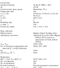
Table 2
Experimental details.
Crystal data
Chemical formula 3C5H12N+HSO4
SO42
Mr 451.60
Crystal system, space group Monoclinic,P21/c
Temperature (K) 100
a,b,c(A˚ ) 10.592 (4), 17.922 (5), 11.161 (4)
() 99.25 (2)
V(A˚3) 2091.1 (12)
Z 4
Radiation type MoK
(mm1) 0.30
Crystal size (mm) 0.200.180.16
Data collection
Diffractometer Rigaku Oxford Xcalibur Atlas
Absorption correction Analytical [CrysAlis PRO(Rigaku Oxford, 2015), based on expressions of Clark & Reid (1995)]
Tmin,Tmax 0.994, 0.996
No. of measured, independent and observed [I> 2(I)] reflections
35669, 5411, 4291
Rint 0.039
(sin/)max(A˚1) 0.691
Refinement
R[F2> 2(F2)],wR(F2),S 0.035, 0.083, 1.03
No. of reflections 5411
No. of parameters 254
H-atom treatment H-atom parameters constrained max,min(e A˚3) 0.33,0.42
supporting information
sup-1 Acta Cryst. (2015). E71, 1444-1446
supporting information
Acta Cryst. (2015). E71, 1444-1446 [https://doi.org/10.1107/S2056989015020538]
Crystal structure of tris(piperidinium) hydrogen sulfate sulfate
Tamara J. Lukianova, Vasyl Kinzhybalo and Adam Pietraszko
Computing details
Data collection: CrysAlis PRO (Rigaku Oxford, 2015); cell refinement: CrysAlis PRO (Rigaku Oxford, 2015); data
reduction: CrysAlis PRO (Rigaku Oxford, 2015); program(s) used to solve structure: SHELXS97 (Sheldrick, 2008);
program(s) used to refine structure: SHELXL2014 (Sheldrick, 2015); molecular graphics: DIAMOND (Brandenburg,
1997); software used to prepare material for publication: OLEX2 (Dolomanov et al., 2009).
Tris(piperidinium) hydrogen sulfate sulfate
Crystal data
3C5H12N+·HSO4−·SO42− Mr = 451.60
Monoclinic, P21/c a = 10.592 (4) Å
b = 17.922 (5) Å
c = 11.161 (4) Å
β = 99.25 (2)°
V = 2091.1 (12) Å3 Z = 4
F(000) = 976
Dx = 1.434 Mg m−3
Mo Kα radiation, λ = 0.71073 Å Cell parameters from 12552 reflections
θ = 2.2–29.4°
µ = 0.30 mm−1 T = 100 K Block, colourless 0.20 × 0.18 × 0.16 mm
Data collection
Rigaku Oxford Xcalibur Atlas diffractometer
Radiation source: fine-focus sealed X-ray tube, Enhance (Mo) X-ray source
Graphite monochromator
Detector resolution: 10.6249 pixels mm-1 ω scans
Absorption correction: analytical
[CrysAlis PRO (Rigaku Oxford, 2015), based on expressions of Clark & Reid (1995)]
Tmin = 0.994, Tmax = 0.996
35669 measured reflections 5411 independent reflections 4291 reflections with I > 2σ(I)
Rint = 0.039
θmax = 29.4°, θmin = 2.7° h = −14→14
k = −23→24
l = −15→15
Refinement
Refinement on F2
Least-squares matrix: full
R[F2 > 2σ(F2)] = 0.035 wR(F2) = 0.083 S = 1.03 5411 reflections 254 parameters 0 restraints
Hydrogen site location: inferred from neighbouring sites
H-atom parameters constrained
w = 1/[σ2(F
o2) + (0.0343P)2 + 1.1863P]
where P = (Fo2 + 2Fc2)/3
(Δ/σ)max = 0.001
Δρmax = 0.33 e Å−3
supporting information
sup-2 Acta Cryst. (2015). E71, 1444-1446
Special details
Geometry. All e.s.d.'s (except the e.s.d. in the dihedral angle between two l.s. planes) are estimated using the full covariance matrix. The cell e.s.d.'s are taken into account individually in the estimation of e.s.d.'s in distances, angles and torsion angles; correlations between e.s.d.'s in cell parameters are only used when they are defined by crystal symmetry. An approximate (isotropic) treatment of cell e.s.d.'s is used for estimating e.s.d.'s involving l.s. planes.
Fractional atomic coordinates and isotropic or equivalent isotropic displacement parameters (Å2)
x y z Uiso*/Ueq
S1 0.66217 (3) 0.57717 (2) 0.56541 (3) 0.01118 (9)
S2 0.39851 (3) 0.75550 (2) 0.40181 (3) 0.01134 (9)
O12 0.57890 (10) 0.58513 (6) 0.65614 (9) 0.0155 (2)
O21 0.39138 (10) 0.67389 (6) 0.42549 (10) 0.0183 (2)
O14 0.57685 (10) 0.58114 (6) 0.43754 (10) 0.0175 (2)
H14 0.5194 0.6134 0.4385 0.026*
O11 0.72505 (10) 0.50505 (6) 0.56684 (10) 0.0174 (2)
O13 0.75266 (10) 0.63911 (6) 0.57133 (10) 0.0180 (2)
O22 0.50129 (11) 0.79014 (7) 0.48587 (10) 0.0222 (3)
C25 0.19519 (15) 0.50258 (8) 0.72404 (14) 0.0154 (3)
H25A 0.2000 0.4735 0.8001 0.018*
H25B 0.1955 0.4669 0.6563 0.018*
N21 0.30210 (12) 0.60129 (7) 0.62212 (11) 0.0127 (3)
H21A 0.3029 0.5718 0.5559 0.015*
H21B 0.3716 0.6318 0.6293 0.015*
O23 0.27443 (10) 0.78993 (6) 0.41375 (9) 0.0142 (2)
O24 0.42147 (10) 0.76415 (6) 0.27366 (9) 0.0135 (2)
N11 0.32098 (12) 0.63977 (7) 0.14102 (11) 0.0131 (3)
H11A 0.3570 0.6768 0.1909 0.016*
H11B 0.3156 0.6560 0.0631 0.016*
N31 0.67984 (12) 0.73600 (7) 0.74774 (12) 0.0159 (3)
H31A 0.6901 0.7023 0.6890 0.019*
H31B 0.5947 0.7394 0.7510 0.019*
C14 0.21075 (15) 0.49250 (8) 0.09492 (14) 0.0164 (3)
H14A 0.2125 0.4710 0.1768 0.020*
H14B 0.1725 0.4551 0.0345 0.020*
C26 0.31146 (14) 0.55327 (8) 0.73257 (13) 0.0138 (3)
H26A 0.3169 0.5851 0.8057 0.017*
H26B 0.3901 0.5226 0.7403 0.017*
C23 0.06623 (15) 0.59747 (9) 0.59190 (15) 0.0176 (3)
H23A 0.0622 0.5662 0.5183 0.021*
H23B −0.0121 0.6285 0.5828 0.021*
C16 0.40461 (14) 0.57238 (8) 0.15854 (14) 0.0152 (3)
H16A 0.4139 0.5557 0.2441 0.018*
H16B 0.4907 0.5850 0.1406 0.018*
C22 0.18310 (14) 0.64782 (8) 0.60349 (14) 0.0155 (3)
H22A 0.1799 0.6782 0.5290 0.019*
H22B 0.1837 0.6821 0.6731 0.019*
supporting information
sup-3 Acta Cryst. (2015). E71, 1444-1446
H24A 0.0655 0.5782 0.7757 0.023*
H24B −0.0027 0.5127 0.6912 0.023*
C12 0.18991 (14) 0.62418 (9) 0.16711 (14) 0.0160 (3)
H12A 0.1374 0.6701 0.1552 0.019*
H12B 0.1946 0.6080 0.2525 0.019*
C33 0.86970 (15) 0.80450 (9) 0.70869 (15) 0.0180 (3)
H33A 0.8828 0.7698 0.6428 0.022*
H33B 0.9023 0.8541 0.6895 0.022*
C35 0.89016 (16) 0.70228 (10) 0.86207 (16) 0.0217 (4)
H35A 0.9040 0.6641 0.8014 0.026*
H35B 0.9357 0.6860 0.9422 0.026*
C15 0.34720 (15) 0.50991 (8) 0.07536 (14) 0.0161 (3)
H15A 0.4007 0.4646 0.0915 0.019*
H15B 0.3466 0.5247 −0.0102 0.019*
C13 0.12870 (15) 0.56320 (9) 0.08232 (15) 0.0169 (3)
H13A 0.1182 0.5812 −0.0026 0.020*
H13B 0.0428 0.5515 0.1015 0.020*
C32 0.72742 (14) 0.81012 (8) 0.71454 (15) 0.0167 (3)
H32A 0.7129 0.8477 0.7758 0.020*
H32B 0.6804 0.8260 0.6348 0.020*
C36 0.74807 (16) 0.70877 (9) 0.86681 (16) 0.0205 (3)
H36A 0.7138 0.6595 0.8854 0.025*
H36B 0.7343 0.7440 0.9318 0.025*
C34 0.94462 (15) 0.77687 (10) 0.82831 (15) 0.0207 (3)
H34A 0.9390 0.8139 0.8930 0.025*
H34B 1.0358 0.7708 0.8205 0.025*
Atomic displacement parameters (Å2)
U11 U22 U33 U12 U13 U23
S1 0.01134 (17) 0.01084 (17) 0.01124 (17) 0.00075 (13) 0.00142 (13) −0.00154 (13)
S2 0.01054 (17) 0.01264 (17) 0.01101 (17) 0.00085 (13) 0.00228 (13) −0.00016 (13)
O12 0.0153 (5) 0.0173 (5) 0.0147 (5) 0.0002 (4) 0.0049 (4) −0.0014 (4)
O21 0.0207 (6) 0.0144 (5) 0.0214 (6) 0.0051 (4) 0.0077 (5) 0.0061 (4)
O14 0.0175 (6) 0.0209 (6) 0.0127 (5) 0.0071 (4) −0.0014 (4) −0.0035 (4)
O11 0.0208 (6) 0.0132 (5) 0.0174 (6) 0.0054 (4) 0.0013 (5) −0.0018 (4)
O13 0.0147 (5) 0.0157 (5) 0.0246 (6) −0.0029 (4) 0.0063 (5) −0.0038 (4)
O22 0.0144 (5) 0.0339 (7) 0.0180 (6) −0.0035 (5) 0.0013 (5) −0.0088 (5)
C25 0.0189 (8) 0.0125 (7) 0.0154 (7) −0.0006 (6) 0.0046 (6) 0.0019 (6)
N21 0.0109 (6) 0.0133 (6) 0.0140 (6) −0.0009 (5) 0.0023 (5) 0.0015 (5)
O23 0.0130 (5) 0.0152 (5) 0.0152 (5) 0.0029 (4) 0.0043 (4) 0.0004 (4)
O24 0.0147 (5) 0.0141 (5) 0.0124 (5) 0.0001 (4) 0.0041 (4) 0.0008 (4)
N11 0.0151 (6) 0.0129 (6) 0.0114 (6) −0.0017 (5) 0.0023 (5) 0.0004 (5)
N31 0.0110 (6) 0.0143 (6) 0.0231 (7) −0.0014 (5) 0.0046 (5) −0.0073 (5)
C14 0.0169 (7) 0.0141 (7) 0.0168 (8) −0.0024 (6) −0.0017 (6) −0.0003 (6)
C26 0.0139 (7) 0.0143 (7) 0.0127 (7) 0.0006 (6) 0.0008 (6) 0.0028 (6)
C23 0.0122 (7) 0.0232 (8) 0.0177 (8) 0.0023 (6) 0.0027 (6) 0.0040 (6)
supporting information
sup-4 Acta Cryst. (2015). E71, 1444-1446
C22 0.0159 (7) 0.0140 (7) 0.0168 (7) 0.0040 (6) 0.0034 (6) 0.0034 (6)
C24 0.0151 (7) 0.0226 (8) 0.0200 (8) −0.0014 (6) 0.0058 (6) 0.0039 (6)
C12 0.0138 (7) 0.0170 (7) 0.0177 (8) −0.0006 (6) 0.0042 (6) −0.0001 (6)
C33 0.0148 (7) 0.0159 (7) 0.0236 (8) −0.0010 (6) 0.0044 (7) 0.0003 (6)
C35 0.0195 (8) 0.0271 (9) 0.0196 (8) 0.0088 (7) 0.0061 (7) 0.0044 (7)
C15 0.0168 (8) 0.0148 (7) 0.0162 (7) 0.0008 (6) 0.0010 (6) −0.0002 (6)
C13 0.0132 (7) 0.0173 (7) 0.0194 (8) −0.0015 (6) 0.0001 (6) 0.0006 (6)
C32 0.0140 (7) 0.0147 (7) 0.0208 (8) 0.0008 (6) 0.0012 (6) −0.0004 (6)
C36 0.0210 (8) 0.0185 (8) 0.0242 (9) 0.0035 (6) 0.0106 (7) 0.0043 (6)
C34 0.0124 (7) 0.0273 (9) 0.0217 (8) −0.0013 (6) 0.0009 (6) −0.0050 (7)
Geometric parameters (Å, º)
S1—O12 1.4529 (12) C23—H23B 0.9900
S1—O14 1.5633 (12) C23—C22 1.520 (2)
S1—O11 1.4530 (11) C23—C24 1.529 (2)
S1—O13 1.4612 (11) C16—H16A 0.9900
S2—O21 1.4904 (12) C16—H16B 0.9900
S2—O22 1.4569 (12) C16—C15 1.518 (2)
S2—O23 1.4773 (11) C22—H22A 0.9900
S2—O24 1.4969 (12) C22—H22B 0.9900
O14—H14 0.8400 C24—H24A 0.9900
C25—H25A 0.9900 C24—H24B 0.9900
C25—H25B 0.9900 C12—H12A 0.9900
C25—C26 1.521 (2) C12—H12B 0.9900
C25—C24 1.523 (2) C12—C13 1.521 (2)
N21—H21A 0.9100 C33—H33A 0.9900
N21—H21B 0.9100 C33—H33B 0.9900
N21—C26 1.4936 (19) C33—C32 1.522 (2)
N21—C22 1.4978 (19) C33—C34 1.522 (2)
N11—H11A 0.9100 C35—H35A 0.9900
N11—H11B 0.9100 C35—H35B 0.9900
N11—C16 1.4920 (19) C35—C36 1.519 (2)
N11—C12 1.4900 (19) C35—C34 1.527 (2)
N31—H31A 0.9100 C15—H15A 0.9900
N31—H31B 0.9100 C15—H15B 0.9900
N31—C32 1.489 (2) C13—H13A 0.9900
N31—C36 1.489 (2) C13—H13B 0.9900
C14—H14A 0.9900 C32—H32A 0.9900
C14—H14B 0.9900 C32—H32B 0.9900
C14—C15 1.528 (2) C36—H36A 0.9900
C14—C13 1.530 (2) C36—H36B 0.9900
C26—H26A 0.9900 C34—H34A 0.9900
C26—H26B 0.9900 C34—H34B 0.9900
C23—H23A 0.9900
O12—S1—O14 107.77 (7) N21—C22—C23 109.68 (12)
supporting information
sup-5 Acta Cryst. (2015). E71, 1444-1446
O12—S1—O13 111.17 (7) N21—C22—H22B 109.7
O11—S1—O14 104.31 (6) C23—C22—H22A 109.7
O11—S1—O13 112.28 (7) C23—C22—H22B 109.7
O13—S1—O14 106.58 (7) H22A—C22—H22B 108.2
O21—S2—O24 106.96 (6) C25—C24—C23 110.44 (13)
O22—S2—O21 110.98 (7) C25—C24—H24A 109.6
O22—S2—O23 110.32 (7) C25—C24—H24B 109.6
O22—S2—O24 110.60 (7) C23—C24—H24A 109.6
O23—S2—O21 108.83 (6) C23—C24—H24B 109.6
O23—S2—O24 109.05 (6) H24A—C24—H24B 108.1
S1—O14—H14 109.5 N11—C12—H12A 109.8
H25A—C25—H25B 108.0 N11—C12—H12B 109.8
C26—C25—H25A 109.3 N11—C12—C13 109.30 (12)
C26—C25—H25B 109.3 H12A—C12—H12B 108.3
C26—C25—C24 111.42 (13) C13—C12—H12A 109.8
C24—C25—H25A 109.3 C13—C12—H12B 109.8
C24—C25—H25B 109.3 H33A—C33—H33B 108.0
H21A—N21—H21B 107.9 C32—C33—H33A 109.4
C26—N21—H21A 109.2 C32—C33—H33B 109.4
C26—N21—H21B 109.2 C34—C33—H33A 109.4
C26—N21—C22 112.22 (11) C34—C33—H33B 109.4
C22—N21—H21A 109.2 C34—C33—C32 111.35 (14)
C22—N21—H21B 109.2 H35A—C35—H35B 108.0
H11A—N11—H11B 107.9 C36—C35—H35A 109.5
C16—N11—H11A 109.2 C36—C35—H35B 109.5
C16—N11—H11B 109.2 C36—C35—C34 110.94 (13)
C12—N11—H11A 109.2 C34—C35—H35A 109.5
C12—N11—H11B 109.2 C34—C35—H35B 109.5
C12—N11—C16 112.01 (11) C14—C15—H15A 109.4
H31A—N31—H31B 107.9 C14—C15—H15B 109.4
C32—N31—H31A 109.1 C16—C15—C14 111.00 (13)
C32—N31—H31B 109.1 C16—C15—H15A 109.4
C32—N31—C36 112.30 (12) C16—C15—H15B 109.4
C36—N31—H31A 109.1 H15A—C15—H15B 108.0
C36—N31—H31B 109.1 C14—C13—H13A 109.4
H14A—C14—H14B 108.1 C14—C13—H13B 109.4
C15—C14—H14A 109.5 C12—C13—C14 111.03 (13)
C15—C14—H14B 109.5 C12—C13—H13A 109.4
C15—C14—C13 110.74 (12) C12—C13—H13B 109.4
C13—C14—H14A 109.5 H13A—C13—H13B 108.0
C13—C14—H14B 109.5 N31—C32—C33 109.20 (12)
C25—C26—H26A 109.6 N31—C32—H32A 109.8
C25—C26—H26B 109.6 N31—C32—H32B 109.8
N21—C26—C25 110.29 (12) C33—C32—H32A 109.8
N21—C26—H26A 109.6 C33—C32—H32B 109.8
N21—C26—H26B 109.6 H32A—C32—H32B 108.3
H26A—C26—H26B 108.1 N31—C36—C35 109.60 (13)
supporting information
sup-6 Acta Cryst. (2015). E71, 1444-1446
C22—C23—H23A 109.4 N31—C36—H36B 109.8
C22—C23—H23B 109.4 C35—C36—H36A 109.8
C22—C23—C24 111.05 (13) C35—C36—H36B 109.8
C24—C23—H23A 109.4 H36A—C36—H36B 108.2
C24—C23—H23B 109.4 C33—C34—C35 109.69 (13)
N11—C16—H16A 109.6 C33—C34—H34A 109.7
N11—C16—H16B 109.6 C33—C34—H34B 109.7
N11—C16—C15 110.24 (12) C35—C34—H34A 109.7
H16A—C16—H16B 108.1 C35—C34—H34B 109.7
C15—C16—H16A 109.6 H34A—C34—H34B 108.2
C15—C16—H16B 109.6
N11—C16—C15—C14 −55.57 (16) C12—N11—C16—C15 59.41 (16)
N11—C12—C13—C14 57.09 (17) C15—C14—C13—C12 −54.79 (17)
C26—C25—C24—C23 54.49 (17) C13—C14—C15—C16 53.79 (17)
C26—N21—C22—C23 −58.61 (16) C32—N31—C36—C35 −59.33 (17)
C16—N11—C12—C13 −59.86 (16) C32—C33—C34—C35 55.93 (17)
C22—N21—C26—C25 57.84 (16) C36—N31—C32—C33 59.01 (17)
C22—C23—C24—C25 −55.39 (18) C36—C35—C34—C33 −55.78 (18)
C24—C25—C26—N21 −55.37 (16) C34—C33—C32—N31 −57.02 (17)
C24—C23—C22—N21 56.89 (17) C34—C35—C36—N31 57.05 (18)
Hydrogen-bond geometry (Å, º)
D—H···A D—H H···A D···A D—H···A
O14—H14···O21 0.84 1.72 2.5603 (16) 173
N21—H21A···O11i 0.91 1.93 2.8226 (18) 166
N21—H21B···O12 0.91 2.32 2.9096 (19) 122
N21—H21B···O24ii 0.91 2.47 3.0964 (18) 127
N11—H11A···O21 0.91 2.59 3.201 (2) 126
N11—H11A···O24 0.91 1.89 2.7904 (17) 171
N11—H11B···O22iii 0.91 2.47 3.0474 (18) 122
N11—H11B···O23iii 0.91 1.92 2.8039 (18) 164
N31—H31A···O12 0.91 2.41 3.0245 (18) 125
N31—H31A···O13 0.91 1.93 2.8240 (18) 167
N31—H31B···O24ii 0.91 1.89 2.7978 (19) 172