Nowadays, probiotics are used in veterinary and human medicine as well as in nutrition with in-creasing frequency. Probiotics are defined as direct feed microbials or microbial cell preparations with a beneficial effect on the health and well-being of the host (Fuller et al., 1989; Nemcová et al., 1997). In contrast to human nutrition, where lactobacilli are predominant probiotic products, in animal nu-trition Enterococcus sp. as well as yeasts from the genus Saccharomyces are the most frequently used as probiotic microorganisms (Simon et al., 2001). Nowadays, rabbit breeding has an increasing impact due to the nutritional value of meat and economic traits of breeding. Development in this animal in-dustry also induces to search for new technologies to improve rabbit health and welfare. The improve-ment of rabbit health and performance by addition of probiotics and organic acids to feed mixtures was already studied (Skřivanová and Marounek, 2002). However, an alternative could also be the application of probiotic microorganisms, especially those isolated from the intestinal ecosystem of
rab-bits. The rabbit caecum is predominantly colonized by strict anaerobes such as members of the genus Bacteroides within the first weeks after birth. The composition of caecal microflora changes with ad-vancing age (Gouet and Fonty, 1979). However, the presence of facultative anaerobic bacteria such as enterococci is also important because enterococci colonize rabbits’ digestive tract and belong to the lactic acid bacteria (LAB) which are frequently of probiotic character. A potentially successful pro-biotic strain is expected to have several desirable properties such as strain origin, acid and bile tol-erance, adherence to the intestinal epithelium and colonization ability, production of antimicrobial substances as well as good technological properties (Salminen et al., 1998). In addition, ureolytic activity is one of the most important metabolic properties especially concerning the enterococci.
The purpose of this study was to select entero-cocci from the digestive tract of rabbits and to test their metabolic properties with the aim to select a suitable probiotic feed additive.
Supported by the Slovak Scientific Agency (Project VEGA 2/5139/25).
Enterococci from rabbits – potential feed additive
M. SIMONOVÁ, A. LAUKOVÁ, I. ŠTYRIAK
Institute of Animal Physiology, Slovak Academy of Sciences, Košice, Slovak Republic
ABSTRACT:Enterococci (58) from faeces of rabbits of various age (from 2 months to 3 years) and 5 different rabbit farms were isolated and tested for survival in the presence of oxgall, lactic acid production, urease activity, resistance to low pH as well as their binding ability was tested. Fifty percent of enterococcal isolates were identi-fied as Enterococcus faecium, 19% as E. faecalis. All strains showed good survival in the presence of 5% oxgall. The urease activity of isolates was in the range from 0.013 to 17.13 nkat/ml, only E. faecalis EE229 strain did not produce any urease. The survival of strains was tested at pH 3.0 and the percentage of their survival ranged between 62.0% and 90.0%. E. faecium EF1819 strain was found to show the best survival ability at low pH. Particle agglutination assay values of selected enterococci expressed only negative (0) or weakly positive (1) binding of heparin, bovine fibrinogen, porcine fibronectin and lactoferrin. Based on the results, most of the selected enterococci could be promising probiotic feed additives.
MATERIAL AND METHODS
Isolation of enterococci.Samples were col-lected from 23 rabbits from five different rabbit farms. Among 23 animals, 6 were New Zealand rabbits, 8 were Californian growing rabbits and the others (9) were crossbred rabbits of various age (from 2 months to 3 years) as follows: 2 months old – 1 rabbit, 4 months old – 1 rabbit, 5 months old – 3 rabbits, 7 months old – 2 rabbits, 12 months old – 7 rabbits, 15 months old – 4 rabbits, 24 months old – 4 rabbits and 36 months old – 1 rabbit). Rabbits on one farm were kept indoors in flat deck batter-ies while rabbits on the other farms were housed in semi-open flat deck batteries. All animals were fed commercial granulated feed (Norm Typ, BIOFER s.r.o. Prešov, Slovak Republic). Additionally, they also received wheat, barley, oats and hay ad libi-tum. To isolate enterococci the samples of fae-ces were diluted by a standard microbiological method in (0.85%) saline buffer. The appropriate dilutions were plated onto M-Enterococcus agar (ME, Becton and Dickinson, Cockeysville, USA) and incubated at 37°C for 24 h in partially CO2/air atmosphere. After incubation, the colony forming units (CFU) were counted. Then, 58 colonies of en-terococci (including each samples) were randomly picked up and maintained on ME agar for further identification and testing. Isolates of enterococci were phenotyped by BBL Crystal ID kits (Becton and Dickinson) and genotyped by PCR followed by agarose electrophoresis according to Woodford et al. (1997).
Resistance to bile.Resistance to bile was tested according to Gilliland and Walker (1990). Brain Heart Infusion broth (BHI, Becton and Dickinson) was prepared by the addition of 5% (w/v) of oxgall
(Becton and Dickinson). The volume 50 µl of an 18-hour culture of each strain was added to 5 ml of BHI broth with oxgall. After incubation at 37°C for 24 h, the bacterial growth of strains was measured spectrophotometrically at 600 nm. Numbers of vi-able cells were estimated at 0 h and after 24 h of in-cubation using M-Enterococcus agar.Enterococcus faecium CCM 4231 was used as a positive control (Lauková et al., 1993).
Urease activity. The method according to Cook (1976) was used to examine the urease activity. It was expressed in nkat/ml.
Resistance to low pH. To test the survival of iso-lates at low pH values, the cells of overnight cultures in BHI (Becton and Dickinson) of selected strains were harvested by centrifugation (2 000 × g for 15 min), resuspended in 0.05 M phosphate buffer of pH 3.0 adjusted with 1 N HCl and kept at 37°C for 1, 2 and 3 h. The CFU were determined on ME agar (Becton and Dickinson).
Particle agglutination assay (PAA).The pro-tein-coated latex beads (20 µl) were mixed on a glass slide with an equal volume of a bacterial cell suspension of 1010 CFU/ml. These two drops were gently mixed and the agglutination reaction was scored after 2 min as a PAA value from strongly positive (3) to negative (0) as was previously de-scribed by Naidu et al. (1998) and Štyriak et al. (1999).
RESULTS
[image:2.595.63.531.590.757.2]The total counts of enterococci from faeces of rabbits ranged from 103 to 105 CFU/ml/g. Among 58 isolates specified by PCR, 29 strains were clas-sified as E. faecium and 11 strains belonged to
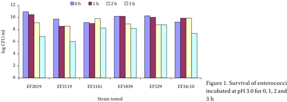
Figure 1. Survival of enterococci incubated at pH 3.0 for 0, 1, 2 and 3 h
0 2 4 6 8 10 12
EF2019 EF2119 EF2119 EF1839 EF529 EF24/10
Strain tested
log
CFU/ml
0.h. 1.h. 2.h. 3.h.
E. faecalis.Strains of E. faecalis were firstly phe-notypically characterized by BBL Crystal ID kits (Becton and Dickinson) and confirmed by PCR method.
Enterococci were able to reach 108–109 CFU/ml in the presence of oxgall in comparison with the control growth (1011–1012 CFU/ml) in oxgall-free broth.
The ureolytic activity of enterococcal isolates was detected in the range from 0.013 ± 0.0 to 17.13 ± 2.25 nkat/ml (Table 1). Fourteen strains of ente-rococci showed low ureolytic activity (0.013 ± 0.0 and 0.98 ± 0.0 nkat/ml) and in 9 strains the highest values of ureolytic activity were measured (in the range from 6.31 ± 2.47 to 17.134 ± 2.25 nkat/ml). The most of strains (37) were detected with mean
TablE1. Urease activity of enterococci
Isolates Urease (nkat/ml ± SD) Isolates Urease (nkat/ml ± SD)
EF1119/23 2.55 ± 0.81 EF419/23 8.72 ± 2.25
EF1129/23 0.98 ± 0.00 EF519/23 2.39 ± 0.44
EF1179/23 1.03 ± 0.32 EF1129/23 13.07 ± 3.76
EF69/23 2.29 ±1.14 EF2139/23 3.95 ± 1.76
11K/1 2.12 ± 1.68 EF929/23 6.31 ± 4.47
EF329/23 1.09 ± 0.11 EF529/23 4.39 ± 1.76
EF429/23 1.78 ± 0.14 EF729/23 0.50 ± 0.17
EF739/23 2.70 ± 0.58 EF629/23 1.27 ± 0.48
EF819/23 3.47 ± 0.10 EF839/23 6.95 ± 0.37
EF939/23 3.68 ± 2.71 EF1839/23 3.95 ± 0.44
EF1819/23 5,21 ± 3,13 EF24/10 12.50 ± 1.87
EF2019/23 0.73 ± 0.55 EF349/23 17.13 ± 2.25
EF2119/23 2.59 ± 0.09 EF639/23 6.40 ± 1.55
EF219/23 4.78 ± 2.10 EF659/23 2.68 ± 0.30
EF319/23 9.11 ± 1.81 EF2129/23 1.04 ± 0.74
EF1219 2.08 ± 0.00 Esp1829 2.20 ± 0.04
EE829 3.87 ± 0.30 Esp1639 1.31 ± 0.07
EE1229 3.13 ± 0.75 Esp1239 1.55 ± 0.04
EE259 8.78 ± 1.34 Esp1339 1.18 ± 0.59
EE339 3.72 ± 0.45 Esp539 1.59 ± 0.21
EE1529 2.83 ± 0.75 Esp249 0.28 ± 0.07
EE2319 3.72 ± 1.04 Esp649 0.45 ± 0.24
EE1539 0.32 ± 0.07 Esp1429 0.55 ± 0.21
EE719 1.65 ± 0.62 Esp2339 0.66 ± 0.04
EE549 1.21 ± 0.45 Esp239 2.21 ± 0.76
EE229 0 ± 0 Esp1519 0.48 ± 0.14
EE1629 0.013 ± 0.00 Esp1619 0.97 ± 0.14
Esp2329 0.89 ± 0.35 Esp1319 0.83 ± 0.41
values of urease expression (1.03 ± 0.32 and 5.21 ± 3.13 nkat/ml). The strain E. faecalis EE229 did not produce any urease.
Six bacteriocin-producing strains (Simonová and Lauková, 2004a) showed good resistance to low pH. As for the survival of tested strains at pH 3.0, it ranged between 62.0 and 90.0% (Figure 1). E. faecium EF1819 strain showed the best ability to survive at low pH (90.0%).
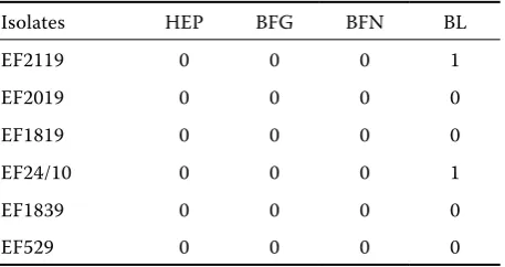
PAA values of 6 selected enterococcal strains from rabbits expressed only the negative (0) bind-ing of heparin (HEP), bovine fibrinogen (BFIB) and porcine fibronectin (PFIB, Table 2). The binding of bovine lactoferrin (BLACT) was negative in four E. faecium strains (EF1839, EF529, EF2019, EF1819) and weakly positive (1) in E. faecium EF24/10 and EF2119.
DISCUSSION
Enterococci are common inhabitants of animal intestines and they are the predominant intestinal microflora during the first 2–3 days of life of many animals (DeVriese et al., 1991; Lauková, 1996). The majority of studies deals with the beneficial effect of probiotic enterococci and/or lactobacilli in poul-try, pigs or dogs (Bomba et al., 1996; Lauková et al., 2003; Strompfová et al., 2004), but not in rabbits. However, lactobacilli are very scarcely found in rabbits’ gastrointestinal tract (Yu and Tsen, 1993). Moreover, until now only little information has been provided concerning the probiotic utiliza-tion in rabbits and no strain originated from the rabbit ecosystem (Simonová and Lauková, 2004b).
For this reason the attention is focused on ente-rococcal strains from rabbits which are present in sufficient numbers there and which are also able to produce antimicrobial substances. In our study, twenty-nine isolates (50%) were identified as E. faecium. On the other hand, Linaje et al. (2004) described the prevalence of E. faecalis (58% out of 24 strains). Only 34% was identified as E. faecium and 8% as E. durans isolated from both the faeces and the caecum content of rabbits. The survival ability of enterococcal strains in 4% bile was also reported by Linaje et al. (2004); which is in accord-ance with our results. These authors also reported good resistance of enterococci to an acid environ-ment (survival after 4 h of incubation in an acid medium). This fact indicates high bile resistance and good survival possibilities of enterococci in the digestive tract of animals in general (Marciňáková et al., 2004).
The rabbit caecal microflora is characterized by several enzymatic activities, e.g. cellulolytic, pec-tinolytic, amylolytic, proteolytic and ureolytic ones (Gidenne, 1997). In general, urease is an important enzyme from the clinical aspect (to determine urea in blood) as well as in nitrogen metabolism in ru-minants (Hausinger, 1986). Moreover, the ureolytic activity of enterococci is an important process from the viewpoint of the basic metabolic properties. In our study, enterococci isolated from rabbits produced lower levels of urease compared to the ureolytic activity of ruminal enterococci (Lauková and Juriš, 1997).
[image:4.595.64.293.128.249.2]Among the criteria for strains that could be used as probiotic feed additive their ability to colonize the mucosal surfaces and to prevent the attachment of pathogens is also included. Enterococcal strains isolated from silage and commercially known pro-biotic strain e.g. E. faecium M74 expressed the simi-lar binding activity as was detected in our isolates (Štyriak et al., 2003). Perhaps the capacity of ente-rococci to bind bovine lactoferrin – iron-binding protein – is correlated with their growth or viru-lence (Schryvers and Gonzales, 1989). In compari-son with the binding of enterococci from rabbits described previously by Štyriak et al. (2004), our isolates possessed the same or lower binding ability with lactoferrin, and also with other extracellular matrix (ECM)-binding proteins. The reason for their lower binding ability with lactoferrin could be explained by their origin from healthy animals without any clinical signs or disease.
Table 2. Binding of extracellular matrix molecules by strains with bacteriocinogenic activity tested by particle agglutination assay (PAA)
Isolates HEP BFG BFN BL
EF2119 0 0 0 1
EF2019 0 0 0 0
EF1819 0 0 0 0
EF24/10 0 0 0 1
EF1839 0 0 0 0
EF529 0 0 0 0
Based on the results, a majority of the studied enterococci possessed “probiotic” properties. The isolates were characterized by good capability to survive in the presence of oxgall and low pH; most of them showed the ureolytic activity, ability to pro-duce lactic acid. Moreover, as previously described by Simonová and Lauková (2004a) the isolates of E. faecium from rabbits also possessed bacterioci-nogenic properties. It can be concluded that ente-rococci with performed characteristics could be selected for further utilization as probiotic feed additives. Of course, further experimental applica-tions are needed.
Acknowledgements
The authors thank to Mrs. Margita Bodnárová for her technical assistance. The authors acknowledge that the results concerning the total counts and identification of isolates were partially mentioned in another manuscript (Bulletin of the Veterinary Institute in Pulawy, 2004, 48, 383–386).
REFERENCES
Bomba A., Kašteľ R., Gancarčíková S., Nemcová R., Herich R., Čížek M. (1996): The effect of lactobacilli inoculation on organic acid levels in the mucosal film and the small intestine contents in gnotobiotic pigs. Berl. Münch. Tierärztl. Wschr., 9, 428–430.
Cook A.R. (1976): Urease activity in the rumen of sheep and the isolation of ureolytic bacteria. J. Gen. Micro-biol., 92, 32–48.
DeVriese L.A., Hommez J., Wijfels R., Halsebrouck E. (1991): Composition of the enterococcal and strepto-coccal intestinal flora of poultry. J. Appl. Bacteriol., 71, 46–50.
Fuller R. (1989): Probiotics in man and animals: A review. J. Appl. Bacteriol.,66, 365–378.
Gidenne T. (1997): Caeco-colic digestion in the growing rabbit: impact of nutritional factors and related distur-bances. Livest. Prod. Sci., 51, 73–88.
Gilliland S.E., Walker D.K. (1990): Factors to consider when selecting a culture of Lactobacillus acidophilus as a dietary adjunct to produce a hypocholesterolemic effect in humans. J. Dairy Sci., 73, 905–911.
Gouet P., Fonty G. (1979): Changes in the digestive mi-croflora of the holoxenic rabbits from birth until adult-hood. Ann. Biol. Biochem. Biophys., 19, 553–566.
Hausinger R.P. (1986): Purification of a nickel-containing urease from the rumen anaerobic Selenomonas rumi-nantium. J. Biol. Chem., 17, 7866–7870.
Lauková A., Guba P., Nemcová R., Vasilková Z. (2003): Reduction of Salmonella in gnotobiotic Japanese quails caused by the Enterocin A-producing EK13 strain of Enterococcus faecium. Vet. Res. Com., 27, 275–280. Lauková A., Juriš P. (1997): Distribution and
characteri-zation of Enterococcus species in municipal sewages. Microbios, 89, 73–80.
Lauková A., Mareková M., Javorský P. (1993): Detection an antimicrobial spectrum of a bacteriocin like sub-stance produced by Enterococcus faecium CCM4231. Appl. Microbiol., 16, 257–260.
Linaje R., Coloma M.D., Pérez-Martínez G., Zúñiga M. (2004): Characterization of faecal enterococci from rabbits for the selection of probiotic strains. J. Appl. Microbiol., 96, 761–771.
Marciňáková M., Strompfová V., Boldižárová K., Lauková A., Gancarčíková S. (2004): Effect of potential probiotic activity of Enterococcus faecium EE3 strain against Sal-monella infection in Japanese quails. Bull. Vet. Inst. Pulawy, 48, 387–390.
Naidu A.S., Paulsson M., Wadström T. (1998): Particle agglutination assays for rapid detection of fibronectin, fibrinogen, and collagen receptors onStaphylococcus aureus. J. Clin. Microbiol., 26, 1549–1554.
Nemcová R. (1997): Criteria for selection of lactobacilli for probiotic use. Vet. Med., 42, 19–27.
Salminen S., Von Wright A., Morelli L., Martenau P., Brassart D., de Vos W.M., Fondeu R., Saxelin M., Col-lins K., Mogensen G., Birkeland S.-E., Mattila-Sand-holm T. (1998): Demonstration of safety of probiotics – a review, Int. J. Food Microbiol., 44, 93–106. Schryvers A.B., Gonzales G.C. (1989): Comparison of
the abilities of different protein sources of iron to en-hance Neisseria meningitidis infection in mice. Infect. Immun., 57, 2425–2429.
Simon O., Jadamus A., Vahjenen W. (2001): Probiotic feed additives – effectiveness and expected modes of action. J. Anim. Feed Sci., 10, 51–67.
Simonová M., Lauková A. (2004a): Isolation of faecal Enterococcus faecium strains from rabbits and their sensitivity to antibiotics and ability to bacteriocin pro-duction. Bull. Vet. Inst. Pulawy, 48, 383–386.
Simonová M., Lauková A. (2004b): Lactic acid bacteria in rabbit’s ecosystem. In: Proceedings of International Probiotic Conference, New Perspectives of Probiotics, Košice, Slovak Republic. 40 pp.
Štyriak I., Lauková A., Fallgreu C., Wadström T. (1999): Binding of selected extracellular matrix proteins to enterococci and Streptococcus bovis of animal origin. Current Microbiol., 39, 327–335.
Štyriak I., Lauková A., Strompfová V., Ljungh A. (2004): Mode of binding of fibrinogen, fibronectin and iron-binding proteins by animal enterococci. Vet. Res. Com., 28, 587–598.
Štyriak I., Nemcová R., Chang Y.-H., Ljungh A. (2003): Binding of extracellular matrix molecules by probiotic bacteria. Lett. Appl. Microbiol., 37, 329–333.
Woodford N., Egelton M.C., Morrison D. (1997): Com-parison of PCR with phenotypic methods for the spe-ciation of enterococci. Plenum Press, New York, 47, 405–409.
Yu B., Tsen H. (1993): Lactobacillus cells in the rabbit digestive-tract and the factors affecting their distribu-tion. J. Appl. Bacteriol., 75, 269–275.
Received: 05–03–09 Accepted after corrections: 05–05–20
Corresponding Author
MVDr. Monika Simonová, Institute of Animal Physiology, Slovak Academy of Sciences, Šoltésovej 4-6, 04001 Košice, Slovak Republic