ORIGINAL RESEARCH
Effect of obesity on oxygen uptake and cardiovascular
dynamics during whole-body and leg exercise in adult
males and females
Simon Green1, Eamon O’Connor2, Catherine Kiely2, Donal O’Shea3& Mikel Egana~ 2
1 School of Science and Health, Western Sydney University, Sydney, Australia 2 School of Medicine, Department of Physiology, Trinity College Dublin, Dublin, Ireland 3 Endocrinology, St. Columcille’s and St. Vincent’s Hospitals, Dublin, Ireland
Keywords
Muscle vasodilation, O2uptake, obesity, submaximal exercise, time constant.
Correspondence
Simon Green, School of Science and Health, Western Sydney University, Sydney, NSW, Australia.
Tel/Fax: 61 2 46203272
E-mail: simon.green@westernsydney.edu.au
Funding Information
This publication has emanated from research conducted with the financial support of Science Foundation Ireland under Grant 08/ RFP/BMT1342.
Received: 28 March 2018; Accepted: 13 April 2018
doi: 10.14814/phy2.13705
Physiol Rep, 6 (9), 2018, e13705, https://doi.org/10.14814/phy2.13705
Abstract
Obesity has been associated with a slowing of V_O2 dynamics in children and adolescents, but this problem has not been studied in adults. Cardiovascular mechanisms underlying this effect are not clear. In this study, 48 adults (18 males, 30 females) grouped according to body mass index (BMI) (lean<25 kgm2, overweight=25–29.9 kgm2, obese ≥30 kgm2) pro-vided a fasting blood sample, completed a maximal graded exercise test and six bouts of submaximal exercise on a cycle ergometer, and performed two protocols of calf exercise. Dynamic response characteristics of V_O2 and leg vascular conductance (LVC) were assessed during cycling (80% ventilatory threshold) and calf exercise (30% MVC), respectively. Dynamic responses of cardiac output, mean arterial pressure and total systemic vascular conductance were also assessed during cycling based on measurements at 30 and 240 sec. The time constant of the second phase of the V_O2 response was significantly greater in obese than lean subjects (39.4 (9.2) vs. 29.1 (7.6) sec); whereas dynamic responses of cardiac output and systemic vascular conductance were not affected by BMI. For calf exercise, the time constant of the second growth phase of LVC was slowed significantly in obese subjects (22.1 (12.7) sec) com-pared with lean and overweight subjects (11.6 (4.5) sec and 13.4 (6.7) sec). These data show that obesity slows dynamic responses of V_O2 during cycling and the slower phase of vasodilation in contracting muscles of male and female adults.
Introduction
Obesity, represented by a body mass index (BMI) greater than 30 kgm2 (Ortega et al. 2016), contributes to a loss of cardiorespiratory fitness. This is based largely on evi-dence of inverse associations between BMI, or other esti-mates of adiposity, and V_O2peak (normalized to body mass) and performance during weight-bearing tests (Wei et al. 2000; Drinkard et al. 2001; Tsiros et al. 2016). However, interpretation of these associations is compli-cated by the fact that, when comparisons are made between individuals of greatly different body masses, these
peak exercise measurements are inherently correlated with body mass (i.e., V_O2peak) or directly influenced by body mass when exercise is weight-bearing, such as treadmill and walk tests (Green et al. 2007; Krachler et al. 2015). Consequently, the physiological effects of obesity, inde-pendent of body mass per se, on cardiorespiratory func-tion andV_O2during exercise are less clear.
moderate submaximal exercise, theV_O2 measured ‘at the lungs’ increases in a biphasic manner (Barstow and Mole 1991). The initial phase of this response is rapid, lasting for 15–40 sec, and is related to the abrupt increase in pul-monary blood flow (cardiac output) and hyperemia in contracting muscles (Whipp et al. 2002). The second phase evolves more slowly, exponentially, and it repre-sents the O2consumption by contracting muscles (Grassi et al. 1996). The time constant of this second (s2) phase provides an estimate of the rate of change of this phase and perhaps best represents the speed of the dynamic response of V_O2 and underlying physiological processes linked to the supply and utilization of O2. In addition, this parameter is theoretically independent of its ampli-tude and, thus, an individual’s body mass.
Evidence from human studies pertaining to the effects of obesity on s2 are limited to studies of girls and boys (Lambrick et al. 2013) and male adolescents (Salvadego et al. 2010). This evidence suggests that a BMI>30 kgm2 is associated with an increase in s2 and slowing of the dynamic response of V_O2 by ~20– 25%. As yet, the effects of BMI on s2 or other aspects of V_O2 dynamics in healthy adults have not been established.
According to the Fick principle applied to V_O2 (V_O2=Q_ 9a-vO2, where Q_ is blood flow, a-vO2 is the arterial-venous difference in blood O2content), any effect of BMI on V_O2 dynamics measured at the lungs might be attributed to effects on dynamic responses of cardiac output (i.e., pulmonary arterial blood flow) and/or the arterial-venous difference for O2measured across the pul-monary circuit. Measurements of these systemic responses might shed light on whether BMI-related changes inV_O2 dynamics are related to systemic rates of O2delivery and/ or O2 utilization. However, as we observed in patients with type 2 diabetes (MacAnaney et al. 2011; O’Connor et al. 2012; Kiely et al. 2014), it is also possible that a slowing in V_O2 dynamics is not explained by such sys-temic measurements but are associated with impaired responses of vasodilation measured in contracting muscles.
Therefore, in the this study, we tested the hypothesis that an increase in BMI is associated with a slowing of
_
VO2 dynamics (i.e., increase in s2) during moderate cycling exercise in apparently healthy men and women. These subjects were not involved in regular physical activ-ity and represented a ‘middle-aged’ cohort. To explore some of the physiological factors underlyingV_O2 dynam-ics, we measured cardiac output during cycling exercise and also the dynamic response characteristics of muscle vasodilation during single-limb exercise with better tem-poral resolution than the cardiovascular measurements during cycling.
Methods
Subjects
Forty eight subjects (18 males, 30 females) participated in this study. Subjects were recruited from the general com-munity of Dublin on the basis of the following inclusion criteria: aged 30–70 years; sedentary, ≤1 h of exercise per week and/or had not participated in a continuous exercise program for the previous 6 months; fasting glucose
<6.1 mmolL1 and HbA1c <6.5%; nonsmoker; systolic and diastolic blood pressures below 170 and 95 mmHg, respectively; and free of cardiovascular disease and other chronic diseases (neurological, respiratory, orthopedic) as assessed by a physician and medical health history ques-tionnaire. Fifteen participants (10 females, 5 males) included in the study were taking 1–3 medications for the treatment of hypertension (n = 3), hypercholesterolemia (n =5), hormone replacement therapy (n =2), hypothy-roidism (n =1), gastrointestinal complaints (n =4), osteoporosis (n= 1), urinary incontinence (n =1) and/or menstrual pain (n=1). Individuals taking beta blockers were excluded from participation. Participants provided written informed consent prior to participation. All stud-ies were approved by the Faculty of Health Science Research Ethics Committee (Trinity College Dublin) and conducted in accordance with the Declaration of Helsinki (2008).
Baseline characteristics and BMI
Prior to exercise testing, each participant was assessed for BMI and waist-to-hip ratio, provided a fasting blood sample, and had their physical activity levels monitored for 5 days using triaxial accelerometry (StayHealthy, Monrovia, CA) (Rowlands et al. 2004). Ankle-brachial blood pressure index (ABI) was also measured in both legs and all subjects had an ABI > 0.9, indicative of an absence of peripheral arterial disease. Baseline characteris-tics for females and males obtained using these techniques are shown in Table 1. For analyses involving BMI as a main effect (see Statistical analyses), participants were stratified into three BMI groups and classified as normal (BMI=20–24.9 kgm2), overweight (BMI=25– 29.9 kgm2) and obese (BMI ≥30 kgm2).
Exercise testing
second session subjects performed constant-load and incremental calf exercise tests, and after 30 min of rest subjects then performed a graded exercise test on a cycle ergometer. During the third session subjects performed six constant-load tests for measurement of V_O2and car-diovascular dynamics. Subjects refrained from consuming caffeine and alcohol in the 24 h prior to testing and lim-ited activity to normal activities of daily living. For pre-menopausal women (n =9), testing occurred during the mid-follicular phase of their menstrual cycle (days 5–12).
Cycling: maximum graded exercise
Exercise testing was performed on an electrically braked cycle ergometer (Excalibur Sport, Lode, Groningen, Netherlands) at a cadence of 60 rpm. Subjects first com-pleted a graded exercise test (GXT) to failure to deter-mine their ventilatory threshold (Tvent),V_O2peakand peak heart rate. The GXT protocol consisted of a 3 min rest period, an initial power output of 40 W for 3 min, and then stepwise increases in power of 20 W (females) or 30 W (males) each 3 min until task failure (Ega~na et al. 2007). Tvent was determined using the V-slope method and V_O2peak was the highest mean V_O2 recorded from consecutive 30 sec intervals during the test (Green and Askew 2018). We acknowledge that the use of the V-slope method applied to incremental tests might overestimate
Tvent in some subjects and contribute to the appearance of a slow phase in the dynamic response during ‘moder-ate’ exercise (see below). Gas exchange variables were measured breath-by-breath (Innocor, Innovision A/S, Odense, Denmark). Heart rate was recorded using a HR monitor (S610i, Polar Electro Oy, Finland) at 5 sec intervals.
Cycling: V_O2and cardiovascular dynamics
during moderate exercise
Subjects performed 6, 9-min bouts of cycling at 80%Tvent, with each bout separated by 12 min of rest. These rest peri-ods were sufficient to allow heart rate (n =48) and blood lactate (n =21) to return to baseline levels. Exercise was performed initially at 10 W (‘unloaded’ cycling) for 3 min before completing the remaining 6 min at 80%Tvent.V_O2 and HR were recorded during the first four bouts, and car-diac output and arterial blood pressure were measured dur-ing the last two bouts. Evidence from others showed that dynamic response characteristics of V_O2during moderate exercise were similar when estimated from repeated trials performed on the same day compared with separate days (Spencer et al. 2011).
_
[image:3.612.70.539.89.315.2]VO2dynamics were assessed during the first four exer-cise bouts. V_O2 was measured breath-by-breath and responses from all four bouts were linearly interpolated to
Table 1. Baseline physical characteristics of subjects grouped by BMI category.
Lean Overweight Obese
Male/Female 6/10 6/10 6/10
Age (year) 51.6 (11.0) 56.4 (8.6) 54.0 (9.2)
Height (m) 1.69 (0.10) 1.67 (0.13) 1.68 (0.11)
Mass (kg) 66.8 (10.5) 76.5 (12.5)1 91.0 (11.9)1,2
BMI (kgm2) 23.3 (1.4) 27.4 (1.3)1 32.0 (2.0)1,2
Waist–hip ratio 0.90 (0.07) 0.96 (0.08)1 0.95 (0.04)1
Glucose (mmolL1) 4.4 (0.7) 4.8 (0.6) 4.9 (0.6)
HbA1c (%) 5.3 (0.2) 5.6 (0.3) 5.5 (0.5)
Cholesterol (mmolL1) 5.4 (1.1) 5.6 (1.4) 4.8 (0.7)
LDL-C (mmolL1) 3.5 (0.8) 3.8 (1.2) 3.2 (0.5)
HDL-C (mmolL1) 1.8 (0.4) 1.6 (0.5) 1.3 (0.4)
Triglycerides (mmolL1) 0.9 (0.4) 1.5 (0.7) 1.5 (0.6)
Systolic blood pressure (mmHg) 124 (8) 125 (11) 128 (19)
Diastolic blood pressure (mmHg) 79 (7) 81 (7) 87 (10)
ABI 1.10 (0.14) 1.15 (0.08) 1.11 (0.08)
Inactivity (hday1) 17.5 (1.9) 16.5 (1.5) 17.5 (2.1)
Light activity (hday1) 5.0 (1.4) 6.1 (1.3) 5.2 (1.5)
Moderate (hday1) 1.1 (0.5) 1.1 (0.5) 1.0 (0.6)
Vigorous (hday1) 0.5 (0.4) 0.4 (0.3) 0.3 (0.2)
Data are means (SD). BMI, body mass index; HbA1c, glycosylated hemoglobin; HDL, high-density lipoprotein; LDL, low-density lipoprotein; ABI, ankle-brachial blood pressure index.
1
Different from lean group (P≤0.05).
2
1 sec intervals, time aligned, averaged (mean) and then smoothed using a 5 sec moving average filter (Keir et al. 2014). This averaged and smoothed response for each participant was fitted to a biexponential or triexponential function as follows:
_
VO2ðtÞ ¼a + A1ð1eðtTD1Þ=s1ÞF1þA2ð1
eðtTD2Þ=s2ÞF2 (1)
_
VO2ðtÞ ¼a + A1ð1eðtTD1Þ=s1ÞF1þA2ð1
eðtTD2Þ=s2ÞF2þA3ð1eðtTD3Þ=s3ÞF3 (2)
where parameterarepresentsV_O2 during unloaded exer-cise, A1-A3 are phase amplitudes, TD1-TD3 are phase delays, and s1-s3 are time constants of each phase. The conditional expressions (F1 and F2) limit the fitting of a particular phase to the period at and beyond the time delay associated with that phase. Parameter estimates of the best-fit function (Reeder and Green 2012) were used and only estimates representing the first two phases are presented. The presence of a third phase was detected in 9 participants, but the amplitude of this phase was small (mean (SD)=98 (48) mLmin1), it was detected in members of each group (lean=4; overweight= 4; obese =1), and its presence does not appear to signifi-cantly affect the parameter estimates of the earlier phases (Wilkerson et al. 2004). V_O2 responses were fitted to both functions using a two-step fitting process where data lying outside the 95% prediction interval during the ini-tial fit were excluded and the fitting was performed a sec-ond time. Fitting was performed using the Levenburg– Marquardt algorithm and a weighted least-squares nonlin-ear regression procedure (TableCurve 2D, Systat).
The V_O2 at the end of moderate exercise (EndA) was calculated as follows:
EndA¼a + A1ð1eð360TD1=s1ÞÞ þA2ð1
eð360TD2=s2ÞÞ (3)
The V_O2 gain (mLmin1W1) was calculated as the difference between EndA andV_O2during unloaded cycling (parametera) normalized to the difference in power out-puts between moderate exercise and unloaded cycling.
The dynamic response of heart rate during the above-mentioned exercise bouts was also estimated. Four time-series of heart rate responses measured at 5 sec intervals were averaged to yield a single time-series of heart rate data for each subject and fitted to the monoexponential function,
HRðtÞ ¼aþAð1eðtTDÞ=sÞ (4)
where parameter a is a baseline heart rate estimated for the 3 min of unloaded cycling (10 W), A is the amplitude
of the heart rate response during exercise at 80% Tvent, TD is the delay in rise of heart rate after exercise onset, and sis the time constant of the response. Fitting proce-dures were identical to that described forV_O2.
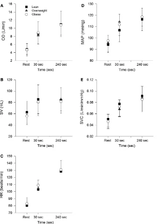
Cardiac output dynamics were assessed during the fifth and six exercise bouts. Cardiac output (CO, Lmin1) was measured using inert gases (sulfur hexafluoride and nitrous oxide) rebreathing technique (Innocor, Innovision A/S, Odense, Denmark) at rest and at 30 sec and 240 sec of exercise. Rebreathing maneuvers lasted ~10 sec. Heart rate was recorded every 5 sec (S610i, Polar Electro Oy, Finland) and along with simultaneous CO measurements used to estimate stroke volume (SV=CO/HR, mLbeat1). Measurements of systolic and diastolic blood pressure (mmHg) were made at the same times as CO measurements using manual sphygmomanometry and used to estimate mean arterial pressure (MAP=SBP/ 3+2.DBP/3; mmHg). Systemic vascular conductance (SVC) was calculated, using these measurements (SVC=CO/MAP, mLmin1mmHg1). Final values for all cardiovascular variables were averaged from responses during the two submaximal exercise bouts.
Calf exercise and haemodynamics
Calf exercise involved plantar flexors of the right leg and was performed in the supine position on a custom-built ergometer (Egana and Green 2005). For both protocols~ contractions were static and performed intermittently. Subjects were secured to the ergometer with a harness that minimized the backward displacement of the body during each contraction. Feet were attached to an immo-bile footplate, connected to a load cell, against which force was applied as subjects attempted to plantar-flex the foot. The measured force was amplified and sampled at 40 Hz before being processed by a PowerLab analog-to-digital converter (ML 795, AD Instruments). Force was displayed on a screen visible to the subject (Chart v6.0, AD Instruments) to help them control their effort and conform to specific instructions given by the investigator.
fatigue over 30 min (Ega~na and Green 2007). On this basis the exercise intensity might be considered to be mild-to-moderate and somewhat consistent with the ‘mild-to-moderate’ intensity of cycling exercise used in this study. Subjects then performed an incremental test to failure (intermittent contractions as above), beginning with contractions at a force of 100 N for 2 min and then stepwise increases in force each 2 min of 200 N for males and 150 N for females until task failure (Kiely et al. 2014). Peak force (Fpeak) achieved during this test was defined as the highest force that a subject could sustain for at least 60 sec.
At rest and during calf exercise, leg blood flow (LBF) and MAP were measured simultaneously and used to calculate leg vascular conductance (LVC=LBF/MAP). LBF was assessed using venous occlusion plethysmography (Ega~na and Green 2005). When compared with Doppler ultra-sound, this technique provides similar estimates of limb blood flow and vascular conductance during incremental and constant-load calf exercise protocols (Green et al. 2011; Murphy et al. 2018), as well as similar parameter esti-mates used to define the dynamic response of LVC (Mur-phy et al. 2018). To measure LBF, a mercury-silastic strain gauge (Hokanson EC-6) was placed around the widest part of the subject’s right calf and connected to the plethysmo-graph (Hokanson EC-6). A cuff (Hokanson) placed around the upper right thigh of the subject was inflated to a con-stant pressure of~50 mmHg for the duration of the exer-cise. The pressure of ~50 mmHg was chosen to occlude venous return without interfering with arterial flow into the leg. LBF during exercise was assessed by measuring the change in leg volume detected by the strain gauge over the 4 sec relaxation period between contractions. Beat-to-beat heart rate (HR) and systolic & diastolic blood pressures were measured at rest and during exercise using either applanation tonometry of the radial artery (COLIN CBM7000, Japan) or the volume clamp method at the level of the finger (Finometer, Finapres Medical Systems B.V., the Netherlands). A pilot reliability study was performed (n=10) to ensure that the readings attained for each of these two methods were accurate when recordings during an incremental calf plantar-flexion exercise were taken using both units simultaneously (intra-correlation coeffi-cient=0.82). MAP was calculated from systolic and dias-tolic pressures (MAP: 0.33 sysdias-tolic BP+0.66 diastolic BP). The plethysmographic estimates of LBF, expressed relative to the resting limb volume (mL100 mL1min1), were converted to millilitres per minute (mLmin1) using an estimate of each subject’s leg volume obtained from anthropometric measurements of the leg (Clarys and Mar-fell-Jones 1986a). LVC was calculated as the ratio of LBF to MAP (LVC= LBF/MAP, mLmin1mmHg1). Leg muscle mass was estimated using an anthropometrical approach (Clarys and Marfell-Jones 1986b).
LVC responses (mLmin1mmHg1) for the three exercise bouts were averaged to yield a temporal profile of LVC for each subject. In accordance with our recent description of the structure of this dynamic response in young healthy subjects (Reeder and Green 2012; Murphy et al. 2018), as well as its verification in older healthy subjects and overweight and obese subjects with type 2 diabetes (Kiely et al. 2014), these averaged, individual responses were fitted to the following function,
LVCðtÞ ¼A0þA1ð1eðtTD1Þ=s1ÞF1A2ð1 eðtTD2Þ=s2ÞF2þA3ð1eðtTD3Þ=s3ÞF3
A4ð1eðtTD4Þ=s4ÞF4 (5)
where A0is the baseline response immediately prior to the initial contraction, A1–A4 are amplitudes, TD1–TD4 are time delays, ands1–s4are time constants of the first (rapid growth), second (rapid decay), third (slow growth), and fourth (slow decay) phases, respectively. The parameters F1–F4 are conditional expressions that limit the fitting of a particular phase to the period at and beyond the time delay associated with that phase. LVC data were fitted using a weighted least-squares nonlinear regression procedure and the Marquardt–Levenberg algorithm (TableCurve 2D, Jan-del Scientific). For all moJan-dels, data that exceeded the 95% prediction intervals during an initial fit of a model were excluded. No more than four data points were removed from the original time-series of data. Some subjects did not display a second decay phase and so only parameters describing the first three phases were analyzed.
The steady-state amplitude of the LVC response, referred to as End A, was calculated as follows:
End A¼a + A1ð1eð360TD1=s1ÞÞ
A2ð1eð360TD2=s2ÞÞ þA3ð1eð360TD3=s3ÞÞ þA4ð1eð360TD4=s4ÞÞ
(6)
Statistical analysis
Data were normally distributed and so effects of BMI (3 levels) were initially tested using one-way ANOVA. A two-way ANOVA was used to explore interactions involv-ing BMI and sex and verify main effects of BMI. The level of significance was set at P≤0.05. All values are expressed as mean and SD. All statistical analyses were performed using SPSS v.24 (IBM SPSS).
Results
BMI
groups (F2,45= 123, P<0.05) were a function of differ-ences in body mass (F2,45=17.1, P<0.05). Other physi-cal characteristics, blood-borne measurements and physical activity levels were similar between groups. Calf skinfold measurements, required for estimation of leg muscle mass, were significantly lower in lean (11.3 (4.1) mm) compared with obese (14.5 (4.2) mm) subjects and intermediate values were observed for overweight subjects (12.4 (4.8) mm).
[image:6.612.79.552.89.231.2]Peak responses during graded exercise are shown in Table 2. V_O2peak normalized to body mass varied as a function of BMI (F2,45= 5.6, P<0.05) and was signifi-cantly different between the three groups. No other peak responses were significantly affected by BMI.
Parameters describing the dynamic response of V_O2 during cycling at 80% Tvent are shown in Table 3. Two
obese subjects failed to complete the required exercise bouts and so the following analyses are based on 46 sub-jects. BMI had a significant effect on s2 (F2,43=5.3,
P<0.05), which was significantly greater in obese than lean groups. By contrast, BMI did not significantly affect any other parameter or the gain of V_O2. Temporal responses of V_O2 from representative lean, overweight and obese individuals are shown in Figure 1.
Cardiovascular responses during cycling at 80% Tvent are shown in Figure 2. Again, two obese subjects failed to complete this testing and so analyses of cardiac output, heart rate and stroke volume are based on 46 rather than 48 subjects. In addition, successful blood pressure record-ings and estimates of MAP and SVC were only obtained in 36 subjects (12 lean, 10 overweight, 14 obese). None of the responses measured at rest, 30 sec and 240 sec were
Table 2. Peak physiological responses measured during maximal graded exercise grouped by BMI category.
Lean (n=16)
Overweight (n=16)
Obese (n=16)
Male/Female 6/10 6/10 6/10
Workratepeak(W) 149 (48) 146 (43) 143 (50)
_
VO2peak(LO2min1) 2.12 (0.88) 2.03 (0.70) 2.00 (0.76)
_
VO2peak(mLO2kg1min1) 30.5 (9.0) 26.0 (5.6) 21.6 (6.2)1,2
_
VEpeak(Lmin1) 74.9 (23.9) 72.0 (18.0) 71.0 (28.3)
HRpeak(beatsmin1) 164 (15) 159 (17) 158 (11)
RERpeak 1.10 (0.05) 1.11 (0.05) 1.10 (0.05)
Tvent(W) 122 (45) 123 (34) 120 (40)
Tvent(LO2min1) 1.55 (0.69) 1.49 (0.59) 1.47 (0.62)
Tvent(%V_O2peak) 73.9 (8.7) 72.7 (10.9) 71.8 (8.7)
Data are means (SD).V_O2, peak oxygen consumption;V_Epeak, peak minute ventilation; HRpeak, peak heart rate; RERpeak, peak respiratory exchange ratio;Tvent, ventilatory threshold.
1
Different from lean group (P≤0.05).
2
[image:6.612.83.545.310.459.2]Different from overweight group (P≤0.05).
Table 3. Parameter estimates related toV_O2dynamics grouped by BMI category.
Lean (n=16)
Overweight
(n=16) Obese (n=14)
Male/Female 6/10 6/10 6/8
Workrate (W) 98 (36) 99 (27) 97 (33)
a(Lmin1) 0.58 (0.14) 0.63 (0.13) 0.64 (0.16)
A1(Lmin1) 0.35 (0.22) 0.39 (0.14) 0.31 (0.17)
TD1(sec) 4.7 (4.0) 5.5 (4.5) 4.4 (3.4)
s1(sec) 13.0 (6.9) 14.7 (5.7) 8.9 (5.6)
A2(Lmin1) 0.62 (0.35) 0.57 (0.24) 0.65 (0.31)
TD2(sec) 32.3 (8.2) 31.0 (6.9) 27.8 (8.9)
s2(sec) 29.1 (7.6) 34.7 (9.0) 39.4 (9.2)1
End A (Lmin1) 1.55 (0.54) 1.59 (0.40) 1.60 (0.51)
_
VO2Gain (mLmin1W1) 10.9 (1.7) 10.8 (0.8) 11.1 (1.3)
Data are means (SD).a,amplitude during unloaded cycling; A1and A2amplitudes, TD1and TD2, time delays ands1ands2, time constants of the first and second phases, respectively.
1
significantly different between groups. Ratios of the change of these responses over the initial 30 sec of exercise relative to responses at 240 sec are shown in Table 4. The ratio of change in MAP was significantly affected by BMI (F2,33=7.2, P<0.05) and significantly different between lean and overweight groups. None of the other ratios were significantly affected by BMI.
The time constant of the heart rate response was also not different between lean (60.9 (10.2) sec), overweight (57.1 (11.9) sec) and obese subjects (66.7 (21.6) sec). None of the other parameters describing the HR response (i.e., baseline, amplitude, time delay) were significantly different between the three groups.
Peak responses during incremental calf exercise, as well as baseline measurements of the leg, grouped by BMI are shown in Table 5. There were significant differences in leg muscle mass (F2,43=9.4, P<0.05) and leg volume (F2,43=4.1, P<0.05) between groups. By contrast, MVC and performance of the exercise task were not signifi-cantly affected by BMI. Peak LBF, LVC and MAP were also not significantly affected by BMI, although the effect on MAP was close to significance (F2,41=3.1,P= 0.06).
LVC responses during submaximal calf exercise (30% MVC) are shown in Figure 3. For technical reasons, data from one overweight subject and one obese subject were not analyzed and so the following analyses are based on a sample of 46 subjects. Estimates of parameters describing the first three phases of this response are shown in Table 6. BMI had significant effects on the time delay of phase 2 (TD2: F2,43=4.5, P<0.05) and time constant of phase 3 (s3: F2,43=6.5, P<0.05), with the latter being significantly greater in the obese group compared with the lean and overweight groups. There was also a more modest effect of BMI on TD3which did not reach signifi-cance (F2,43=2.4, P=0.10). None of the phase ampli-tudes or total amplitude were affected by BMI.
BMI and sex
[image:7.612.66.292.58.517.2]For variables identified in Tables 1–6, two-way ANOVA was performed to test for main and interactive effects involving sex and BMI. A large number of variables were significantly affected by sex (main effect), as shown in Table 7. By contrast, only two variables were associated with significant BMI-by-sex interactions and which related to timing of the onsets of the second phase (TD2) and third phase (TD3) of the LVC response. For both TD2 (sex-by-BMI: F2,40=6.0) and TD3 (sex-by-BMI: F2,40=5.4), a positive effect of BMI on this parameter was only observed in females and not males. By contrast, weak and nonsignifi-cant interactions (F2,40≤1.3) were observed for the time constants of the three phases of the LVC response.
Correlations with blood-borne markers
Significant associations (P≤0.05) between baseline physi-cal characteristics (Table 1), V_O2s2 and LVCs3 were explored using the Pearson correlation coefficient (n=44–46). Both V_O2s2 and LVCs3 were significantly correlated with BMI (r =0.35, 0.36) and the correlation
between them was close to significance (r= 0.28,
P=0.07). No other significant correlations were observed forV_O2s2. For LVCs3, there were significant correlations with body mass (r =0.40), HDL (r= 0.31) and triglyc-erides (r= 0.30), and the correlation with total choles-terol was close to significance (r= 0.28,P=0.07).
Discussion
This study tested the hypothesis that obesity, represented by a BMI >30 kgm2, slows V_O2 dynamics during
[image:8.612.154.470.66.516.2]moderate cycling exercise in a cohort of middle-aged and apparently healthy adults. The present findings support this hypothesis and show that the time constant of the second phase (s2) was significantly greater in obese com-pared with lean subjects, with intermediate s2 values for overweight subjects. By contrast, estimates of the rates of change in cardiac output and systemic vascular conduc-tance were not significantly affected by BMI. For calf exercise, BMI was associated with a slowing of the second growth phase of the leg vascular conductance response (i.e., s3), a phase which coincides with phase 2 of the
_
VO2 response. These findings show that BMI impairs _
VO2 dynamics and vasodilation in contracting muscles of apparently healthy but sedentary adults.
BMI andV_O2dynamics during cycling
This study provides the first evidence that BMI affects _
VO2 dynamics (s2) in apparently healthy adults and that this effect was similar between males and females. This effect also appeared to be proportional to BMI across the three categories (Table 3) and was greatest for a level of obesity which can be considered to be mild (BMI 32 mkg2). The potency of this effect of mild obesity – an average of 35% increase in s2-when com-pared with lean subjects is similar to the effect of type 2 diabetes on s2 in males (36%) and females (34%) aver-aged from several studies (Green et al. 2015). These observations are also similar to the effects of mild obesity on s2 observed in girls and boys (Lambrick et al. 2013) and male adolescents (Salvadego et al. 2010) of ~20–25%. Collectively, these findings suggest that increases in body mass leading to obesity induce a considerable slowing of the dynamic response of V_O2 in inactive males and females and across a wide range of ages.
Cardiovascular dynamics during cycling
Mechanisms underlying any effect of BMI on V_O2 dynamics must influence the rates of O2 delivery to and/ or utilization by contracting muscles. There is evidence from animal studies that obesity impairs control of mus-cle vasodilation and perfusion (Frisbee 2003; Ribiero et al. 2005; Xiang et al. 2005, 2006; Frisbee et al. 2009), although how this relates to the present findings pertain-ing to exercispertain-ing humans is not clear. The measurement in human subjects of the ‘dynamic’ responses of O2 deliv-ery and utilization at a temporal resolution consistent with breath-by-breath gas analysis is a considerable chal-lenge (Grassi et al. 1996). This is further complicated by the need to describe behaviors of Fick variables for V_O2 (blood flow,a-vO2) measured both at the lungs and for a large number of contracting muscles. In this study, we used two approaches to shed light on aspects of cardio-vascular dynamics at a systemic level during whole-body exercise and on vascular dynamics during intermittent contractions in an isolated limb.
First, under the same exercise conditions used to assess _
VO2 dynamics, we estimated the dynamic response of cardiac output based on the ratio of change in cardiac output during the early period of exercise (30 sec) relative to its change after 4 min (Table 4). Previously, we showed that this ratio and s2 (V_O2) were increased by training in individuals with type 2 diabetes (MacAnaney
et al. 2012), suggesting that this ratio was sensitive to changes in cardiac output and V_O2 dynamics. However, neither this estimate of the initial rate of change nor the absolute values of cardiac output during exercise were affected by BMI. In addition, BMI did not affect the dynamic responses of stroke volume and heart rate (Table 4) or estimates of heart rate dynamics obtained from empirical modelling (Eq. 4). This evidence suggests that cardiac output dynamics during moderate exercise are not affected by obesity and do not contribute to the slowing ofV_O2dynamics with increasing BMI.
Large changes in cardiac output during exercise relative to much smaller changes in mean arterial pressure reflect an increase in systemic vascular conductance. During exercise at a moderate intensity, the increase in systemic vascular conductance is affected mainly by an increase in vascular conductance in contracting muscles (Mortensen et al. 2005). Thus, estimates of the dynamic response of systemic vascular conductance might provide insight into haemodynamic effects of BMI in contracting muscles. In this study, the average change in systemic vascular con-ductance from baseline to the fourth minute of moderate exercise was similar between lean (81%), overweight (90%) and obese (80%) subjects, consistent with the simi-lar power outputs at which these groups exercised. Approximately 50–65% of this change in systemic vascu-lar conductance was achieved during the first 30 sec of exercise and the ratio which reflects this estimate (Table 4) was also not different between these groups. This suggests that BMI does not affect the dynamic response of systemic vascular conductance during cycling and possibly the underlying response (vasodilation) in contracting muscles.
to a measurement frequency of 2 min or more and the challenge of optimizing the timing of the first exercise measurement.
Vasodilation during isolated muscle contractions
Ideally, establishing the role of muscle blood flow and O2 supply in BMI-related effects on V_O2 dynamics requires these variables to be measured during cycling. This, how-ever, is not possible with available techniques and proba-bly requires adaptation of a technique, such as Doppler ultrasound, with the required temporal resolution of mea-surement applied in the correct location (e.g., aortic-iliac bifurcation) to ‘sample’ all primary muscles involved. We chose a lesser alternative but one which enabled an accu-rate description of the structure of the dynamic response
of vasodilation in contracting muscles (Reeder and Green 2012), represented by the measurement of LVC.
Using a model of isolated limb (calf) exercise and intermittent contractions, we previously showed that the response of LVC in contracting muscles consisted of 3–4 phases which alternate between growth and decay in vas-cular conductance. This dynamic structure was observed in younger, healthy individuals (Reeder and Green 2012; Donnelly and Green 2013; Murphy et al. 2018), older healthy individuals (Reeder and Green 2011), as well as middle-aged subjects with type 2 diabetes (Kiely et al. 2014). This same structure was observed in the present cohort and, because the fourth phase was not observed in all subjects, our analyses were restricted to the first three phases.
[image:10.612.74.544.99.199.2]The first and third phases of the leg vascular conduc-tance response are ‘growth’ phases which represent an
Table 4. Estimates of dynamic responses of cardiovascular variables based on changes from rest over the initial 30 and 240 sec of exercise expressed as ratios (D30/D240).
Lean (n=16)
Overweight (n=16)
Obese (n=14)
Male/Female 6/10 6/10 6/8
HR 0.29 (0.11) 0.30 (0.10) 0.34 (0.26)
SV 0.40 (0.32) 0.44 (0.31) 0.49 (0.34)
CO 0.62 (0.14) 0.58 (0.19) 0.66 (0.15)
MAP 0.56 (0.25) 0.86 (0.17)1 0.73 (0.20)
SVC 0.65 (0.14) 0.49 (0.29) 0.66 (0.15)
A higher ratio indicates a faster response. For example, the first estimate (mean=0.29) indicates that~30% of the change in heart rate over 240 sec of exercise was achieved in the first 30 sec of exercise. Data are means (SD). HR, heart rate; SV, stroke volume; CO, cardiac output; MAP, mean arterial pressure; SVC, systemic vascular conductance. Note: sample sizes for MAP and SVC were 12 (lean), 10 (overweight) and 14 (obese).
1
Different from lean group (P≤0.05).
Table 5. Characteristics of leg anthropometry, strength, and responses to incremental calf exercise according to BMI category.
Lean (n=16)
Overweight (n=15)
Obese (n=15)
Male/Female 6/10 6/9 6/9
Leg muscle mass (kg) 1.64 (0.31) 1.91 (0.28)1 2.12 (0.33)1
Leg volume (mL) 2437 (339) 2685 (363) 2833 (467)1
MVC (N) 737 (224) 805 (333) 819 (317)
MVC (Nkg1leg muscle mass) 443 (83) 410 (125) 379 (105)
Exercise time (sec) 416 (110) 434 (125) 440 (87)
Forcepeak(N) 525 (203) 567 (224) 557 (161)
Forcepeak(%MVC) 71.0 (13.8) 70.8 (9.5) 71.6 (15.3)
LBFpeak(mLmin1) 644 (231) 677 (344) 716 (350)
MAPpeak(mmHg) 101 (14) 106 (12) 113 (14)
LVCpeak(mLmin1mmHg1) 6.76 (2.93) 6.52 (3.27) 6.27 (3.03)
Data are means (SD). MVC, maximal voluntary contraction; LBFpeak, peak leg blood flow; MAPpeak, peak mean arterial pressure LVCpeak, peak leg vascular conductance.
1
[image:10.612.86.544.291.441.2]initial and rapid vasodilation followed by a smaller and slower rate of vasodilation, respectively. The second of these growth phases begins at a time (time delay20– 30 sec) similar to the onset of phase 2 of the V_O2 response. In the present study, this second growth phase of LVC was significantly slower (higher s3) in obese
subjects compared with lean and overweight subjects. Although we did not analyze the dynamic response char-acteristics of leg (muscle) blood flow, with this experi-mental model the leg vascular conductance and blood flow responses are nearly identical (Reeder and Green 2012; Donnelly and Green 2013; Murphy et al. 2018). These observations suggest that obesity impairs the rate of the secondary phase of vasodilation and hyperemia in contracting muscles.
The similar time delays between the second phase of the V_O2 response during cycling and second growth phase (phase 3) of the LVC response during calf exercise suggest that these phases represent the same general phase of an overall dynamic physiological response to exercise. The positive correlation between the time constants of these two phases, albeit not quite significant (P= 0.07), supports the prospect of a causal link between vasodila-tion and V_O2 dynamics. This, combined with the rela-tively larger effect of BMI on LVC dynamics (phase 3) compared withV_O2dynamics raises the possibility that a slowing of vasodilation in contracting muscles contributes to the BMI-related slowing of V_O2 dynamics observed during cycling.
Becoming overweight
A question arises about the timing of effects of BMI on _
VO2and cardiovascular dynamics as subjects gain weight, and whether or not any of these effects appear ‘early’ in overweight subjects. Although V_O2 dynamics during cycling were not significantly slowed in overweight subjects, their BMI and s2 (V_O2) values were intermediate and equidistant between lean and obese values. This suggests that there is a proportional effect of BMI on s2 and that important effects might appear in overweight subjects.
With respect to LVC dynamics during calf exercise, the time delays of the second (decay) and third (growth) phase were greater in overweight subjects compared with lean subjects but similar between lean and obese subjects. These effects, however, were observed only in female sub-jects. These data raise the possibility that an increase in body mass and transition from lean to overweight cate-gories results in changes to the timing of later phases of vascular responsiveness and which precede the slowing of vasodilation seen with larger increases in BMI, at least in females.
Lipids and vasodilation
[image:11.612.67.292.61.529.2]Correlations between several plasma lipid markers (total cholesterol, HDL, triglycerides) and s3 of the LVC response, but not s2 of the V_O2 response, are broadly consistent with the detrimental effects of some lipid
species on vascular reactivity in human limbs (Steinberg et al. 1997) and skeletal muscle (Clerk et al. 2002; Stein-berg and Baron 2002). These correlations raise the possi-bility that changes in blood lipids which accompany weight gain might affect vascular dynamics in contracting muscles and possibly contribute to the present findings. Further research is needed to clarify this.
Limitations
Despite its limitations, BMI is recognized as a useful measurement of obesity (Ortega et al. 2016). Leg skinfold estimates (Results) along with muscle mass and volume measurements (Table 1) show that increasing BMI was associated with increasing skinfold values and a relatively smaller muscle mass per limb volume, measurements
[image:12.612.83.545.89.251.2]consistent with increasing adiposity. Whether the effects of BMI on V_O2 and cardiovascular dynamics can be attributed to physiological effects of obesity per se or related factors (e.g., age, blood pressure, inactivity) requires clarification. Nevertheless, with the exception of BMI and body mass, none of these other factors (see characteristics in Table 1) were correlated with BMI or parameters of V_O2 dynamics despite their wide and nor-mal distributions. The use of nor-males and fenor-males to explore effects of BMI might be criticized on the basis of observations of sex differences in V_O2 dynamics in over-weight and obese individuals with type 2 diabetes (Regen-steiner et al. 2015). However, these observations are not consistent with our findings (O’Connor et al. 2012) or a review of all studies of males and females with type 2 diabetes (Green et al. 2015). Moreover, the weak effects
Table 6. Dynamic response characteristics of leg vascular conductance during calf exercise at 30%MVC according to BMI category.
Lean (n=16)
Overweight (n=15)
Obese (n=15)
Male/Female 6/10 6/9 6/9
A0(mLmin1mmHg1) 0.63 (0.29) 0.86 (0.51) 0.60 (0.29)
A1(mLmin1mmHg1) 2.17 (1.23) 2.29 (2.07) 1.91 (1.15)
TD1(sec) 1.2 (0.9) 1.3 (1.0) 0.8 (0.9)
s1(sec) 4.1 (1.7) 5.4 (4.1) 3.4 (3.0)
A2(mLmin1mmHg1) 0.93 (0.88) 0.67 (0.60) 0.47 (0.39)
TD2(sec) 7.8 (5.1) 15.1 (9.8)1 10.2 (4.6)
s2(sec) 7.1 (3.5) 9.6 (5.3) 11.1 (11.3)
A3(mLmin1mmHg1) 1.13 (0.94) 0.96 (0.85) 1.10 (0.75)
TD3(sec) 19.8 (8.5) 27.3 (11.4) 23.9 (8.5)
s3(sec) 11.6 (4.5) 13.4 (6.7) 22.1 (12.7)1,2
EndA (mLmin1mmHg1) 3.45 (1.71) 3.47 (2.12) 2.98 (1.49)
Data are means (SD). A0baseline amplitude; A1, A2and A3, amplitudes, TD1, TD2and TD3, time delays ands1,s2ands3, time constants of the first (rapid growth), second (rapid decay) and third (slow growth) phases, respectively.
1
Different from lean group (P≤0.05).
2
Different from overweight group (P≤0.05).
Table 7. Variables identified in Tables 1-6 which were significantly different (ANOVAP<0.05) between males (♂:n=18) and females (♀:
n=28). Unless indicated, values for males were greater than values for females.
Physical GXT V_O2dynamics Leg
Height Workratepeak(W) a Leg mass
Mass V_O2peak(mLkg1min1) A1 Leg volume
FPG V_O2peak(Lmin1) A2 MVC (N)
HbA1C EndA MVC (Nkg1)
ABI VEpeak(Lmin1) ExTime (sec)
DBP Tvent(W) Fpeak(N)
HDL: (♂<♀) MAPpeak
[image:12.612.77.545.353.486.2]of sex (F <1.0) on parameters describing V_O2 dynamics and most LVC variables supports our use of males and females in this study. We acknowledge that a larger and more even sample of males and females is required for studies of sex effects.
Perspective
Obesity reduces cardiorespiratory fitness but it is not clear whether this effect is a function of increasedweight per se on estimates of fitness or reflects direct effects underlying physiological function (see Introduction). Assessment of
_
VO2 dynamics during submaximal exercise that is not weight-bearing, combined with the absence of normaliza-tion of parameter estimates to body mass, helps overcome the confounding problem of body mass. The speed with which V_O2 and underlying cardiorespiratory function adjusts to the onset of submaximal exercise is probably as important an indicator of cardiorespiratory ‘fitness’ as a peak or maximum exercise response. Present findings show that obesity impairs such dynamic responses and, given practical problems with the assessment of peak and maxi-mum responses (Poole and Jones 2017; Green and Askew 2018), has implications for further study of effects of obe-sity on cardiorespiratory fitness.
Conclusion
In conclusion, the present findings show that BMI is asso-ciated with a slowing of the dynamic responses of V_O2 (s2) during cycling and leg vascular conductance during isolated limb exercise in apparently healthy adults of both sexes. Further study is required to verify these effects and clarify the mechanisms involved.
Conflicts of Interest
All authors have no conflicts of interest to declare.
References
Barstow, T. J., and P. A. Mole. 1991. Linear and nonlinear characteristics of oxygen uptake kinetics during heavy exercise. J. Appl. Physiol. 71:2099–2106.
Clarys, J. P., and M. J. Marfell-Jones. 1986a. Anatomical segmentation in humans and the prediction of segmental masses from intra-segmental anthropometry. Hum. Biol. 58:771–782.
Clarys, J. P., and M. J. Marfell-Jones. 1986b. Anthropometric prediction of component tissue masses in the minor limb segments of the human body. Hum. Biol. 58:761–769. Clerk, L. H., S. Rattigan, and M. G. Clark. 2002. Lipid
infusion impairs physiologic insulin-mediated capillary
recruitment and muscle glucose uptake in vivo. Diabetes 51:1138–1145.
Donnelly, J., and S. Green. 2013. Effect of hypoxia on the dynamic response characteristics of hyperaemia in the contracting human calf muscle. Exp. Physiol. 98(1):81–93. Drinkard, B., J. McDuffie, S. McCann, G. I. Uwaifo, J.
Nicholson, and J. A. Yanovski. 2001. Relationships between walk/run performance test and cardiorespiratory fitness in adolescents who are overweight. Phys. Ther. 81:1889–1896. Ega~na, M., and S. Green. 2005. Effect of body tilt on calf
muscle performance and blood flow in humans. J. Appl. Physiol. 98:2249–2258.
Ega~na, M., and S. Green. 2007. Intensity-dependent effect of body tilt angle on calf muscle fatigue in humans. Eur. J. Appl. Physiol. 99:1–9.
Ega~na, M., S. Smith, and S. Green. 2007. Revisiting the effect of posture on high-intensity constant-load cycling performance in men and women. Eur. J. Appl. Physiol. 99:495–501.
Frisbee, J. C. 2003. Impaired skeletal muscle perfusion in obese Zucker rats. Am. J. Physiol. Regul. Integr. Comp. Physiol. 285: R1124–R1134.
Frisbee, J. C., J. M. Hollander, R. W. Brock, H. G. Yu, and M. A. Boegehold. 2009. Integration of skeletal muscle resistance arteriolar reactivity for perfusion responses in the metabolic syndrome. Am. J. Physiol. Regul. Integr. Comp. Physiol. 296:R1771–R1782.
Grassi, B., D. C. Poole, R. S. Richardson, D. R. Knight, B. K. Erickson, and P. D. Wagner. 1996. Muscle O2uptake kinetics in humans: Implications for metabolic control. J. Appl. Physiol. 80:988–998.
Green, S., and C. D. Askew. 2018.V_O2peakis an acceptable estimate of cardiorespiratory fitness but notV_O2max. J. Appl. Physiol.. https://doi.org/10.1152/japplphysiol.00850. 2017.
Green, S., C. D. Askew, and P. J. Walker. 2007. Effect of type 2 diabetes mellitus on exercise intolerance and the physiological responses to exercise in peripheral arterial disease. Diabetologia 50:859–866.
Green, S., R. Thorp, E. Reeder, J. Donnelly, and G. Fordy. 2011. Venous occlusion plethysmography versus Doppler ultrasound in the assessment of leg blood flow during calf exercise. Eur. J. Appl. Physiol. 111:1889–1900.
Green, S., M. Ega~na, J. C. Baldi, R. Lamberts, and J. G. Regensteiner. 2015. Cardiovascular control in type 2 diabetes. J. Diabetes Res. 1: 1–11.
Hughson, R. L., M. E. Tschakovsky, and M. E. Houston. 2001. Regulation of oxygen consumption at the onset of exercise. Exerc. Sport Sci. Rev. 29:129–133.
Kiely, C., E. O’Connor, D. O’Shea, S. Green, and M. Egana.~ 2014. Hemodynamic responses during graded and constant-load plantar flexion exercise in middle-aged men and women with type 2 diabetes. J. Appl. Physiol. 117:755–764.
Krachler, B., K. Savonen, P. Komulainen, M. Hassinen, T. A. Lakka, and R. Rauramaa. 2015. VO2max/kg is expected to be lower in obese individuals!. Int. J. Cardiol. 189:234. Lambrick, D., J. Faulkner, N. Westrupp, and M. McNarry.
2013. The influence of body weight on the pulmonary oxygen uptake kinetics in pre-pubertal children during moderate- and heavy intensity treadmill exercise. Eur. J. Appl. Physiol. 113:1947–1955.
MacAnaney, O., J. Malone, S. Warmington, D. O’Shea, S. Green, and M. Ega~na. 2011. Cardiac output responses are not related to the slowed oxygen uptake kinetics in type 2 diabetes. Med. Sci. Sports Exerc. 43:935–942.
MacAnaney, O., D. O’Shea, S. Warmington, S. Green, and M. Ega~na. 2012. Gymnasium-based unsupervised exercise maintains benefits in VO2kinetics obtained following supervised training in type 2 diabetes. Appl. Physiol. Nutr. Metab. 37:599–609.
Mortensen, S. P., E. A. Dawson, C. C. Yoshiga, M. K. Dalsgaard, R. Damsgaard, N. H. Secher, et al. 2005. Limitations to systemic and locomotor limb muscle oxygen delivery and uptake during maximal exercise in humans. J.Physiol. Lond. 566:273–285.
Murphy, E., J. Rocha, N. Gildea, S. Green, and M. Ega~na. 2018. Venous occlusion plethysmography vs. Doppler ultrasound in the assessment of leg blood flow kinetics during different intensities of calf exercise. Eur. J. Appl. Physiol. 118:249–260.
O’Connor, E., C. Kiely, D. O’Shea, S. Green, and M. Ega~na. 2012. Similar level of impairment in exercise tolerance and VO2kinetics in middle aged men and women. Am. J. Physio. (Regulatory and Integrative Physiology) 303: R70–R76.
Ortega, F. B., C. J. Lavie, and S. N. Blair. 2016. Obesity and cardiovascular disease. Circ. Res. 118:1752–1770. Poole, D. C., and A. M. Jones. 2017. Measurement of the
maximum oxygen uptakeV_O2max:V_O2peakis no longer acceptable. J. Appl. Physiol. 122:997–1002.
Reeder, E., and S. Green. 2011. Effect of age on the dynamic response of muscle hyperaemia during exercise. In: Proceedings of the Australian Physiological Society. Perth; p. 109P. Reeder, E., and S. Green. 2012. Dynamic response
characteristics of muscle hyperaemia: effect of exercise intensity and relation to electromyographic activity. Eur. J. Appl. Physiol. 112:3997–4013.
Regensteiner, J. G., T. A. Bauer, A. G. Huebschmann, L. Herlache, H. D. Weinberger, E. E. Wolfel, et al. 2015. Sex
differences in the effects of type 2 diabetes on exercise performance. Med. Sci. Sports Exerc. 47:58–65.
Ribiero, M., A. G. Silva, N. S. Santos, I. Guazzelle , L. N. J. Matos, I. C. Trombetta, et al. 2005. Diet and exercise training restore blood pressure and vasodilatory responses during physiological maneuvers in obese children. Circulation 111:1915–1923.
Rowlands, A. V., P. W. Thomas, R. G. Eston, and R. Topping. 2004. Validation of the RT3 triaxial accelerometer for the assessment of physical activity. Med. Sci. Sports Exerc. 36:518–524.
Salvadego, D., S. Lazzer, C. Busti, R. Galli, F. Agosti, C. Lafortuna, et al. 2010. Gas exchange kinetics in obese adolescents. Inferences on exercise tolerance and
prescription. Am. J. Physiol. Regul. Integr. Comp. Physiol. 299: R1298–R1305.
Spencer, M. D., J. M. Murias, H. P. Lamb, J. M. Kowalchuk, and D. H. Paterson. 2011. Are the parameters ofV_O2, heart rate and muscle deoxygenation kinetics affected by serial moderate-intensity exercise transitions in a single day? Eur. J. Appl. Physiol. 111:591–600.
Steinberg, H. O., and A. D. Baron. 2002. Vascular function, insulin resistance and fatty acids. Diabetologia 45:623–634. Steinberg, H. O., M. Tarshoby, R. Monestel, G. Hook, J.
Cronin, A. Johnson, et al. 1997. Elevated circulating free fatty acid levels impair endothelium-dependent vasodilation. J. Clin. Invest. 100:1230–1239.
Tsiros, M. D., A. M. Coates, P. R. C. Howe, J. Walkley, A. P. Hills, R. E. Wood, et al. 2016. Adiposity is related to decrements in cardiorespiratory fitness in obese and normal-weight children. Pediatr. Obes. 11:144–150.
Wei, M., L. W. Gibbons, J. B. Kampert, M. Z. Nichaman, and S. N. Blair. 2000. Low cardiorespiratory fitness and physical inactivity as predictors of mortality in men with type 2 diabetes. Ann. Intern. Med. 132:605–611.
Whipp, B. J., H. B. Rossiter, and S. A. Ward. 2002. Exertional oxygen uptake kinetics: a stamen of stamina? Biochem. Soc. Trans. 30:237–247.
Wilkerson, D. P., K. Koppo, T. J. Barstow, and A. M. Jones. 2004. Effect of work rate on the functional ‘gain’ of Phase II pulmonary O-2 uptake response to exercise. Respir. Physiol. Neurobiol. 142:211–223.
Xiang, L., J. Naik, and R. L. Hester. 2005. Exercise-induced increase in skeletal muscle vasodilatory responses in obese Zucker rats. Am. J. Physiol. Regul. Integr. Comp. Physiol. 288: R987–R991.