©2013
Landes
Bioscience.
Do
not
distribute.
Cancer Biology & Therapy 14:6, 537–545; June 2013; © 2013 Landes Bioscience
ReseaRCh papeR ReseaRCh papeR
*Correspondence to: Patricia M. McGowan; Email: [email protected] Submitted: 01/07/13; Revised: 03/05/13; Accepted: 03/18/13
http://dx.doi.org/10.4161/cbt/24349
Introduction
PARP1 is a multifunctional enzyme, best known for its role in
the repair of single strand DNA breaks.1,2 In recent years, several
low molecular weight PARP inhibitors have been investigated as
potential anticancer agents.3 As anticancer drugs, PARP
inhibi-tors can potentially be used in several different ways, such as monotherapy or as potentiators of the effects of specific cytotoxic drugs. To date, the efficacy of PARP inhibitors as monotherapy has been mostly confined to patients with BRCA1/2
mutation-associated breast and ovarian cancer.4-7 In these patients, the
efficacy of the PARP inhibitor, olaparib appears to relate to a synthetic lethal interaction between defective BRCA1/2 function
and inhibition of PARP1.8,9
Synthetic lethality occurs between two genes when loss of one gene is compatible with cell viability but loss of both results in cell
death.8,9 The implication of synthetic lethality is that targeting of
one such gene in a cancer (e.g., PARP1), where the other is non-functioning (e.g., in patients with BRCA1 or BRCA2 germline
paRp inhibitors, both as monotherapy and in combination with cytotoxic drugs, are currently undergoing clinical trials in several different cancer types. In this investigation, we compared the antiproliferative activity of two paRp/putative paRp inhibitors, i.e., olaparib and iniparib, in a panel of 14 breast cancer cell lines (seven triple-negative and seven non-triple-negative). In almost all cell lines investigated, olaparib was a more potent inhibitor of cell growth than iniparib. Inhibition by both drugs was cell line-dependent and independent of the molecular subtype status of the cells, i.e., whether cells were triple-negative or non-triple negative. although the primary target of paRp inhibitors is paRp1, no significant association was found between baseline levels of paRp1 activity and inhibition with either agent. similarly, no significant correlation was evident between sensitivity and levels of CDK1, BRCa1 or miR-182. Combined addition of olaparib and either the CDK1 inhibitor, RO-3306 or a pan heR inhibitor (neratinib, afatinib) resulted in superior growth inhibition to that obtained with olaparib alone. We conclude that olaparib, in contrast to iniparib, is a strong inhibitor of breast cancer cell growth and may have efficacy in breast cancer irrespective of its molecular subtype, i.e., whether heR2-positive, estrogen receptor (eR)-positive or triple-negative. Olaparib, in combination with a selective CDK1 inhibitor or a pan heR inhibitor, is a potential new approach for treating breast cancer.
Comparative antiproliferative effects of iniparib
and olaparib on a panel of triple-negative
and non-triple-negative breast cancer cell lines
aisling pierce,1,2,† patricia M. McGowan,1,2,†,* Maura Cotter,1,2 Maeve Mullooly,1,2 Norma O’Donovan,3 sweta Rani,4
Lorraine O’Driscoll,4 John Crown5 and Michael J. Duffy2,6
1education and Research Centre; st. Vincent’s University hospital; Dublin, Ireland; 2UCD school of Medicine and Medical science; Conway Institute of Biomolecular
and Biomedical Research; University College Dublin; Dublin, Ireland; 3National Institute for Cellular Biotechnology; Dublin City University; Dublin, Ireland; 4school of pharmacy
and pharmaceutical sciences; panoz Institute; Trinity College; Dublin, Ireland; 5Department of Medical Oncology; st Vincent’s University hospital; Dublin, Ireland; 6UCD Clinical
Research Centre; st Vincent’s University hospital; Dublin, Ireland
†These authors contributed equally to this work.
Keywords: PARP inhibitors, olaparib, iniparib, breast cancer, triple-negative, CDK1
mutations), should in theory, be selectively lethal to tumor cells
but not affect normal cells.8,9
Germline BRCA1/2 mutations however, are responsible for only a minority of all breast and ovarian cancers, constituting < 10% of these malignancies. Although phase I/II clinical trials have shown promising results with the PARP inhibitor olaparib,
in patients with germline defects in these genes,4-7 it remains to be
shown if PARP inhibitors have efficacy in patients with sporadic breast cancer.
A subgroup of patients with sporadic breast cancer possessing biological and clinical similarities with BRCA1/2-related malig-nancies is those with triple-negative (TN) disease, i.e., patients negative for estrogen receptors (ER), progesterone receptors and HER2. These similarities include a tendency for high tumor grade, expression of the basal cytokeratins 5 and 6, aberrant
DNA repair and related gene expression signatures.10-12 These
©2013
Landes
Bioscience.
Do
not
distribute.
inhibitors, as mentioned above for BRCA1/2 mutation related cancers. Although a phase II clinical trial showed that combina-tions of the putative PARP inhibitor, iniparib with cisplatin and gemcitabine was superior to cisplatin and gemcitabine alone in
patients with advanced TNBC,13 a follow-up phase III trial failed
to meet the pre-specified criteria for significance with respect to
progression free survival and overall survival.14
Although iniparib has undergone evaluation in phase II/III
clinical trials,15 relatively little preclinical work appears to have
been published on this agent prior to the initiation of these tri-als.15-17 We therefore decided to carry out a detailed preclinical
study on the antiproliferative effects of iniparib in a panel of estrogen receptor-positive, HER2-positive and TN breast can-cer cell lines and to compare response to this agent with that of olaparib. We also show that combined treatment with olaparib and either a CDK1 or a pan HER inhibitor was superior to olapa-rib in a cell line-dependent manner.
Results
Comparative effects of olaparib and iniparib on cell growth in triple-negative and non-triple-negative breast cancer cell lines.
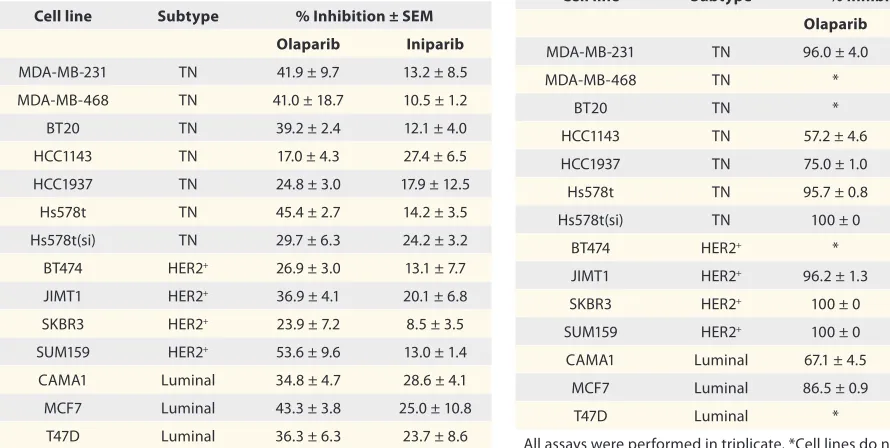
Tables 1 and 2 compare the relative growth inhibitory effects of
5 µM olaparib and iniparib on a panel of breast cancer cell lines.
As can be seen, using both the MTT (Table 1) and colony
forma-tion assays (Table 2), olaparib was a more effective growth
inhib-itor in almost all the cell lines investigated. Overall, the observed growth inhibition for both olaparib and iniparib was greater with the colony formation assay than with the MTT assay.
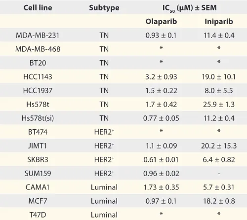
Tables 3 and 4 compare the IC50 values for olaparib and
iniparib in the same panel of cell lines. Consistent with above
findings, IC50 values for olaparib were considerably lower than
those found for iniparib. Thus, with the MTT assay, IC50
val-ues for olaparib across the cell lines, varied from 4.2 to 19.8 µM
(Table 3), whereas the corresponding IC50 values for iniparib
were all > 10 µM. Using the colony formation assays, IC50 values
were lower than those found with the MTT assay, ranging from 0.6 to 3.2 µM with olaparib and from 5.7 to > 20 µM with
ini-parib (Table 4). No significant correlation however, was found
between the IC50 values obtained with the MTT and colony
for-mation assays.
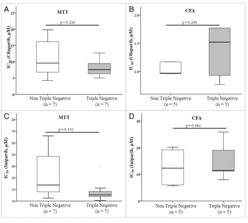
Since TN breast cancers have been shown to exhibit similar molecular and histological characteristics to BRCA1/2 associated cancers (see above), we compared response to both olaparib and iniparib in TN and non-TN cell lines. However, using both the MTT and colony formation assay, no significant difference in response was found between the 2 groups of cell lines, i.e., TN and non-TN cell lines exhibited similar responses to olaparib and
iniparib (Fig. 1).
Attempts to identify biomarkers predictive of iniparib and
olaparib sensitivity. Since response to both olaparib and
inipa-rib was variable from cell line to cell line and independent of the TN status of the cells, we attempted to identify potential markers of sensitivity. As preliminary data had previously
sug-gested that CDK1,18 BRCA1 protein and miR-182 levels19 were
associated with response to PARP inhibitors, we investigated these as potential predictors of response to olaparib and inipa-rib. However, BRCA1, CDK1 and miR-182 levels were unrelated
to olaparib (Figs. S2 and S3) or iniparib sensitivity (Figs. S4
and S5). Similar results were obtained when BRCA1, CDK1 and
miR-182 levels were expressed as continuous variables.
Since PARP1 is the primary target for PARP inhibitors, we also investigated if a relationship existed between PARP1 activity and sensitivity to iniparib and olaparib. Although PARP1 activ-ity varied widely in the cell lines investigated, no relationship was found between basal activity levels, and response to either
[image:2.612.271.555.494.725.2]olaparib (Figs. S2A and S3A) or iniparib (Figs. S4A and S5A).
Table 1. Comparative antiproliferative effects of olaparib and iniparib at 5 µM concentration, using the MTT assay
Cell line Subtype % Inhibition ± SEM
Olaparib Iniparib
MDa-MB-231 TN 41.9 ± 9.7 13.2 ± 8.5
MDa-MB-468 TN 41.0 ± 18.7 10.5 ± 1.2
BT20 TN 39.2 ± 2.4 12.1 ± 4.0
hCC1143 TN 17.0 ± 4.3 27.4 ± 6.5
hCC1937 TN 24.8 ± 3.0 17.9 ± 12.5
hs578t TN 45.4 ± 2.7 14.2 ± 3.5
hs578t(si) TN 29.7 ± 6.3 24.2 ± 3.2
BT474 heR2+ 26.9 ± 3.0 13.1 ± 7.7
JIMT1 heR2+ 36.9 ± 4.1 20.1 ± 6.8
sKBR3 heR2+ 23.9 ± 7.2 8.5 ± 3.5
sUM159 heR2+ 53.6 ± 9.6 13.0 ± 1.4
CaMa1 Luminal 34.8 ± 4.7 28.6 ± 4.1
MCF7 Luminal 43.3 ± 3.8 25.0 ± 10.8
T47D Luminal 36.3 ± 6.3 23.7 ± 8.6
all assays were performed in triplicate.
Table 2. Comparative antiproliferative effects of olaparib and iniparib at 5 µM concentration, using the colony formation assay
Cell line Subtype % Inhibition ± SEM
Olaparib Iniparib
MDa-MB-231 TN 96.0 ± 4.0 11.6 ± 5.5
MDa-MB-468 TN * *
BT20 TN * *
hCC1143 TN 57.2 ± 4.6 17.4 ± 8.7
hCC1937 TN 75.0 ± 1.0 60.7 ± 24.8
hs578t TN 95.7 ± 0.8 36.1 ± 0.9
hs578t(si) TN 100 ± 0 0 ± 0
BT474 heR2+ * *
JIMT1 heR2+ 96.2 ± 1.3 55.4 ± 9.8
sKBR3 heR2+ 100 ± 0 22.4 ± 2.4
sUM159 heR2+ 100 ± 0 0.99 ± 0.8
CaMa1 Luminal 67.1 ± 4.5 33.3 ± 11.9
MCF7 Luminal 86.5 ± 0.9 28.4 ± 5.9
T47D Luminal * *
[image:2.612.55.500.505.729.2]©2013
Landes
Bioscience.
Do
not
distribute.
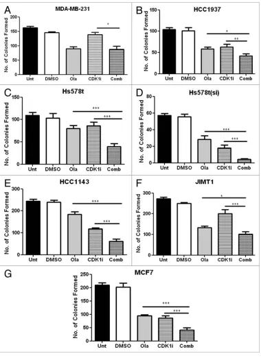
Effect of combined treatment with olaparib and a CDK1
inhibitor on cell growth. As olaparib was the more potent cell
growth inhibitor, we decided to investigate its effects in combina-tion with novel drugs. Since reduced CDK1 activity was previously shown to impair both BRCA1 function and homologous
recombi-nation repair,18 we investigated combined inhibition with olaparib
and the selective CDK1 inhibitor, RO-3306 (Merck). As shown
in Figure 2, combined addition of both olaparib and RO-3306
significantly enhanced growth inhibition compared with either olaparib or RO-3306 alone in 6/7 cell lines investigated.
Effect of combined treatment with olaparib and pan HER
inhibitors on cell growth. The combined effects of olaparib and
neratinib as well as olaparib and afatinib on colony formation
were investigated in four cell lines. As shown in Figure 3, the
effects of the dual treatment were cell line-dependent. Thus, in both the MDA-MB-231 and JIMT1 cells, the anti-growth effects of 1 µM of olaparib and either neratinib or afatinib were significantly more potent than either agent alone. In HCC1143 cells on the other hand, the combined treatment was more potent than olaparib alone but not significantly stronger than that of neratinib or afatinib alone. In contrast, using the MCF7 cells, combined treatment with olaparib and neratinib did not enhance cytotoxicity over either drug alone. The reason for this may relate to sensitivity of this cell line to neratinib. In the MCF7 cells, combined treatment with olaparib and afatinib gave significantly stronger inhibition of colony formation than afatinib alone but
not compared with olaparib alone (Fig. 3).
Discussion
Olaparib and iniparib are among two of the PARP/putative PARP inhibitors most widely investigated in clinical trials in
patients with breast cancer.4-7,13,14 To our knowledge, this is one
of the first in vitro studies to compare cytotoxicity of these agents in a broad panel of breast cancer cell lines, including TNBC, HER2-positive and ER-positive cells. Our findings suggest that response to both olaparib and iniparib is variable from cell line to cell line and independent of molecular subtype. Despite this
variability, and in agreement with recent reports,20,21 we found
that olaparib was a more potent inhibitor of cell growth than ini-parib in almost all the cell lines investigated. Our results are thus consistent with recent studies showing that iniparib was a weak
inhibitor of PARP1.20-22
Although MTT and colony formation assays are widely used to evaluate in vitro antiproliferative activities of investigational
drugs, we showed that the IC50 values obtained with the colony
formation assay were consistently lower than those obtained with the MTT assays. Furthermore, no significant correlation
was found between the IC50 values obtained with the two assays
across the panel cell lines investigated. Thus, the specific IC50
values determined, depended not only on the cell line but also on the specific viability assay used. It remains to be shown which assays better predict antitumor activity in vivo.
Because of the similarities between triple-negative and
BRCA1/2 associated tumors,10-12 it might be expected that PARP
inhibitors would be more effective in breast cancer cells, negative
for ER, PR and HER2. Indeed, Hastak et al.23 reported that TN
breast cancer cell lines were more sensitive than luminal cell lines to the experimental PARP inhibitor, PJ34 (EMD Biosciences). In contrast, our preclinical study using a larger panel of breast
can-cer cell lines than investigated by Hastak et al.,23 found no
[image:3.612.313.565.83.306.2]sig-nificant relationship between response to olaparib or iniparib and TN status. Indeed, of all the cell lines investigated in the current study, the most sensitive was the HER2-positive cell line, SKBR3
Table 3. Comparative IC50 concentrations for olaparib and iniparib, using the MTT assay
Cell line Subtype IC50 (µM) ± SEM
Olaparib Iniparib
MDa-MB-231 TN 6.9 ± 1.1 13.5 ± 1.4
MDa-MB-468 TN 5.0 ± 1.4 39.7 ± 13.3
BT20 TN 7.7 ± 0.85 15.3 ± 0.9
hCC1143 TN 11.1 ± 0.24 15.4 ± 7.9
hCC1937 TN 12.6 ± 2.0 21.1 ± 3.2
hs578t TN 5.6 ± 0.66 14.4 ± 3.0
hs578t(si) TN 7.5 ± 0.59 10.5 ± 1.9
BT474 heR2+ 19.8 ± 2.6 280 ± 18.5
JIMT1 heR2+ 7.7 ± 0.41 12.4 ± 0.5
sKBR3 heR2+ 11.1 ± 2.2 66.0 ± 39.5
sUM159 heR2+ 4.2 ± 1.0 19.6 ± 4.4
CaMa1 Luminal 15.8 ± 5.3 23.8 ± 1.4
MCF7 Luminal 5.8 ± 1.6 16.3 ± 3.5
T47D Luminal 9.6 ± 1.7 30.7 ± 16.7
all assays were performed in triplicate. IC50 values > 20 µM were extrap-olated findings.
Table 4. Comparative IC50 concentrations for olaparib and iniparib using the colony formation assay
Cell line Subtype IC50 (µM) ± SEM
Olaparib Iniparib
MDa-MB-231 TN 0.93 ± 0.1 11.4 ± 0.4
MDa-MB-468 TN * *
BT20 TN * *
hCC1143 TN 3.2 ± 0.93 19.0 ± 10.1
hCC1937 TN 1.5 ± 0.22 8.0 ± 5.5
hs578t TN 1.7 ± 0.42 25.9 ± 1.3
hs578t(si) TN 0.77 ± 0.05 11.2 ± 0.4
BT474 heR2+ * *
JIMT1 heR2+ 1.1 ± 0.09 20.2 ± 15.3
sKBR3 heR2+ 0.61 ± 0.01 6.4 ± 0.82
sUM159 heR2+ 0.96 ± 0.02
-CaMa1 Luminal 1.73 ± 0.35 5.7 ± 0.31
MCF7 Luminal 0.97 ± 0.1 18.2 ± 0.8
T47D Luminal * *
[image:3.612.313.563.379.602.2]©2013
Landes
Bioscience.
Do
not
distribute.
with the colony formation assay. Consistent with this finding,
Nowsheen et al.24 recently reported that HER2 overexpression
conferred sensitivity to two different PARP inhibitors.
As mentioned in the Introduction, the main success to date with PARP inhibitor monotherapy has been in BRCA1/2
associ-ated breast and ovarian cancer.4-7 In these malignancies,
defec-tive BRCA1/2 impairs homologous recombination (HR) which in turn results in synthetic lethality in the presence of a PARP
inhibitor.8,9 Although mutations in BRCA1 and 2 are rare in
spo-radic breast cancer, decreased expression of these proteins, pos-sibly mediated by promoter methylation, gene loss or increased levels of negatively activating transcriptional factors, may occur. If such a decrease occurs, it might be expected, like that of muta-tion, to impair HR and thus confer sensitivity to PARP inhibitors.
Evidence for this was recently obtained when Moskwa et al.19
reported that overexpression of miR-182 in the breast cancer cell line, MDA-MB-231 suppressed expression of BRCA1 and con-ferred sensitivity to two different PARP inhibitors. Conversely, antagonizing miR-182 increased BRCA1 levels and resulted in resistance to PARP1 inhibition. Based on these findings, either increased miR-182 levels or decreased BRCA1 protein might be expected to confer sensitivity to PARP inhibitors. In our study however, using a panel of cell rather than just the MDA-MB-231 cell line, neither miR-182 nor BRCA1 baseline levels correlated with response to olaparib or iniparib. A previous study using a larger panel of cell lines showed a non-significant trend for a cor-relation between low BRCA1 mRNA levels and sensitivity to
[image:4.612.58.558.53.503.2]olaparib.19
©2013
Landes
Bioscience.
Do
not
distribute.
One of the cell lines inves-tigated in this study, i.e., HCC 1937 is known to harbor a BRCA1 mutation and thus might have been expected to be highly sensitive to the PARP inhibitors used. However, simi-lar to previous reports (Drew
et al.28; Lehmann et al.29), we
also found that this cell line was poorly sensitive to olaparib. Thus, while BRCA1 mutations may be necessary for high sen-sitivity to PARP inhibitors, it alone is insufficient. Consistent with this in vitro finding, early clinical trial data show that only some patients with BRCA mutated tumors responded to
PARP inhibitors.5,6
Since PARP1 is the primary target of PARP inhibitors, we also investigated if a relationship existed between PARP1 activity levels and response to olaparib and iniparib. As with, miR-182 and BRCA1, no significant relationship was found between the basal levels of PARP1 activ-ity and response to either of the inhibitors. A previous study showed a lack of correlation between PARP1 mRNA and
response to olaparib.25 PARP1
catalytic activity, as measured in the current investigation might however, be expected to be a more meaningful measure of active PARP1 than PARP1 mRNA levels.
Although several preclinical studies have investigated com-bined treatment with PARP inhibitors and specific cytotoxic agents, until recently, few had analyzed combinations of PARP inhibitors and non-cytotoxic agents. Here we show that
com-bined treatment with olaparib and either of the pan HER inhibi-tors, neratinib or afatinib enhanced growth inhibition. Indeed, combined treatment with olaparib and either neratinib or afa-tinib augmented growth inhibition over either agents alone in two of the four cell lines studied. While this work was in
prepa-ration, Nowsheen et al.26 reported a synthetic lethal interaction
between PARP inhibitors and lapatinib in TN breast cancer,
while Ibrahim et al.27 showed that PI3K inhibition sensitized TN
breast cancer cells to PARP inhibition. All of these findings when taken together suggest that combinations of PARP inhibitors and specific tyrosine kinase inhibitors may have efficacy in TN breast cancer.
Since reduced CDK1 activity was previously shown to result in increased anti-tumor response to PARP inhibitors in a mouse
model of lung cancer,18 we investigated combined treatment with
[image:5.612.186.566.56.578.2]the CDK1 inhibitor, RO-3306 and olaparib. In agreement with
©2013
Landes
Bioscience.
Do
not
distribute.
the previously reported results from the lung cancer model,18 we
found that combined treatment with olaparib and RO-3306 was superior to either drug alone in a cell line-dependent manner.
Based on this finding and previous data,18 combinations of PARP
inhibitors and CDK1 inhibitors might now be considered for clinical trials in sporadic breast cancer.
[image:6.612.98.517.51.636.2]In addition to our study, a number of other preclinical stud-ies have investigated PARP inhibitors in a panel of breast cancer
©2013
Landes
Bioscience.
Do
not
distribute.
cell lines. Drew et al.28 evaluated AG-014699 (rucaparib) (Pfizer
GRD) in 9 human cell lines (most of which were breast cancer derived) and concluded that this agent was cytotoxic in cells with mutated BRCA1/2 genes, epigenetically silenced BRCA1 cells as well as in cells with XRCC3 mutations. In that study, unlike ours, no distinction was made between TN and non-TN breast cancer cell lines. Furthermore, unlike the present study, AG-014699 was not investigated in combination with other agents.
In a second in vitro study, Hastak et al.23 compared the effect
of the PARP inhibitor, PJ34 (EMD Biosciences) on the in vitro growth of 4 TN and 3 luminal breast cancer cell lines. As men-tioned above, the TN cell lines were found to be more sensitive to PJ34 than the luminal cell lines investigated. Furthermore, synergy was observed between PJ34 and gemcitabine and cisplat-inum in the TN cell lines.
In a third study, Chung et al.,21 investigated four PARP
inhibitors in three triple-negative cell lines. Of the four exam-ined, AG-014699 (rucaparib) was the most potent, followed by olaparib, ABT-888 (veliparib) and iniparib. Potency was related to their effects on G2/M arrest and extent of DNA damage. Of potential significance was the finding that AG-014699 sup-pressed STAT3 phosphorylation, suggesting that this anti-PARP agent may also act as a potential inhibitor of cell signaling.
A fourth study identified a seven-gene panel whose mRNA
levels were associated with response to olaparib.25 This panel
included five genes, i.e., BRCA1, XPA, TDG, NBS1 and MRE11A, whose mRNAs were associated with resistance and two genes, i.e., MK2 and CHEK2, whose mRNAs correlated with sensitivity. However, as mentioned above, levels of BRCA1 mRNA, when treated as a single variable, did not show a statisti-cally significant relation (i.e, at the 0.05 level) with sensitivity to olaparib.
Our findings when combined with results from the recent reports indicating that iniparib does not appear to act as a
competitive PARP inhibitor19 and indeed has little inhibitory
impact on PARP1,19-21 may explain the lack of efficacy observed
in patients with advanced triple-negative breast cancer.14 These
negative results reported with iniparib14 however, may not apply
to competitive-type PARP inhibitors, which constitute the vast majority of PARP inhibitors currently undergoing clinical tri-als. We therefore suggest that trials of bona-fide PARP inhibi-tors in triple-negative breast cancer should not be abandoned at this point in time because of the negative phase III trial results with iniparib. Furthermore, our results suggest that bona fide PARP inhibitors such as olaparib may have antitumor activity in HER2-positive and ER-positive breast cancers.
In conclusion, we showed that olaparib is more potent than iniparib in the inhibition of breast cancer cell line growth in vitro. Our results also show that sensitivity to olaparib is cell line-dependent and that this anticancer agent inhibits growth of both TN and non-TN cell lines. Olaparib may thus have benefit in at least some sporadic breast cancers. Furthermore, combined treatment involving olaparib and RO-3306 or olaparib and a pan HER inhibitor (neratinib or afatinib) was more potent than either agent alone in at least some of the cell lines investigated. These results should now be confirmed in appropriate animal
models of breast cancer. Pending the outcome of these preclini-cal experiments, clinipreclini-cal trials involving olaparib and selective CDK1 inhibitors or olaparib and a pan HER inhibitor, may be warranted in breast cancer.
Materials and Methods
Cell culture. The following panel of breast cancer cell lines
were used: MDA-MB-231, MDA-MB-468, BT20, HCC1143, HCC1937, Hs578t, Hs578t(si) (all TN), BT474, JIMT1, SKBR3, SUM159 (all HER2-positive), CAMA1, MCF7 and T47D (ER-positive/luminal). Further details relating to these
cell lines are listed in Table 1 in supplementary data. All
were obtained from the American Tissue Culture Collection, apart from Hs578t(si) cells which were supplied by Dr Susan McDonnell, University College, Dublin. This cell line was derived from the parental Hs578t cell line by sequential
selec-tion through in vitro invasive chambers.30 Cell lines were
main-tained in RPMI 1640, supplemented with 10% fetal bovine serum (FBS) (Invitrogen Life Technologies), 1% penicillin/ streptomycin (Invitrogen Life Technologies) and 1% Fungizone
(Invitrogen Life Technologies) and maintained in a 37°C CO2
-humidified incubator.
Olaparib and iniparib were obtained from Selleck Chemicals; RO-3306 from Merck and neratinib and afatinib from Sequoia Research Products.
Cell viability assays. Cell proliferation was assessed using the MTT Cell Proliferation Kit I (Roche Applied Science) as
pre-viously described.31 To test the effect of iniparib or olaparib on
proliferation, cells were plated at a density of 1 × 10³ per well in
96-well flat-bottomed plates (Corning Costar, Sigma-Aldrich). Following overnight incubation, quadruplicate wells were treated with varying concentrations of compounds alone or in combina-tion, for 5 d.
Clonogenic assays. Cells were seeded in 6-well plates
(Corning Costar, Sigma-Aldrich) at a density of 1 × 10³ cells/
well in quadruplicate and treated with compound for 14 d. Cells were fixed in 1% glutaraldehyde (Sigma) and stained with 0.1% crystal violet (Pro-Lab Diagnostics). The mean colony count and standard error of the mean were calculated.
Detection of miR-182. Following RNA isolation,32 reverse
©2013
Landes
Bioscience.
Do
not
distribute.
Expression of miR-182 (002334, Applied Biosystems) was normalized to the endogenous control, RNU44 (001094, Applied Biosystems) for RNA from both the cells and
condi-tioned medium. The comparative CT method was used for data
analysis. For miR-182 in conditioned medium, fold changes were
calculated using their respective cells CT values as calibrator.
Western blotting. CDK1 and BRCA1 levels were determined
using western blotting analysis. Total protein was extracted from cell lines using 50 mmol/L TRIS-HCl (pH 7.4) containing a cocktail of protease and phosphatase inhibitors (Roche Applied Science) and Triton X-100 (1%), under agitation at 4°C for 1 h. Equal amounts of protein were separated on 10% SDS-PAGE and transferred to PVDF membranes (Millipore). Membranes were blocked in 5% low-fat dry milk (Marvel instant dried skimmed milk) in PBS-T and then stained for CDK1 with a monoclonal mouse antibody (1:200) (MAB8878, Millipore) for 1 h at room temperature or with the anti-BRCA1 (Ab-1) mouse monoclonal antibody (1:100) (MS110, Calbiochem) overnight at 4°C.
Following three washes for 10 min in PBS-T, the membrane was incubated with 1:2,000 horseradish peroxidase-conjugated anti-mouse secondary antibody (Cell Signaling) for 1 h at room temperature before incubation with chemiluminescence reagent (Luminol, Santa Cruz Biotechnology) for 1 min. Membranes were then exposed to X-ray film (Fujifilm). The intensity of the protein bands observed was semiquantified using the Autochemi
UVP Bioimaging System, with normalization against β-actin
(Sigma).
PAR ELISA. PARP1 catalytic activity was measured by
moni-toring the formation of PAR polymer by ELISA (Trevigen, R&D Systems). Manufacturer’s instructions were followed for all steps, including protein extraction.
Statistical analysis. The Student’s paired t-test was used to
compare the effects of the PARP inhibitors alone versus their combinations with other agents. Data was analyzed using PASW Statistics Version 18 (SPSS Inc.) and Prism version 5.0b
soft-ware (GraphPad Softsoft-ware). The IC50 (concentration required to
inhibit growth by 50%) for each inhibitor was determined using CalcuSyn software (Biosoft).
Disclosure of Potential Conflicts of Interest
A.P., P.M.M, M.C. M.M., N.O., S.R., L.O. and M.J.D. have no conflict of interest to report. J.C. has received speaker’s fees and research support from GlaxoSmithKline, Roche, Novartis, Sanofi-Aventis and Pfizer.
Acknowledgments
The authors wish to thank Science Foundation Ireland, Strategic Research Cluster Award (08/SRC/B1410) to Molecular Therapeutics for Cancer Ireland, the Health Research Board Clinician Scientist Award (CSA/2007/11) and the Cancer Clinical Research Trust for funding this work.
Supplemental Materials
Supplemental materials may be found here:
www.landesbioscience.com/journals/cbt/articles/24349
References
1. Krishnakumar R, Kraus WL. The PARP side of the nucleus: molecular actions, physiological out-comes, and clinical targets. Mol Cell 2010; 39:8-24; PMID:20603072; http://dx.doi.org/10.1016/j.mol-cel.2010.06.017.
2. Rouleau M, Patel A, Hendzel MJ, Kaufmann SH, Poirier GG. PARP inhibition: PARP1 and beyond. Nat Rev Cancer 2010; 10:293-301; PMID:20200537; http://dx.doi.org/10.1038/nrc2812.
3. Yap TA, Sandhu SK, Carden CP, de Bono JS. Poly(ADP-ribose) polymerase (PARP) inhibitors: Exploiting a synthetic lethal strategy in the clinic. CA Cancer J Clin 2011; 61:31-49; PMID:21205831; http://dx.doi.org/10.3322/caac.20095.
4. Fong PC, Boss DS, Yap TA, Tutt A, Wu P, Mergui-Roelvink M, et al. Inhibition of poly(ADP-ribose) polymerase in tumors from BRCA mutation carriers. N Engl J Med 2009; 361:123-34; PMID:19553641; http://dx.doi.org/10.1056/NEJMoa0900212. 5. Tutt A, Robson M, Garber JE, Domchek SM, Audeh
MW, Weitzel JN, et al. Oral poly(ADP-ribose) poly-merase inhibitor olaparib in patients with BRCA1 or BRCA2 mutations and advanced breast cancer: a proof-of-concept trial. Lancet 2010; 376:235-44; PMID:20609467; http://dx.doi.org/10.1016/S0140-6736(10)60892-6.
6. Audeh MW, Carmichael J, Penson RT, Friedlander M, Powell B, Bell-McGuinn KM, et al. Oral poly(ADP-ribose) polymerase inhibitor olaparib in patients with BRCA1 or BRCA2 mutations and recurrent ovarian cancer: a proof-of-concept trial. Lancet 2010; 376:245-51; PMID:20609468; http://dx.doi.org/10.1016/ S0140-6736(10)60893-8.
7. Gelmon KA, Tischkowitz M, Mackay H, Swenerton K, Robidoux A, Tonkin K, et al. Olaparib in patients with recurrent high-grade serous or poorly differ-entiated ovarian carcinoma or triple-negative breast cancer: a phase 2, multicentre, open-label, non-randomised study. Lancet Oncol 2011; 12:852-61; PMID:21862407; http://dx.doi.org/10.1016/S1470-2045(11)70214-5.
8. Ashworth A. Drug resistance caused by reversion mutation. [Review]. Cancer Res 2008; 68:10021-3; PMID:19074863; http://dx.doi.org/10.1158/0008-5472.CAN-08-2287.
9. Banerjee S, Kaye SB, Ashworth A. Making the best of PARP inhibitors in ovarian cancer. Nat Rev Clin Oncol 2010; 7:508-19; PMID:20700108; http:// dx.doi.org/10.1038/nrclinonc.2010.116.
10. Turner N, Tutt A, Ashworth A. Hallmarks of ‘BRCAness’ in sporadic cancers. Nat Rev Cancer 2004; 4:814-9; PMID:15510162; http://dx.doi.org/10.1038/ nrc1457.
11. Anders CK, Winer EP, Ford JM, Dent R, Silver DP, Sledge GW, et al. Poly(ADP-Ribose) polymerase inhibition: “targeted” therapy for triple-negative breast cancer. Clin Cancer Res 2010; 16:4702-10; PMID:20858840; http://dx.doi.org/10.1158/1078-0432.CCR-10-0939.
12. Hartman AR, Kaldate RR, Sailer LM, Painter L, Grier CE, Endsley RR, et al. Prevalence of BRCA mutations in an unselected population of triple-negative breast cancer. Cancer 2012; 118:2787-95; PMID:22614657; http://dx.doi.org/10.1002/cncr.26576.
13. O’Shaughnessy J, Osborne C, Pippen JE, Yoffe M, Patt D, Rocha C, et al. Iniparib plus chemotherapy in metastatic triple-negative breast cancer. N Engl J Med 2011; 364:205-14; PMID:21208101; http://dx.doi. org/10.1056/NEJMoa1011418.
14. O’Shaughnessy JS, Schwartzberg LS, Danso MA, et al. A randomized phase III study of iniparib (BSI-201) in combination with gemcitabine/carboplatin (G/C) in metastatic triple-negative breast cancer (TNBC). J Clin Oncol 2011;29:107.
15. Ossovskaya V, Li L, Broude E, Lim CU, Roninson I, Bradley C, et al. (2009) BSI-201 enhances the activity of multiple classes of cytotoxic agents and irridation in triple negative breast cancer [abstract]. In: Proceedings of the 100th Annual Meeting of the American Association for Cancer Research; 2009 Apr 18-22, Denver, CO. Philadelphia: AACR. Abstract nr 5552.
16. Ji J, Lee MP, Kadota M, et al. (2011) Pharmacodynamic and pathway analysis of three presumed inhibitors of poly (ADP-ribose) polymerase: ABT-888, AZD2281, and BSI201 [abstract]. In: Proceedings of the 101st Annual Meeting of the American Association for Cancer Research; Apr 17–21; Washington, DC. Philadelphia (PA): AACR; 2011. Abstract nr 4527. 17. Maegley KA, Bingham P, Dalvie D, Bergqvist S,
Tatlock J, Kania R. (2010) An in vitro mechanistic comparison of clinical PARP inhibitors. PARP 2010. In: Proceedings of the 18th International Conference on ADP-ribose metabolism; Aug 18–21; Zurich, Switzerland. Abstract nr 72.
18. Johnson N, Li YC, Walton ZE, Cheng KA, Li D, Rodig SJ, et al. Compromised CDK1 activity sensi-tizes BRCA-proficient cancers to PARP inhibition. Nat Med 2011; 17:875-82; PMID:21706030; http:// dx.doi.org/10.1038/nm.2377.
©2013
Landes
Bioscience.
Do
not
distribute.
20. Liu X, Shi Y, Maag DX, Palma JP, Patterson MJ, Ellis PA, et al. Iniparib nonselectively modifies cysteine-con-taining proteins in tumor cells and is not a bona fide PARP inhibitor. Clin Cancer Res 2012; 18:510-23; PMID:22128301; http://dx.doi.org/10.1158/1078-0432.CCR-11-1973.
21. Patel AG, De Lorenzo SB, Flatten KS, Poirier GG, Kaufmann SH. Failure of iniparib to inhibit poly(ADP-Ribose) polymerase in vitro. Clin Cancer Res 2012; 18:1655-62; PMID:22291137; http://dx.doi. org/10.1158/1078-0432.CCR-11-2890.
22. Chuang HC, Kapuriya N, Kulp SK, Chen CS, Shapiro CL. Differential anti-proliferative activities of poly(ADP-ribose) polymerase (PARP) inhibitors in triple-negative breast cancer cells. Breast Cancer Res Treat 2012; 134:649-59; PMID:22678161; http:// dx.doi.org/10.1007/s10549-012-2106-5.
23. Hastak K, Alli E, Ford JM. Synergistic chemosensitivity of triple-negative breast cancer cell lines to poly(ADP-Ribose) polymerase inhibition, gemcitabine, and cispla-tin. Cancer Res 2010; 70:7970-80; PMID:20798217; http://dx.doi.org/10.1158/0008-5472.CAN-09-4521. 24. Nowsheen S, Cooper T, Bonner JA, LoBuglio AF,
Yang ES. HER2 overexpression renders human breast cancers sensitive to PARP inhibition independently of any defect in homologous recombination DNA repair. Cancer Res 2012; 72:4796-806; PMID:22987487; http://dx.doi.org/10.1158/0008-5472.CAN-12-1287.
25. Daemen A, Wolf DM, Korkola JE, Griffith OL, Frankum JR, Brough R, et al. Cross-platform pathway-based analysis identifies markers of response to the PARP inhibitor olaparib. Breast Cancer Res Treat 2012; 135:505-17; PMID:22875744; http://dx.doi. org/10.1007/s10549-012-2188-0.
26. Nowsheen S, Cooper T, Stanley JA, Yang ES. Synthetic lethal interactions between EGFR and PARP inhibi-tion in human triple negative breast cancer cells. PLoS One 2012; 7:e46614; PMID:23071597; http://dx.doi. org/10.1371/journal.pone.0046614.
27. Ibrahim YH, García-García C, Serra V, He L, Torres-Lockhart K, Prat A, et al. PI3K inhibition impairs BRCA1/2 expression and sensitizes BRCA-proficient triple-negative breast cancer to PARP inhibition. Cancer Discov 2012; 2:1036-47; PMID:22915752; http://dx.doi.org/10.1158/2159-8290.CD-11-0348. 28. Drew Y, Mulligan EA, Vong W-T, Thomas HD, Kahn
S, Kyle S, et al. Therapeutic potential of poly(ADP-ribose) polymerase inhibitor AG014699 in human can-cers with mutated or methylated BRCA1 or BRCA2. J Natl Cancer Inst 2011; 103:334-46; PMID:21183737; http://dx.doi.org/10.1093/jnci/djq509.
29. Lehmann BD, Bauer JA, Chen X, Sanders ME, Chakravarthy AB, Shyr Y, et al. Identification of human triple-negative breast cancer subtypes and pre-clinical models for selection of targeted therapies. J Clin Invest 2011; 121:2750-67; PMID: 21633166; http:// dx.doi.org/10.1172/JCI45014.
30. Hughes L, Malone C, Chumsri S, Burger AM, McDonnell S. Characterisation of breast cancer cell lines and establishment of a novel isogenic subclone to study migration, invasion and tumourigenicity. Clin Exp Metastasis 2008; 25:549-57; PMID:18386134; http://dx.doi.org/10.1007/s10585-008-9169-z. 31. McGowan PM, Mullooly M, Caiazza F, Sukor S,
Madden SF, Maguire AA, et al. ADAM-17: a novel therapeutic target for triple negative breast cancer. Ann Oncol 2013; 24:362-; PMID:22967992; http://dx.doi. org/10.1093/annonc/mds279.