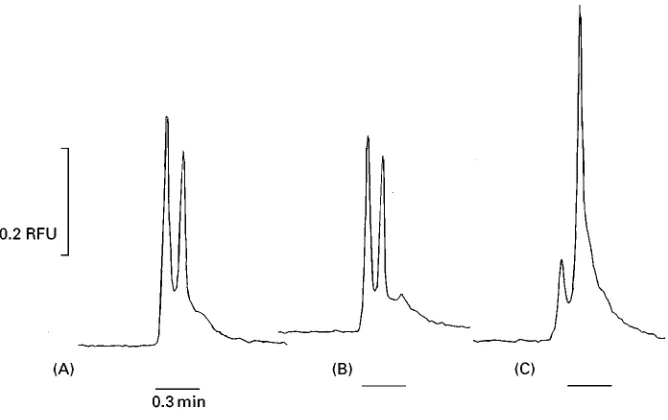
Figure 7 Representative electropherograms of (A) antibody control; (B) buffy coat from a scrapie-negative sheep; (C) buffy coat from a scrapie-positive sheep.
samples extracted from buffy coats of a normal sheep and from a buffy coat of a scrapie-infected sheep.
Concluding Remarks
The capillary electrophoresis assay described in this study is reproducible, more sensitive and faster than other analytical tests. The samples used in the capil-lary electrophoresis assay were obtained from brain and the lymphoid system of the animals. The sensitiv-ity of this assay made it possible to test samples from other tissues that contain much less abnormal prion protein than brain samples. This assay has the poten-tial to use tissues and Suids from live animals and diagnose animals prior to the onset of clinical signs of disease. Automation of this test could lead to more economical and efRcient methods for testing for ab-normal prion protein.
Further Reading
Altria KD (ed.) (1996) Capillary Electrophoresis Guide-book:Principles,Operation and Applications. Totowa, New Jersey: Humana Press.
Landers JP (ed.) (1997) Handbook of Capillary Electrophoresis, 2nd ed. London: CRC Press.
Prusiner SB (ed.) (1996) Prions, Prions, Prions. Current Top. Microbiol. Immunol. vol. 207. Berlin: Springer.
Prusiner SB (1996) Prion biology and diseases}laughing cannibals, mad cows, and scientiRc heresy.Medical Re-search Review16: 487}505.
Prusiner SB (1997) Prion diseases and the BSE crisis. Science278: 245}251.
Schmerr MJ and Jenny AL (1998) A diagnostic test for scrapie-infected sheep using a capillary electrophoresis immunoassay with Suorescent-labelled peptides, Elec-trophoresis19: 409}414.
Schmerr MJ et al. (1999) Use of capillary eletrophoresis andSuorescent labeled peptides to detect the abnormal prion protein in the blood of animals that are infected with a transmissible spongiform en-cephalopathy. Journal of Chromatography A 853: 207}214.
Weissmann C (1996) The ninth Datta lecture. Molecular biology of transmissible spongiform encephalopathies. FEBS Letters289: 3}11.
Field Flow Fractionation
R. Hecker and H. Coilfen, Max-Planck-Institut fuRr Kolloid und GrenzflaRchenforschung (Kolloidchemie), Am MuRhlenberg, Golm, Germany
Copyright^ 2000 Academic Press
Introduction
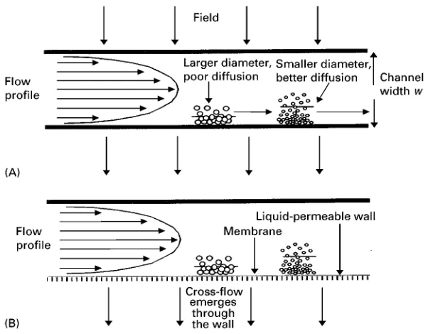
Figure 1 (A) Schematic of the mechanism of FFF separation of proteins. The smaller protein, with greater diffusivity, competes more successfully with the applied field and occupies a mean position further from the accumulation wall. Samples occupying the higher mean position is subject to more rapid flow laminae, and elutes earlier. The particle sizes represented, the channel thickness and the extent of back-diffusion are not to scale. (B) For the subtechnique flow FFF, the accumulation wall is a liquid-permeable porous material, typically ceramic. A membrane exists over the accumulation wall to prevent the samples from leaving the cell through this wall. The upper wall may or may not be porous as well, depending whether the symmetrical or asymmetrical variant is used.
and viruses. FFF is based on the differential transport rates of solutes in a ribbon-like channel when interac-ting with an applied Reld. The type of Reld may be chosen from a wide range, for example an electrical potential, sedimentation, a hydrodynamic cross-Sow, a thermal gradient and so forth. A schematic of this is shown inFigure 1. The solute will therefore occupy a region above the sample wall, with a mean position determined by the balance between the solute’s diffu-sion and the sample}applied Reld interaction. Al-though there exist further complications for solutes greater than&0.5m diameter, they are not relevant given the small hydrodynamic diameter of proteins. Positioned at the outlet of the channel is a sample detector of some sort, typically a traditional high-performance liquid chromatography (HPLC) spectrophotometric detector, although a signiRcant development has been with the application of a num-ber of detectors providing complementary informa-tion about the sample. Such detectors include spectophotometric and refractive index types, and more recently light scattering for molecular mass, electrospray}mass spectrometry, and inductively coupled plasma, although the last two have not yet been applied to protein studies.
There are a number of advantages offered by the FFF methods over other contemporary protein analy-sis methods. FFF is often more rapid than analytical
ultracentrifugation, and the range ofRelds available provide FFF with greater versatility. In comparison with gel-permeation chromatography, FFF is not im-peded by a size exclusion limit, the low exposed surface area limits sample loss through adsorption on to the exposed surface, and the availability of
Reld programming allows a wide range of materials to be analysed in a single channel. The open channel geometry usually allows FFF to characterize samples without need for pretreatment, such asRltration, and provides a very high upper limit to the protein size range. Similarly, the open channel allows the theoret-ical basis of FFF to provide direct access to funda-mental physical constants of proteins, often without the need for calibration. Finally, both FFF and gel electrophoresis may separate a protein mixture, but sample collection is simpler in FFF.
Flow FFF
Table 1 Compilation of flow FFF physicochemical data relevant for selected common proteins and with comparison to commercial polystyrene latexes
Sample Molecular mass (Da) Diffusion coefficient
(;1011m2s\1,;107cm2s\1)
Cytochrome c (bovine heart) 13 400 11.4
Ovalbumin (chicken egg) 45 000 8.71
Bovine serum albumin 64 000 6.89
Catalase (horse liver) 221 000 4.30
Apoferritin 450 000 3.84
Urease 483 000 3.46
Ferritin 622 000 2.91
Tobacco mosaic virus +40 000 000 0.46
Polystyrene latex,H0.090m 0.45
Polystyrene latex,H0.311m 0.22
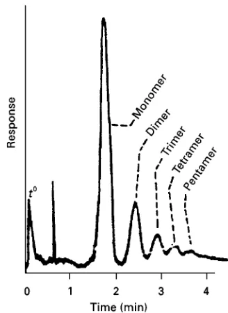
Figure 2 Separation of a monoclonal antibody from its higher clusters showing separable peaks up to pentameter aggregation. (Reproduced with permission from Giddings (1993) Science 260: 1456, Copyright the American Association for the Advance-ment of Science.)
In both cases, the separation is a direct function of the diffusion coefRcient, where the most highly diffusive components are the least retained. A compilation of common biological samples and their diffusion coefR -cients are provided inTable 1.
Fl-FFF is capable of separating proteins with only a 15% size difference within 3}10 min. Reported results for animal proteins and biopolymers include albumins (human and bovine serum, egg), globulins (-globulin, haemoglobin, thyroglobulin), ferritin, apoferritin, lysozyme, casein, blood products (human and rat blood plasmas, lipoproteins) and nucleic acids. Proteins from an industrial perspective are rep-resented by a growing body of work emerging on the characterization of proteins from Sours used for bread-making purposes.
In all of the above cases, no sample treatment is needed prior to injection, such as exhaustive dialysis orRltration. This is to be expected, as the permeable membrane acts as a dialysis cell, and the open channel will not become clogged and require aRlter. Since the sample is not manipulated beforehand, the presence of aggregate structures remains unaltered. Figure 2 shows baseline resolution of a biological mixture. Protein dimers elute as satellite peaks at&1.4 reten-tion times of the monomer, followed similarly by the higher aggregates eluting later. Most signiRcantly, the entire separation takes place in only four minutes.
The asymmetric Sow FFF variant does not inject the sample directly into the inlet line. Rather, a sample pump introduces the sample into the cell and opposing Sows from both ends of the cell hy-drodynamically focus the sample into a narrow band across the channel before elution. This allows for remarkably well-resolved and efRcient protein separ-ations.Figure 3illustrates the sensitivity of the tech-nique. Two plasmid fragments were injected at low concentration (0.1gL\1) and volume (1L) while
exhibiting both baseline resolution and elution in less
than 15 min. One further advantage of the focusing method is the immobilization of the sample prior to elution. For a very dilute sample, multiple injections subject to these opposingSows produce an on-chan-nel concentrating effect, where the protein is retained on the membrane at the focus point.
[image:3.568.312.476.436.661.2]Figure 3 Separation of (1) 2390 bp and (2) 4320 bp plasmids by asymmetrical FFF. (Reproduced with permission from LitzeHn A and Wahlund KG (1989)Journal of Chromatography 476: 413 Copyright Elsevier Science BV.)
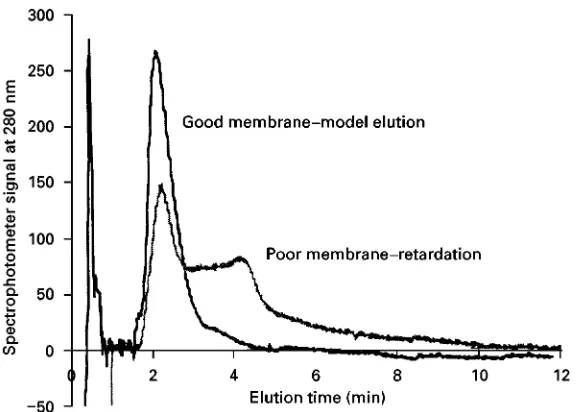
Figure 4 Superposition of two elution profiles for cytochromec (0.82 mg mL\1, 25L) in 0.05 mol L\1 2-[N-morpholino]propanesul-fonic acid (‘mops’) buffer at pH 6.2. The membranes are both regenerated cellulose, with cited 10 000 molecular weight nominal pore sizes from different suppliers. From Hecker, unpublished results.
contemporary practice. Both symmetrical and asym-metrical variants have been successfully applied to proteins. Of special interest is the frit inlet}frit outlet modiRcation. These methods in tandem enhance de-tectability and aid fractionation stability. The combi-nation of frit inlet and outlet has been reported as recently as 1999, for the automation of wheat protein fractionation.
One rarely discussed drawback to the Fl-FFF method is the requirement of a membrane for sample retention. For adhesive protein samples, this demands compatibility between the sample, membrane and the carrier solution. Biopolymers can strongly adsorb on to particular membranes and at modest ionic strengths may be completely adsorbed. The simplest method to test this is to inject samples over a range of concentrations and/or volumes and ensure there is
proportionality between detected signal size and the amount of sample. A partial, reversible adsorption leads to an increased retention and this would indi-cate that the sample is erroneously large, or induce a number of fractionation proRle artefacts. Clearly the chemistry of the system, between the sample, membrane and carrier, must be known before any statements may be made.
[image:4.568.143.434.468.674.2]Figure 5 Elution profiles of the components of a protein} poly-mer ligand mixture, immunoglobulin IgG and polyglutamic acid, and their covalent conjugate. The fractionation of the conjugate suggests that a quantity of the polyglutamic acid remains un-bound, and offers a method of determining the binding constants of such mixtures. (Reproduced with permission from Giddings JC et al. (1992) Journal of Liquid Chromatography 15: 1729 Copy-right Marcel Dekker.)
presence of the membrane therefore determines the smallest-sized species capable of being retained in a Fl-FFF channel.
Such membrane effects have been used to advant-age, however. Proteins have been characterized with a separation based on both standard FFF principles and enhanced retention for some species by sample}membrane interactions. This offers a remark-ably wide scope for characterizing systems with subtle differences in physical sizes but dissimilar chemistries, but assigning peaks in the fractionation proRle calls for a number of pure standards and calibration processes.
Of particular interest to protein science is the ob-servation and quantiRcation of protein}ligand or pro-tein}protein interactions. Such an example is provided inFigure 5for the interaction between im-munoglobulin IgC and an interacting ligand, poly-glutamic acid, with the conjugate peak showing a small amount of free ligand. Quantifying such an interaction to measure the binding constant is a more difRcult task. It is necessary to be able to produce fractionation proRles of the components as a function of concentration, implying that sample loss on to the membrane must be prevented. Furthermore, at least two from the protein, ligand or complex peaks must be well separated for quantiRcation if the stochiometry is known prior to the experiment, otherwise all three must be resolved. This precludes
many simple systems, for example bovine serum al-bumin (BSA)/anti-BSA, or ovalbumin/concavalin A, where the hydrodynamic sizes of these species are too similar for reliable quantiRcation.
The application for protein interaction studies is limited to processes in which the interaction time is insigniRcant compared to the transport time, effec-tively making protein studies with a kinetic barrier to interaction difRcult. Further, the use of FFF to investi-gate sample}sample interactions has been criticized, in that during transport dilution will occur so equilib-rium in the FFF channel will be different to that of the mixing conditions. These limitations are clearly not relevant for rapid, near-irreversible interactions.
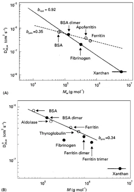
The opportunity for the study of protein shape by Fl-FFF is possible. Like other hydrodynamic methods, the information available from these methods renders them primarily as complementary methods to high resolution crystallography or mag-netic resonance. Nevertheless, both theory and prac-tice, discussed by CoKlfen and Pauck, demonstrate that retention is a function of molecular shape (Figure 6), with the retention decreasing with the degree of asymmetry.
All these examples show that Fl-FFF is a powerful technique for protein characterization, as it is both very rapid and requires only microgram or smaller amounts of sample. Future potential can be seen in the quantiRcation of interactions between proteins. However, potential factors affecting the results and possibly producing artefacts, such as membrane} sample interaction or sample shape, must be considered when interpreting the results.
Sedimentation FFF
Figure 6 Temperature-corrected diffusion coefficients for a variety of proteins, using both analytical ultracentrifugation (A) and asymmetric FFF (B). The molecular weight}diffusion coefficient relationship is linear for the globular proteins, represented as open circles. Less spherical samples (filled circles) show a deviation from the linearity, with increasing deviation with eccentricity. (Reproduced with permission from Pauck T and CoKlfen H (1998)Analytical Chemistry 70: 3886 Copyright the American Chemical Society.)
reported examples include DNA, proteoglycans,
Rbrinogen and myohemerythrin.
Thermal FFF
Thermal FFF, employing the Soret effect, is also suit-able for the separation of biomolecules. Unfortunate-ly, the thermodiffusion effect is extremely poor in water. The use of organic solvents restricts statements about the native state in aqueous-based buffer, and furthermore extensive conformational changes and even denaturation may occur which signiRcantly re-strict the range of applicable samples. Reported uses of thermal FFF for biological samples have been lim-ited to the polysaccharides, dextrans, Rcolls, pul-lulans and cellulose, and the starch polymers amylose
and amylopectin, in dimethylsulfoxide as carrier liquid. Partially aqueous carriers have been investi-gated but it is the fraction in organic solvent that explicitly determines retention.
Electrical and Magnetic FFF
Figure 7 The coating of streptavidin on to a standard 165 nm diameter polystyrene (PS) latex bead affects the elution of the latex substrate by electrical FFF. Under pH 7.2 fractionation conditions the latex has a negative surface charge while the protein is isoelectric. The lower net surface potential is reflected in the poorer retention of the coated bead (A). The magnitude of this peak shift quantifies the degree of surface coating, as shown by the correlation in retention with the protein adsorption isotherm (B). (Reproduced with permission from Schimpf and Caldwell (1995)American Laboratory 27: 64I68.)
electrical FFF for the separation of albumin, ly-sozyme, haemoglobin and-globulin in buffer solu-tions at different pH.
Later, the performance of an electrical FFF channel withSexible membranes, a channel with rigid mem-branes and a circular channel for the separation of proteins was described. In these studies, human and bovine serum albumin, bovine -globulin, cyto-chromec, egg white lysozyme and soluble ribonucleic acid (t-RNA) as well as denatured proteins were suc-cessfully separated. Unfortunately, the electricalReld induces charge polarization of carrier liquid species, such that they migrated adjacent to the electrodes and then screen the electrical Reld. These early
experi-mental conRgurations of electrical FFF utilized ion-permeable membranes separating the channel volume from the electrode compartments. These conditions led to difRculties in forming a homogeneous electric
Reld, and from the late 1970s the technique entered a period of quiescence. Results published in the early to mid 1990s using conductive, rigid walls of either graphite or gold-plated glass, have allowed reproduc-ible separations, while the addition of a redox couple in the carrier liquid, such as quinone-hydroquinone, reduced the polarization effects. Due to these delays in experimental development, electrical FFF is less mature than other FFF techniques.
Electrical FFF is also well suited to measuring pro-tein adsorption on to surfaces. The thin layer provides only subtle differences to the hydrodynamic size and net density, makingSow or sedimentation FFF analy-sis difRcult. However, the adsorption dramatically inSuences the surface charge and thereby inSuences both sample}Reld interaction and retention, as shown inFigure 7.
Although not formally FFF, dielectrophoresis in combination withSuidSow through an open chamber with interdigitated sinusoidally corrugated electrodes has been used for the separation of proteins and DNA. A minor method, magnetic FFF, has been applied to study the retention behaviour of BSA in the pres-ence and abspres-ence of nickel nitrate. In the prespres-ence of nickel ions, the retention time of the BSA sample was 6% higher with the magneticReld than it was without theReld. Retention times reported for BSA samples both with and without a magneticReld did not differ in the absence of Ni (II). However, the application range of magnetic FFF for protein separations is very limited, and the method can only be applied in excep-tional conditions.
Micropreparative FFF Applications
A variant of the FFF apparatus, the split-Sow thin cell (SPLITT) permits continuous separation of milli- or even gram quantities of material. The apparatus is similar to a FFF cell equipped with both frit inlets and outlets. Initial conRgurations fed a mixture of large particles into the upper wall and carrier liquid into the base, while at the other end of the cell the liquid
electrophoretic mobility to pass the splitting plane. The separation of a mixture of model proteins by such a method has been reported. The relatively high throughput reported (15 mg h\1) makes
this an interesting development for routine puriR ca-tion, but it requires a difference in protein pI
of about two units as a necessary precondition for separation.
Miniaturization of FFF
There is a drive to produce the equivalent of hand-held devices for sample analysis based on the FFF principles, the chip laboratory. Advantages of such methods include the ability to analyse freshly sampled, or to undertake a number of simulta-neous parallel analyses. For such miniaturized devices the injection volume is a signiRcant propor-tion of the channel volume, with commensurate band-broadening problems, while theory predicts that some quantities, such as retention ratio and plate height, degrade with decreasing size. None the less, the reported developments for microfabricated electrical and dielectrophoretic FFF show healthy progression.
Concluding Remarks
The early development of FFF was hindered by the experimental complexity of the method and a focus on theory over practice. Over the last ten years, a number of simplifying experimental features such as the frit inlet}outlet system, and a fuller understand-ing of the theoretical background have led to a dramatic worldwide rise in the number of applica-tions. It seems unlikely that more novel Relds will be introduced into this family of techniques, but the subtlety of application is increasing. Methods and procedures are developing, from the analysis of simple proteins and mixtures, to protein aggregates,
proteins in complex matrices and increasingly fragile samples such as liposomes, where the open channel has few, if any, real analytical competitors.
The other exciting branch of development is increased commercial application, where the FFF method becomes a ‘black box’ technique. Leading the way is the Fl-FFF method, but with the recent innovations in electrical FFF, the dominance of gel electrophoresis for protein analysis may be passing.
See also: III/Proteins: Centrifugation.
Further Reading
CoKlfen H and Antionetti M (2000) Field-Sow-fractionation techniques for polymer and colloid analysis.Advances in Polymer Science150: 67}187.
Giddings JC (1991)UniTed Separation Science. New York: John Wiley.
Giddings JC (1993) Field-Sow fractionation: analysis of macromolecular, colloidal, and particulate materials. Science260: 1456}1465.
Janca J (1988)Field-Flow Fractionation. Chromatographic Science Series vol. 39. New York: Marcel Dekker. Liu MK, Li P and Giddings JC (1993) Rapid protein
separ-ation and diffusion coefRcient measurement by frit inlet Sow Reld-Sow fractionation. Protein Science 2: 1520}1531.
Martin M (1998) Theory of Reld-Sow fractionation. Advances in Chromatography39: 1}138.
Myers MN (1997) Overview of Reld-Sow fractionation. Journal of Microcolumn Separations9(3): 151}162. Schimpf ME and Caldwell KD (1995) ElectricalReld-Sow
fractionation for colloid and particle analysis.American Laboratory27(6): 64}68.
Wahlund K-G and LitzeHn A (1989) Application of an asymmetric Sow Reld-Sow fractionation channel to the separation and characterisation of proteins, plasmids, plasmid fragments, polysaccharides, and unicellular algae. Journal of Chromatography 461: 73}87.