Direct interaction of iron-regulated surface
determinant IsdB of
Staphylococcus aureus
with
the GPIIb/IIIa receptor on platelets
Helen Miajlovic,
1Marta Zapotoczna,
1Joan A. Geoghegan,
1Steven W. Kerrigan,
2Pietro Speziale
3and Timothy J. Foster
1Correspondence Timothy J. Foster [email protected]
1Department of Microbiology, Moyne Institute of Preventive Medicine, Trinity College Dublin, Dublin 2, Ireland
2Department of Clinical Pharmacology, Royal College of Surgeons in Ireland, Dublin 2, Ireland
3Department of Biochemistry, Viale Taramelli 3/b, 27100 Pavia, Italy
Received 24 November 2009 Accepted 9 December 2009
The interaction of bacteria with platelets is implicated in the pathogenesis of endovascular infections, including infective endocarditis, of whichStaphylococcus aureusis the leading cause. SeveralS. aureussurface proteins mediate aggregation of platelets by fibrinogen- or fibronectin-dependent processes, which also requires specific antibodies. In this studyS. aureuswas grown in iron-limited medium to mimicin vivoconditions in which iron is unavailable to pathogens. Under such conditions, aS. aureusmutant lacking the known platelet-activating surface proteins adhered directly to platelets in the absence of plasma proteins and triggered aggregation. Platelet adhesion and aggregation was prevented by inhibiting expression of iron-regulated surface determinant (Isd) proteins. Mutants defective in IsdB, but not IsdA or IsdH, were unable to adhere to or aggregate platelets. Antibodies to the platelet integrin GPIIb/IIIa inhibited platelet adhesion by IsdB-expressing strains, as did antagonists of GPIIb/IIIa. Surface plasmon resonance demonstrated that recombinant IsdB interacts directly with GPIIb/IIIa.
INTRODUCTION
Staphylococcus aureus is an opportunistic pathogen that colonizes the desquamated epithelium of the anterior nares. At sites of infection such as wounds, ulcers or abscesses, S. aureus can gain access to the bloodstream, resulting in bacteraemia. Increased use of intravascular devices and invasive procedures in hospitals has led to an increased incidence of bacteraemic infections (Moreillon & Que, 2004). Several endovascular infections, including infective endocarditis, result from the interaction of bacteria with platelets. S. aureus is the leading cause of infective endocarditis, which is characterized by the build-up, on heart-valve surfaces, of vegetative bodies consisting of bacteria, fibrin and aggregated platelets (Moreillon & Que, 2004; Mylonakis & Calderwood, 2001).
The ability ofS. aureusand other bacteria to adhere to and aggregate platelets is thought to contribute to the development of endovascular infections. Aggregation of platelets by bacteria is the result of a multi-step process. Bacteria interact with receptors on the platelet surface either directly or indirectly through bridging molecules such as fibrinogen. Initial adhesion of bacteria to platelets
can result in subsequent platelet activation, which is characterized by signalling events and calcium oscillations within the platelet. Upon activation, the major platelet integrin GPIIb/IIIa undergoes conformational changes that allow it to bind avidly to fibrinogen and fibronectin in solution (Calvete, 1999). Aggregation of platelets occurs when adjacent platelets interact with the c-chain of the bivalent fibrinogen molecule, cross-linking platelets into aggregates.
The interaction betweenS. aureusand platelets is complex and involves multiple factors (O’Brienet al., 2002). Several surface-expressed proteins can stimulate platelet aggrega-tion. These include the fibrinogen-binding proteins clumping factors A and B (ClfA and ClfB), and the bifunctional fibronectin-fibrinogen binding proteins FnBPA and FnBPB (Fitzgerald et al., 2006b; Loughman
et al., 2005; Miajlovic et al., 2007). Recent studies have shown that, under high shear rates such as those seen in small arteries and arterioles, protein A and ClfA are crucial for platelet aggregation (Kerriganet al., 2008; Pawaret al., 2004).
Binding of fibrinogen or fibronectin by surface proteins effectively coatsS. aureuswith these two ligands, allowing it to engage the low-affinity form of the GPIIb/IIIa on resting platelets. Activation of platelets requires specific antibodies Abbreviations:GFP, gel-filtered platelets; PRP, platelet-rich plasma; WP,
to the bacterial surface proteins to engage platelet receptor FccRIIa and trigger intracellular signalling events (Loughman et al., 2005; Miajlovic et al., 2007). A slower complement-dependent mechanism of activation was detected under circumstances where the major proaggre-gatory surface proteins were missing or defective (Fordet al., 1996; Loughmanet al., 2005).
All studies on the interaction of S. aureus with platelets have been performed with bacterial cells grown in rich laboratory media replete with iron. In vivo, S. aureus has restricted access to iron and expresses iron-regulated surface determinant (Isd) proteins to capture haem from haemoglobin and transport it into the cell (Skaar et al., 2004; Skaar & Schneewind, 2004). Two of the Isd proteins, IsdA and IsdH, are known to have other biological functions. IsdA interacts with an array of host proteins, and expression of IsdA byS. aureusalso confers resistance to the innate defences of the human skin (Clarke et al., 2004, 2007, 2009). IsdH has been shown to play a role in evasion of phagocytosis as a result of accelerated degrada-tion of the serum opsonin C3b (Visai et al., 2009). It is reasonable to predict that these major surface components of bacteria grownin vivomight also interact with platelets. This study investigated platelet adhesion and aggregation mediated by S. aureus strain Newman grown in an iron-deficient medium. Using S. aureus mutants defective in various surface proteins, it was shown that IsdB binds to platelets by a direct interaction with the platelet integrin GPIIb/IIIa.
METHODS
Bacterial strains and growth conditions.The bacterial strains and plasmids used in this study are listed in Table 1.S. aureusstrains were grown in at 37uC with shaking (200 r.p.m.) in RPMI 1640 (Sigma) to create iron-poor conditions. Lactococcus lactiswas grown in GM17 medium with 3.2 ng nisin ml21. The following antibiotics (Sigma) were added to the media as required: chloramphenicol (Cm) at 10mg ml21,
erythromycin (Em) at 10mg ml21and tetracycline (Tc) at 2mg ml21.
Construction of S. aureus strains. A frameshift mutation was constructed in theisdBgene by replacing bases 10–14 with aBamHI restriction site. An upstream isdB sequence of 745 bp was PCR amplified with primers 59 -GTCCTGCAGATTCTTACATTAGCT-GACGCA-39 and 59 -GGCGGATCCTTTGTTCATGTTGTAGAAA-CA-39 and a downstream sequence of 904 bp was amplified with primers 59-GAAGGATCCAAAAGAATTTAAATCATTTTA-39and 59 -CGCTCTAGAATCTTGAATTTTATTTAATTC-39. When the two fragments were ligated at the BamHI site, a +1 frameshift was created. The ligated fragments were cloned between thePstI andXbaI sites of plasmid pBluescript II-SK. This construct was then ligated with pTSermC using theXbaI sites in both plasmids to form pHM1. The temperature-sensitive shuttle plasmid bearing theisdBmutation was transduced with phage 85 into NewmanclfAand NewmanclfA isdA. Growth of these strains at a restrictive temperature of 44uC resulted in integration of the pHM1 plasmid into the chromosomal
[image:2.595.52.561.458.733.2]isdB gene. Plasmid excision was encouraged by growth of single integrants without selection at 28uC followed by three cycles of growth in drug-free broth alternately at 44uC and at 28uC to enrich for plasmid-free excisants. Single colonies were isolated on drug-free agar and screened for sensitivity to erythromycin. Mutants were validated by PCR amplifying 1.6 kb isdBgenomic DNA, which was subsequently digested with BamHI. Strains containing the desired mutation had a novelBamHI restriction site present inisdB. Western
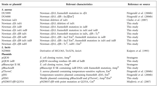
Table 1. Bacterial strains and plasmids
Strain or plasmid Relevant characteristics Reference or source
S. aureus
DU5999 NewmanclfA5, frameshift mutation inclfA Fitzgeraldet al.(2006b)
DU6000 NewmanclfA5, clfB: :lacZ[Emr] Fitzgeraldet al.(2006b)
NewmanisdA Newman deletion ofisdA Clarkeet al.(2007)
NewmanclfA isdA NewmanclfA5,deletion ofisdA This study
NewmanclfA isdB NewmanclfA5,frameshift mutation inisdB This study
NewmanclfA isdA isdB NewmanclfA5,frameshift mutation inisdAandisdB This study NewmanclfA clfB isdA NewmanclfA5, frameshift mutation in isdA,clfB: : TcR This study
NewmanclfA clfB isdB NewmanclfA5, clfB: :lacZEmR, frameshift mutation inisdB This study NewmanclfA clfB isdA isdB NewmanclfA5, clfB: :lacZEmR, frameshift mutation inisdAandisdB This study
NewmanclfA clfB isdH NewmanclfA5,clfB: : TcR,isdH: : EmR This study
L. lactis
NZ9800 Derivative of MG1363, Tn5276,DnisA Kuiperset al.(1993)
Plasmids
pQE30 E. colicloning vector, AmpR Stratagene
pQE30isdB pQE30 encoding residues 48–480 of IsdB This study
pBluescript II SK E. colicloning vector, AmpR Stratagene
pBlueDisdB pBluescript II SK containingisdBDNA with frameshift mutation, AmpR This study
pTSermC S. aureusplasmid containing temperature-sensitive replicon, EmR Fitzgeraldet al.(2006b)
pJH1 Temperature-sensitive plasmid containing frameshiftclfA5, EmR Fitzgeraldet al.(2006b) pHM1 Shuttle plasmid containing pBlueDisdBand pTSermC, AmpR/EmR This study
immunoblotting was also carried out to validate strains. Similarly, a frameshift mutation was introduced into the clfA gene in strain NewmanisdAusing pJH1, resulting in strain NewmanclfA isdA.
To construct NewmanclfA clfB isdAaclfA: : EmRmutation and a
clfB: : TcRmutation were transduced into strain NewmanisdA. Strain NewmanclfA clfB isdB was constructed by transducingclfB: : EmR into strain Newman clfA isdB. The clfB: : EmR mutation was transduced into NewmanclfA isdA isdBto create NewmanclfA clfB isdA isdB. Finally, strain NewmanclfA clfB isdH was constructed by transducingisdH: : EmRandclfB: : TcRinto strain NewmanclfA.
Western immunoblotting.Cultures ofS. aureusgrown in RPMI for 18 h at 37uC were washed twice in PBS and adjusted to an OD600of
10 in 250ml of 20 mM Tris (pH 8), 10 mM MgCl2containing 30 %
(w/v) raffinose. Complete EDTA-free protease inhibitor cocktail (Roche) and lysostaphin (200mg ml21; AMBI) were added to the cells
and incubated at 37uC for 10 min. Protoplasts were sedimented by centrifugation at 5000 r.p.m. for 10 min (Hartfordet al., 2001). The cell-wall fraction was separated on 7.5 % (w/v) polyacrylamide gels, transferred onto PVDF membranes (Roche) and blocked in 10 % (w/v) skimmed milk (Marvel). Membranes were probed with polyclonal anti-IsdB and anti-IsdA at a dilution of 1 : 5000 and with polyclonal anti-IsdH antibodies at a dilution of 1 : 10 000.
Preparation of platelet-rich plasma.Platelet-rich plasma (PRP) was prepared as described previously (Loughmanet al., 2005). Briefly, human blood was drawn into a syringe containing 3.2 % (w/v) sodium citrate. Whole blood was centrifuged at 150gfor 10 min. The top layer, consisting of PRP, was removed. Platelet aggregation was measured using light transmission aggregometry. Aggregation of platelets occurred after a variable period of time referred to as the lag time. This time reflects the time taken for aggregation to occur after bacteria and platelets come into contact. Overall percentage aggregation was also measured.
Preparation of washed platelets.Blood was drawn into a syringe containing acid-citrate-glucose (ACD, 25 mM citric acid, 75 mM sodium citrate, 135 mMD-glucose). Following preparation of PRP the pH of the platelets was adjusted to 6.5 using ACD. Prostaglandin E1 (1mM; Sigma) was added to prevent activation of platelets during centrifugation. PRP was centrifuged at 720g for 10 min. The supernatant was carefully removed and discarded. The platelet pellet was resuspended in 1 ml fresh JNL buffer (6 mMD-glucose, 130 mM NaCl, 9 mM NaHCO3, 10 mM sodium citrate, 10 mM Tris, 3 mM
KCl, 0.8 mM KH2PO4and 0.9 mM MgCl2; pH 7.4) and diluted to
obtain a platelet count of 36108platelets ml21.
Preparation of gel-filtered platelets.Washed platelets (WP) were passed through a 10 ml Sepharose 2B column (Sigma). Gel-filtered platelets (GFP) were collected and diluted to obtain a platelet count of 36108platelets ml21. WP and GFP were supplemented with 2 mM CaCl2.
Removal of IgG from commercial supplies of fibrinogen and from human serum.IgG present in commercial fibrinogen and in serum was removed by passage through a column of protein A coupled to Sepharose (Amersham Biosciences). Depletion of IgG was confirmed by ELISA using protein A-peroxidase (Sigma). Human serum was diluted 1 : 20 and analysed by SDS-PAGE to confirm loss of IgG.
Inactivation of complement proteins in human serum.
Complement components were inactivated in human serum by heating to 56uC for 30 min.
Platelet adhesion assay.Cultures of bacterial strains were pelleted by centrifugation, washed in PBS and resuspended to an OD600of 1.
Wells of microtitre plates were coated with 100ml bacteria and incubated for 16 h at 4uC, washed with PBS and blocked with 1 % BSA (w/v; 100ml) for 90 min at 37uC. Wells were washed with JNL buffer and 50ml WP was added. Plates were incubated at 37uC for 40 min and washed subsequently with JNL buffer. Adherent platelets were measured with a lysis buffer containing a substrate for acid phosphatase [100 mM sodium acetate, 0.1 % (v/v) Triton X-100, 10 mM p-nitrophenol phosphate (Sigma)]. Absorbance at 405 nm was read in an ELISA plate reader. Equal coating of plates withS. aureusstrains was confirmed by fixing adherent cells with 25 % (v/v) formaldehyde and staining with of 0.5 % (w/v) crystal violet.
Platelet aggregation. Bacterial cells were washed with PBS, resuspended to an OD600of 1.6 and 25ml washed cells was added
to 225ml PRP in siliconized flat-bottom glass cuvettes (BioData). PRP and bacteria were incubated with stirring (900 r.p.m.) in an aggregometer (Bio-Data) at 37uC. Light transmission was monitored for 25 min. Inhibitors tested were anti-GPIIb/IIIa (Abciximab, Eli Lily) antibodies at a 1/100 dilution, anti-FccRIIa (Medarex) antibodies at a 1/50 dilution and anti-GPIb monoclonal antibody (AN51, Dako) at a 1/50 dilution. RGD peptide and tirofiban (Merck) were used at 10 mM and 2mM respectively. GFP were supplemented with purified fibrinogen at a final concentration of 0.5 mg ml21. Pooled human IgG (Baxter) was added to GFP to a final concentration of 2mg ml21
. Twenty-five microlitres of human serum was added to a final volume of 225ml GFP.
GPIIb/IIIa-binding assay.Microtitre plates were coated for 16 h with 100mg ml21of GPIIb/IIIa (Calbiochem). Plates were washed with PBS and blocked with 1 % (w/v) BSA for 2 h at 37uC. Bacteria were stained with a fluorescent stain (Cyber), adjusted to an OD600of
0.5 and added to GPIIb/IIIa-coated plates. Following a 40 min incubation at 37uC, plates were washed with PBS and fluorescence units read at 485–535 nm. Values were adjusted by subtracting binding to BSA.
Purification of recombinant IsdB.DNA encoding residues 48–480 of IsdB was amplified from the genomic DNA of strain Newman using primers FisdB 59 -GGCCATGGATCCACAAATACAGAAG-CACAACCAAA-39 and RisdB 59 -GGCCATCCTGCAGAGTAGC-TTCCTTCTTAGCTGA-39. The isdB coding sequence was cloned between the BamHI and PstI sites in pQE30. pQE30 isdB was transformed intoEscherichia colistrain TOPP3 and IsdB expression was induced with 1 mM IPTG. Recombinant rIsdB 48–480 was purified by Ni2+ affinity chromatography as described previously (O’Connellet al., 1998).
Surface plasmon resonance.Surface plasmon resonance (SPR) was performed using the BIAcore X100 system (GE Healthcare). GPIIb/IIIa (Enzyme Research Laboratories) was covalently immobi-lized on CM5 sensor chips using amine coupling. This was performed using 1-ethyl-3-(3-dimethylaminopropyl) carbodiimide hydro-chloride (EDC), followed by N-hydroxysuccinimide (NHS) and ethanolamine hydrochloride, as described by the manufacturer. GPIIb/IIIa (100mg ml21) was dissolved in 10 mM sodium acetate (with 1 mM MgCl2 and 1 mM CaCl2) at pH 4.5 and then
immobilized on the flow cell at a flow rate of 30ml min21
in HEPES-buffered saline. On another flow cell, the dextran matrix was treated as described above but without GPIIb/IIIa present to provide an uncoated reference flow cell. Increasing concentrations of rIsdB were flowed over immobilized GPIIb/IIIa and the reference flow cell at a rate of 5ml min21.
version 3.0. The equilibrium dissociation constant (KD) was obtained
by globally fitting the data using the Langmuir 1 : 1 binding model.
Statistical analysis.The data presented by this study represent the means±SD of three experiments unless otherwise stated. The unpairedt-test was used to determine the significance of differences in aggregation between strains, with significance defined asP,0.05.
RESULTS
Platelet adhesion and aggregation mediated by iron-starvedS. aureus
S. aureusNewman mediates adhesion to and aggregation of platelets using surface proteins ClfA and ClfB. In strain Newman the FnBPA and FnBPB surface proteins are not expressed on the cell surface due to nonsense mutations at the 39ends of the genes and therefore do not contribute to platelet aggregation (Grundmeieret al., 2004). In order to investigate the possibility that Isd proteins adhere to and aggregate platelets in the absence of known proaggregatory surface proteins, strain Newman with mutations in clfA
andclfBwas studied.
Microtitre dishes were coated with Newman clfA clfBthat had been grown in the iron-restricted medium RPMI. Washed platelets (WP) adhered strongly to the bacteria at a level twofold higher than to control wells coated with immobilized fibrinogen (P,0.0001, Fig. 1a). Adherence of platelets to bacteria occurred without the addition of exogenous fibrinogen. Growth of bacteria in RPMI with 50 mM FeCl3 eliminated adhesion of WP to immobilized
bacteria (P,0.0001). These results suggest that direct adherence of strain Newman to platelets is dependent on proteins induced by iron starvation.
Platelet aggregation by RPMI-grown NewmanclfA clfBwas measured by light transmission with an aggregometer. Aggregation occurred with a mean lag time of 5.6±0.67 min at an overall percentage aggregation of 60±4 %. Growth of Newman clfA clfB in RPMI supple-mented with 50mM FeCl3 eliminated platelet aggregation
(Fig. 1b). These results indicate that proteins induced by iron starvation can also trigger platelet aggregation possibly by the direct interaction shown in Fig. 1(a).
Newmanisdmutants
In order to determine whether the surface-exposed Isd proteins IsdA, IsdB or IsdH interact with platelets, mutants of NewmanclfA clfBthat lacked each of these proteins were constructed. The mutants were studied by Western immunoblotting to ensure that each lacked the relevant Isd protein and still produced the other Isd proteins.
The 38 kDa IsdA protein could not be detected in solubilized cell-wall extracts of Newman clfA clfB isdA
whereas IsdB and IsdH proteins could be detected (Fig. 2). Strain Newman clfA clfB isdBexpressed 38 kDa IsdA and 150 kDa IsdH proteins, whereas IsdB was absent. Newman
clfA clfB isdA isdBlacked IsdA and IsdB, whereas IsdH was detected. IsdH was not expressed by NewmanclfA clfB isdH
whereas IsdA and IsdB were present (Fig. 2).
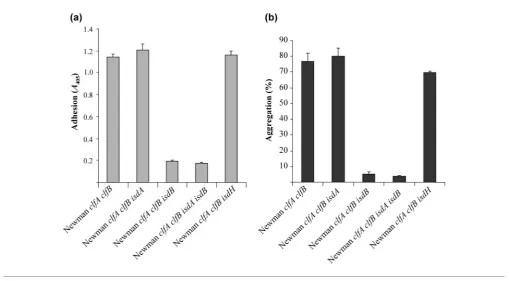
Platelet adhesion and aggregation mediated by Newmanisdmutants
[image:4.595.48.565.483.742.2]Bacterial strains were grown in RPMI for 16 h at 37uC. Cells were washed and added to microtitre plates and tested for their ability to support binding of WP. Platelets adhered avidly to strain Newman clfA clfB. Mutations in
Fig. 1. Platelet adhesion and aggregation mediated by RPMI-grownS. aureus. Newman
clfA clfB was grown in RPMI with or without the addition of 50mM FeCl3. (a) Washed
eitherisdAor isdH had little or no effect on adhesion of platelets (Fig. 3a). In contrast, strain NewmanclfA clfB isdB
was unable to support adherence of WP (P,0.0001). Eliminating IsdA did not significantly lower adhesion (P50.3609; Fig. 3a). These results suggest that IsdB is the protein responsible for adhesion to platelets under iron-limiting conditions.
Newman clfA clfBcells that had been grown under iron-restricted conditions caused platelet aggregation in PRP with a lag time of 5.6±0.67 min. Mutants lacking either IsdA or IsdH were able to promote aggregation of platelets, with no significant differences in the percentage aggrega-tion or lag time to aggregaaggrega-tion (P50.46 and P50.68, respectively). In contrast, theisdB mutant failed to cause platelet aggregation (P50.0002; Fig. 3b). The mutant lacking both IsdB and IsdA was also unable to support platelet aggregation. IsdB appears to be crucial for both adhesion to and aggregation of platelets by iron-starvedS. aureuscells.
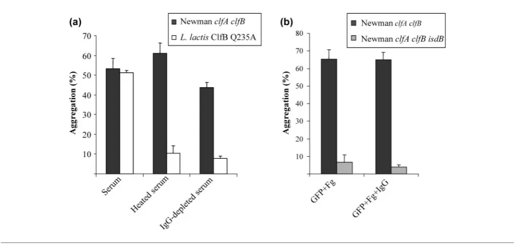
Plasma proteins required for platelet aggregation mediated by IsdB
[image:5.595.47.274.64.302.2]Lag times to aggregation for Newman clfA clfBgrown in RPMI varied with plasma donor, with a mean lag time of 5.6±0.67 min. Longer lag times are often associated with complement-mediated platelet activation (Fordet al., 1996; Loughman et al., 2005). To further investigate the
Fig. 2.Characterization of Newman isd mutants. The strains indicated were grown to stationary phase in RPMI. Cell-wall proteins were solubilized by lysostaphin digestion, separated on 7.5 % SDS-PAGE gels and electroblotted onto PVDF membranes. Membranes were probed with polyclonal anti-IsdA, anti-IsdB and anti-IsdH and bound antibodies were detected with HRP-conjugated protein A-peroxidase.
[image:5.595.39.549.405.686.2]mechanism of platelet aggregation, complement was inactivated in human serum by heat treatment. Serum depleted of IgG was also utilized to determine the necessity for antibodies. L. lactis NZ9800(pNZ8037 clfB Q235A), expressing ClfB Q235A, was used as negative control in this assay. ClfB Q235A lacks the ability to bind to fibrinogen and therefore cannot activate platelets in a fibrinogen-dependent manner. Activation of platelets byL. lactisClfB Q235A is complement dependent and requires IgG. This strain does not cause aggregation of gel-filtered platelets (GFP) in the presence of fibrinogen and complement-inactivated serum or IgG-depleted serum (Miajlovicet al., 2007).
Both NewmanclfA clfBandL. lactisClfB Q235A were able to stimulate aggregation of GFP supplemented with fibrinogen and 10 % human serum (Fig. 4a). Adding heated serum or IgG-depleted serum to GFP did not support platelet aggregation mediated byL. lactisClfB Q235A. In contrast, Newman clfA clfB was able to cause aggregation in GFP supplemented with heated serum and IgG-depleted serum. No significant difference in the percentage aggregation was observed between GFP supplemented with serum, heated serum or IgG-depleted serum (P50.1155 and P50.2758, respectively). Likewise, no significant difference was observed in the lag time to aggregation. Thus platelet aggregation mediated by NewmanclfA clfBgrown in iron-limiting conditions is not complement dependent and IgG does not seem to be necessary.
Further experiments were carried out to identify the mechanism of platelet aggregation. Iron-starved bacterial
cells were added to GFP supplemented with purified fibrinogen lacking contaminating IgG present in commer-cial supplies. Strain Newman expressing IsdB was able to stimulate aggregation of GFP supplemented with fibrino-gen alone (Fig. 4b). The addition of exofibrino-genous IgG decreased the mean lag time to aggregation from 4.33±1.86 min to 2.33±0.88 min. However, this differ-ence was not statistically significant (P50.121) due to considerable variations in lag time between donors. Strain Newman clfA clfB isdBwas unable to cause activation of GFP supplemented with fibrinogen alone or with fibrino-gen and IgG together.
Inhibition of platelet adhesion
Washed platelets were incubated with antibodies that are known to block receptors GPIIb/IIIa, FccRIIa and GP1b prior to their addition to microtitre plates coated with bacteria. The anti-GPIIb/IIIa monoclonal antibody abcix-imab inhibited adherence of WP to RPMI-grown Newman
clfA clfB (P,0.0001, Fig. 5a). Monoclonal antibodies IV3 and AN51, which block the FccRIIa and GP1b, respectively, did not significantly inhibit adhesion of WP to Newman
clfA clfB(Fig. 5a). This indicates that adherence of platelets to iron-starved S. aureus occurs via GPIIb/IIIa and does not involve GP1b or FccRIIa.
[image:6.595.50.555.444.683.2]In order to confirm that IsdB interacts with GPIIb/IIIa, inhibition experiments were carried out with GPIIb/IIIa antagonists. GPIIb/IIIa recognizes an RGD motif present in fibrinogen and fibronectin. Synthetic RGD peptide and tirofiban, a specific inhibitor of GPIIb/IIIa, were tested for
Fig. 4. Role of complement and IgG in platelet aggregation. (a) StrainsL. lactisClfB Q235A and NewmanclfA clfBwere washed and added to GFP supplemented with fibrinogen (Fg) and 10 % human serum (heated to 566C or IgG depleted). (b)
their ability to inhibit adhesion of platelets to iron-starved Newman clfA clfB. Both RGD peptide and tirofiban inhibited adhesion of platelets (P,0.0001 in both cases, Fig. 5b).
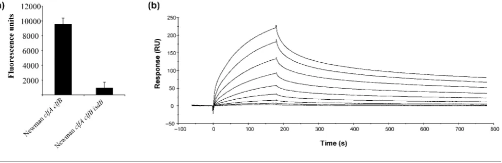
Direct interaction of IsdB and GPIIb/IIIa
Purified GPIIb/IIIa was coated onto microtitre plates. The ability of iron-starved NewmanclfA clfBand NewmanclfA clfB isdB to adhere to the immobilized integrin was measured. Newman clfA clfB adhered to immobilized GPIIb/IIIa but the isdB mutant failed to do so (Fig. 6a). Eliminating IsdB from NewmanclfA clfBinhibited binding to GPIIb/IIIa (P50.0089), confirming the direct inter-action between these proteins.
Surface plasmon resonance was also used to demonstrate a direct interaction between IsdB and GPIIb/IIIa (Fig. 6b). Increasing concentrations of recombinant IsdB were flowed over a chip that had been coated with GPIIb/IIIa. The approximate dissociation constant (KD) of the interaction
was determined to be 405±73.7 nM, indicating that IsdB directly interacts with GPIIb/IIIa with a high affinity.
DISCUSSION
The ability ofS. aureusto adhere to and activate platelets is believed to be an important factor in the pathogenesis of cardiovascular infections, including infective endocarditis. Previous studies have established that S. aureus surface proteins ClfA, ClfB, FnBPA and FnBPB can promote activation of platelets (Fitzgerald et al., 2006a). These
studies were carried out with bacteria grown in complex iron-containing media. This study investigated platelet adhesion and aggregation mediated by bacteria that were grown in an iron-deficient medium to mimic more closely thein vivoenvironment.
Initial adhesion of bacteria to platelets is required to trigger subsequent platelet activation. A common mechanism for platelet activation has been identified for bacterial proteins capable of binding to the blood glycoproteins fibrinogen and fibronectin. ClfA and ClfB of S. aureus mediate adhesion to platelets via a fibrinogen bridge to platelet receptor GPIIb/IIIa (Loughman et al., 2005; Miajlovic et al., 2007). Under iron-restricted growth conditions the adhesion ofS. aureuslacking these proteins to platelets was direct and did not require fibrinogen (Fig. 1a). Elimination of IsdB from the cell surface by growth in the presence of iron or by mutation of the isdB gene resulted in loss of platelet adhesion (Figs 1a and 3a). IsdH or IsdA were not required to promote platelet adhesion, as strains lacking these proteins were able to adhere at wild-type levels (Fig. 3a).
[image:7.595.107.539.66.287.2]In order to identify the receptor for IsdB on the platelet surface, inhibitors of the major platelet receptors were utilized. Adhesion to platelets was only inhibited by function-blocking antibodies to receptor GPIIb/IIIa (Fig. 5a). GPIIb/IIIa is the most abundant receptor found on the platelet surface. It recognizes RGD motifs present in multiple ligands, including fibrinogen and fibronectin, to promote platelet adhesion at sites of thrombus formation. Tirofiban, a specific inhibitor of GPIIb/IIIa function, completely inhibited adhesion of platelets (Fig. 5b).
Fig. 5. Effect of platelet receptor inhibitors on adhesion. (a) WP were incubated for 15 min at 376C with inhibitory antibodies
to platelet receptors and added to microtitre plates coated with RPMI-grown NewmanclfA clfB.(b) WP were incubated for 15 min at 376C with RGD peptide or tirofiban and added to microtitre plates coated with RPMI-grown NewmanclfA clfB.
Incubating platelets with soluble RGD peptide also inhibited adhesion of platelets to bacteria expressing IsdB, indicating that IsdB binds to the GPIIb/IIIa receptor on platelets.
To demonstrate that IsdB interacts directly with GPIIb/IIIa, it was shown that S. aureus strain Newman clfA clfB
adhered to purified GPIIb/IIIa in an IsdB-dependent fashion (Fig. 6a). The direct nature of the interaction between IsdB and GPIIb/IIIa was confirmed by surface plasmon resonance (Fig. 6b). IsdB bound to GPIIb/IIIa with a high affinity (approximate KD 405±73.7 nM).
Previous studies carried out with IsdH have shown that the residues in the fuctional NEAT I domain were crucial for both haemoglobin binding and evasion of phagocytosis (Visai et al., 2009). The NEAT domain of IsdB does not have an integrin-binding RGD motif. Further studies will be carried out in this laboratory to characterize the GPIIb/ IIIa-binding epitope in IsdB by substituting residues in the NEAT domains.
Aggregation of platelets following adhesion was dependent on expression of IsdB and occurred after a lag time of 5.6±0.67 min. Longer lag times to activation are some-times associated with complement-dependent platelet activation and reflect the time taken for complement fixation to occur on the bacterial cell surface. However, in this case aggregation was not complement dependent and occurred when complement proteins were lacking or inactivated (Fig. 4).
Bacteria expressing either clumping factors or fibronectin-binding proteins require specific IgG to trigger platelet aggregation. The only plasma factor necessary for platelet aggregation mediated by IsdB was fibrinogen, to allow cross-linking of platelets into aggregates (Fig. 4b). IgG did not seem to be directly required for aggregation since the addition of fibrinogen alone to GFP was sufficient to allow
aggregation. However, there was a trend towards a faster lag time in the presence of IgG. This needs to be investigated further with more donors. It is possible that engagement of the Fc receptors on platelets by IgG will promote faster aggregation by triggering a second signal-ling event. The primary event is outside-in signalsignal-ling mediated by IsdB binding to the low-affinity form of GPIIb/IIIa. To our knowledge, this is the first report of direct interaction of aS. aureussurface protein with GPIIb/ IIIa. However, recent studies have shown that the platelet adhesion protein (PadA) of Streptococcus gordonii can directly bind to GPIIb/IIIa on platelets to promote adhesion but not aggregation (Petersen et al., 2009). Other direct mechanisms of platelet aggregation have also been identified. Streptococcus sanguis expressing SrpA can mediate aggregation of platelets through direct binding to GP1b without the requirement for IgG (Kerrigan et al., 2002).
IsdB-mediated platelet aggregation may be of relevance in vivo, particularly if other proaggregatory molecules are expressed at lower levels than when bacteria are grown in rich broth. Although ClfB was expressed at constant high levels from S. aureus grown in RPMI, expression of ClfA was dramatically reduced compared to that seen from cells grown in rich broth (data not shown). Induction of IsdB expression in vivo is likely to provide a mechanism of platelet aggregation that is independent of clumping factors and FnBPs. It will be interesting to determine if the Isd-promoted mechanism of platelet aggregation is of signific-ance in bacteria that express clumping factors and FnBPs.
[image:8.595.59.554.66.227.2]This study has identified a novel mechanism of platelet aggregation forS. aureusexpressing Isd proteins, in which IsdB binds directly to the resting form of platelet integrin GPIIb/IIIa. Proteins such as IsdB whose expression is induced by the harsh growth environment in vivomay be
Fig. 6. Direct interaction of IsdB with GPIIb/IIIa. (a) Bacterial strains were fluorescently stained and incubated at 376C in
important in mediating platelet aggregation and thrombus formation in the human host.
ACKNOWLEDGEMENTS
This work was supported by the Health Research Board of Ireland.
REFERENCES
Calvete, J. J. (1999).Platelet integrin GPIIb/IIIa: structure-function correlations. An update and lessons from other integrins.Proc Soc Exp Biol Med222, 29–38.
Clarke, S. R., Wiltshire, M. D. & Foster, S. J. (2004). IsdA of
Staphylococcus aureusis a broad spectrum, iron-regulated adhesin.
Mol Microbiol51, 1509–1519.
Clarke, S. R., Mohamed, R., Bian, L., Routh, A. F., Kokai-Kun, J. F., Mond, J. J., Tarkowski, A. & Foster, S. J. (2007).TheStaphylococcus aureussurface protein IsdA mediates resistance to innate defenses of human skin.Cell Host Microbe1, 199–212.
Clarke, S. R., Andre, G., Walsh, E. J., Dufrene, Y. F., Foster, T. J. & Foster, S. J. (2009).Iron-regulated surface determinant protein A (IsdA) mediates adhesion of Staphylococcus aureus to human corneocyte envelope proteins.Infect Immun77, 2408–2416.
Fitzgerald, J. R., Foster, T. J. & Cox, D. (2006a).The interaction of bacterial pathogens with platelets.Nat Rev Microbiol4, 445–457.
Fitzgerald, J. R., Loughman, A., Keane, F., Brennan, M., Knobel, M., Higgins, J., Visai, L., Speziale, P., Cox, D. & Foster, T. J. (2006b).
Fibronectin-binding proteins of Staphylococcus aureus mediate activation of human platelets via fibrinogen and fibronectin bridges to integrin GPIIb/IIIa and IgG binding to the FccRIIa receptor.Mol Microbiol59, 212–230.
Ford, I., Douglas, C. W., Heath, J., Rees, C. & Preston, F. E. (1996).
Evidence for the involvement of complement proteins in platelet aggregation byStreptococcus sanguisNCTC 7863.Br J Haematol94, 729–739.
Grundmeier, M., Hussain, M., Becker, P., Heilmann, C., Peters, G. & Sinha, B. (2004). Truncation of fibronectin-binding proteins in
Staphylococcus aureusstrain Newman leads to deficient adherence and host cell invasion due to loss of the cell wall anchor function.Infect Immun72, 7155–7163.
Hartford, O. M., Wann, E. R., Hook, M. & Foster, T. J. (2001).
Identification of residues in the Staphylococcus aureus fibrinogen-binding MSCRAMM clumping factor A (ClfA) that are important for ligand binding.J Biol Chem276, 2466–2473.
Kerrigan, S. W., Douglas, I., Wray, A., Heath, J., Byrne, M. F., Fitzgerald, D. & Cox, D. (2002).A role for glycoprotein Ib inStreptococcus sanguis -induced platelet aggregation.Blood100, 509–516.
Kerrigan, S. W., Clarke, N., Loughman, A., Meade, G., Foster, T. J. & Cox, D. (2008).Molecular basis forStaphylococcus aureus-mediated
platelet aggregate formation under arterial shear in vitro.Arterioscler Thromb Vasc Biol28, 335–340.
Kuipers, O. P., Beerthuyzen, M. M., Siezen, R. J. & De Vos, W. M. (1993). Characterization of the nisin gene cluster nisABTCIPR of
Lactococcus lactis.Requirement of expression of the nisA and nisI
genes for development of immunity.Eur J Biochem216, 281–291.
Loughman, A., Fitzgerald, J. R., Brennan, M. P., Higgins, J., Downer, R., Cox, D. & Foster, T. J. (2005).Roles for fibrinogen, immunoglobulin and complement in platelet activation promoted byStaphylococcus aureusclumping factor A.Mol Microbiol57, 804–818.
Miajlovic, H., Loughman, A., Brennan, M., Cox, D. & Foster, T. J. (2007). Both complement- and fibrinogen-dependent mechanisms contribute to platelet aggregation mediated byStaphylococcus aureus
clumping factor B.Infect Immun75, 3335–3343.
Moreillon, P. & Que, Y. A. (2004).Infective endocarditis.Lancet363, 139–149.
Mylonakis, E. & Calderwood, S. B. (2001).Infective endocarditis in adults.N Engl J Med345, 1318–1330.
O’Brien, L., Kerrigan, S. W., Kaw, G., Hogan, M., Penades, J., Litt, D., Fitzgerald, D. J., Foster, T. J. & Cox, D. (2002).Multiple mechanisms for the activation of human platelet aggregation by Staphylococcus aureus: roles for the clumping factors ClfA and ClfB, the serine-aspartate repeat protein SdrE and protein A.Mol Microbiol44, 1033– 1044.
O’Connell, D. P., Nanavaty, T., McDevitt, D., Gurusiddappa, S., Hook, M. & Foster, T. J. (1998).The fibrinogen-binding MSCRAMM (clumping factor) of Staphylococcus aureus has a Ca2+-dependent
inhibitory site.J Biol Chem273, 6821–6829.
Pawar, P., Shin, P. K., Mousa, S. A., Ross, J. M. & Konstantopoulos, K. (2004).Fluid shear regulates the kinetics and receptor specificity of
Staphylococcus aureusbinding to activated platelets.J Immunol173, 1258–1265.
Petersen, H. J., Keane, C., Jenkinson, H. F., Vickerman, M. M., Jesionowski, A., Waterhouse, J. C., Cox, D. & Kerrigan, S. W. (2009).
Human platelets recognize a novel surface protein PadA on
Streptococcus gordonii through a unique interaction involving fibrinogen receptor GPIIbIIIa.Infect Immun78, 413–422.
Skaar, E. P. & Schneewind, O. (2004). Iron-regulated surface determinants (Isd) ofStaphylococcus aureus: stealing iron from heme.
Microbes Infect6, 390–397.
Skaar, E. P., Humayun, M., Bae, T., DeBord, K. L. & Schneewind, O. (2004). Iron-source preference of Staphylococcus aureus infections.
Science305, 1626–1628.
Visai, L., Yanagisawa, N., Josefsson, E., Tarkowski, A., Pezzali, I., Rooijakkers, S. H., Foster, T. J. & Speziale, P. (2009). Immune evasion byStaphylococcus aureusconferred by iron-regulated surface determinant protein IsdH.Microbiology155, 667–679.