For Peer Review Only
The effect of culture medium on the propagation andphenotype of corneal stroma-derived stem cells
Journal: Cytotherapy
Manuscript ID: CYTH-2015-0114.R1 Article Type: Original Paper Date Submitted by the Author: n/a
Complete List of Authors: Sidney, Laura; University of Nottingham, Academic Ophthalmology Branch, Matthew; University of Nottingham, Academic Ophthalmology Dua, Harminder; University of Nottingham, Academic Ophthalmology Hopkinson, Andrew; University of Nottingham, Academic Ophthalmology Keywords: Cornea, Corneal Stromal Stem Cells, Keratocytes, Mesenchymal Stromal
For Peer Review Only
Title: The effect of culture medium on the propagation and phenotype of corneal stroma-derived stem cells
Running Title: Culture of corneal stroma-derived stem cells
Authors: Laura E Sidney, Matthew J Branch, Harminder S Dua, Andrew Hopkinson
Author Affiliation: Academic Ophthalmology, Division of Clinical Neuroscience, Queen’s Medical Centre Campus, University of Nottingham, UK
Corresponding Author: Dr Laura Sidney, Academic Ophthalmology, B Floor, Eye and ENT Building, Queen’s Medical Campus, University of Nottingham, NG7 2UH, UK.
Tel: +44 (0)115 924 9924 Ext:62025 Email: laura.sidney@nottingham.ac.uk 3
For Peer Review Only
Abstract
Background: The limbal area of the corneal stroma has been identified as a source of
mesenchymal-like stem cells, which have potential for exploitation as a cell therapy.
However, the optimal culture conditions are disputed and few direct media comparisons have
been performed. In this report, we evaluated several media types to identify the optimal for
inducing an in vitro stem cell phenotype.
Methods: Primary human corneal stroma-derived stem cells (CSSC) were extracted from
corneoscleral rims. Culture in seven different media types was compared: Dulbecco’s
modified Eagle’s medium (DMEM) with 10% foetal bovine serum (FBS); M199 with 20%
FBS; DMEM-F12 with 20% serum replacement, basic fibroblast growth factor and leukaemia
inhibitory factor (SCM); endothelial growth medium (EGM); semi-solid MethoCult™;
serum-free keratinocyte medium (K-SFM); and StemPro®-34. Effect on proliferation,
morphology, protein and mRNA expression were evaluated.
Results: All media supported proliferation of CSSC with the exception of K-SFM and
StemPro-34. Morphology differed between media: DMEM produced large cells whereas
EGM produced very small cells. Culture in M199 produced a typical mesenchymal stem cell
phenotype with high expression of CD105, CD90 and CD73 but not CD34. Culture in SCM
produced a phenotype more reminiscent of a progenitor cell type with expression of CD34,
ABCG2, SSEA-4 and PAX6.
Discussion: Culture medium can significantly influence CSSC phenotype.SCM produced a
cell phenotype closest to that of a pluripotent stem cell, and we considerate to be the most
appropriate for development as a clinical grade medium for the production of CSSC
phenotypes suitable for cell therapy. 3
For Peer Review Only
Keywords:Cornea, Corneal Stroma, Corneal Stromal Stem Cells, Keratocytes, Mesenchymal
Stromal Cells, Culture Medium
Abbreviations
α-SMA α-smooth muscle actin
ALDH Aldehyde dehydrogenase
bFGF Basic fibroblast growth factor
BSA Bovine serum albumin
CD Cluster of differentiation
CSSC Corneal stroma-derived stem cells
DAPI 4’,6-diamidino-2-phenylindole
DMEM Dulbecco’s modified Eagle’s medium
ECM Extracellular matrix
EGF Epidermal growth factor
EGM Endothelial growth medium
FBS Foetal bovine serum
hESC Human embryonic stem cells
iPSC Induced pluripotent stem cells
ISCT International Society for Cellular Therapy
K-SFM Keratinocyte-serum free medium
KSR Knockout serum replacement
LIF Leukaemia inhibitory factor
M199 Medium 199
MSC Multipotent mesenchymal stromal cells
PBS Phosphate buffered saline 3
For Peer Review Only
PDGF Platelet-Derived Growth Factor
RT Room temperature
RT-qPCR Quantitative reverse transcription polymerase chain reaction
SCM Stem cell medium
TGF Transforming growth factor 3
For Peer Review Only
Introduction
Corneal blindness is one of the leading causes of treatable vision loss worldwide [1]. Trauma
to the cornea can occur from a wide range of environmental factors including chemical
agents, thermal and mechanical injuries, surgical intervention, and microbial infection [2, 3].
Specialised cellular and structural organization is responsible for corneal transparency
essential for normal vision [4-6]. Due to this, the cornea poses unique therapeutic challenges.
Chronic donor shortages, tissue quality issues, and complications with immune rejection have
propelled the development of regenerative medicine strategies for the cornea. These new
treatments include the development of stem cell therapies for the treatment of ocular surface
disorders.
The stroma of the cornea contains a population of cells known as keratocytes [7], which
under normal healthy conditions remain quiescent and exhibit a dendritic morphology with
extensive intercellular contacts [8, 9]. Keratocytes act to maintain the structure and
transparency of the stroma by producing and maintaining extracellular matrix (ECM)
proteins, such as collagen and proteoglycans [10-12]. Markers traditionally used to identify
the keratocyte phenotype include aldehyde dehydrogenase (ALDH), keratocan, CD133 and,
as originally identified by our group, CD34 [13-15].
Keratocytes can be isolated from the stroma using collagenase treatment for ex vivo culture.
However, once transferred to tissue culture plastic, the cells differentiate, and alternative cell
populations emerge, dependent on the culture environment [16-18]. The extracted stromal
cells “activate” and take on a fibroblastic phenotype [19, 20]. In vivo, this “activation” is
associated with response to injury, as the keratocytes adjacent to the wound begin to exhibit
morphological characteristics of fibroblasts and commence tissue remodelling [5, 8]. In
severe injuries or later stages of remodelling, a myofibroblast phenotype is adopted, which 3
For Peer Review Only
actively secretes contractile ECM components, such as α-smooth muscle actin (α-SMA). This
can cause scar formation and loss of corneal transparency [8, 21].
In vitro, keratocytes extracted from the limbus, have been shown to display characteristics of
multipotent mesenchymal stromal cells (MSC) [20, 22], that after several passages in a
certain medium, conform to a criteria stipulated by the International Society for Cellular
Therapy (ISCT) [18, 23]. The extracted corneal stroma-derived stem cells (CSSC) express
MSC-associated cell surface markers such as CD29, CD73, CD90 and CD105, and possess
the ability to differentiate down the osteogenic, chondrogenic and adipogenic lineages in
vitro [18, 24]. Therefore, it has been hypothesized that the limbal keratocyte is an MSC
progenitor found in the corneal stroma [18, 25]. A specific population of CSSC that can be
identified by side population isolation has also been described that is believed to be a
keratocyte progenitor [26].
Cells extracted from the corneal stroma have previously been cultured under a number of
different conditions, with the intention of either retaining keratocyte phenotype or promoting
a stem cell phenotype. Traditionally, keratocytes have been cultured in Dulbecco’s modified
Eagle’s medium (DMEM) supplemented with 10% foetal bovine serum (FBS) [20, 27, 28],
however this has been shown to produce sub-optimal culture conditions for the production of
MSC [24]. Researchers have now expanded the repertoire of media that has been used for the
culture of corneal stromal cells but few comparative studies between media have been
performed. Culture in medium containing serum, of varying concentrations, is usually
preferred. Serum has advantages as it provides a source of attachment and growth factors,
allowing proliferation and rapid expansion of the cells. However, the presence of serum, or
more specifically growth factors present in serum, such as transforming growth factor-β1
(TGF-β1), are reported to cause fibroblastic or myofibroblastic differentiation, characterized
by a fusiform morphology and protein markers such as α-SMA [29, 30] and CD90 [31] along 3
For Peer Review Only
with the loss of keratocyte markers [5, 16]. Moreover, the addition of serum has been used to
deliberately generate corneal fibroblasts rather than keratocytes [32], however, most
researchers would prefer an undifferentiated/inactive phenotype. Cultures that contain a
lower percentage of serum, such as 2% (v/v), retain a keratocyte phenotype more effectively,
but have much lower proliferation rates [33]. It has also been suggested that serum-free
growth media developed for other cell types might also be suitable, such as the use of
keratinocyte serum free medium (K-SFM) [34]. Changing the basal medium from DMEM to
medium 199 (M199) or DMEM/F12, which contain a greater proportion of other
components such as amino acids and nucleotides leads to the generation of a population of
MSC or MSC-like cells [23, 35]. The use of M199 with the addition of 20% FBS to culture
keratocytes, generates MSC that adhere to ISCT criteria [18]. Some researchers have cultured
cells in media containing recombinant growth factors such as epidermal growth factor (EGF),
basic fibroblast growth factor (bFGF), platelet-derived growth factor (PDGF) and leukaemia
inhibitory factor (LIF), in lieu of serum [32, 36, 37], in order to get a cell phenotype more
indicative of a pluripotent stem cell. While this may prove more easily translatable to the
clinic compared to serum, the addition of recombinant growth factors, both in research and
clinically, can be costly. In addition, keratocytes express CD34 in vivo [14, 15], a marker
commonly associated with haematopoietic stem cells, which might suggest that the use of
culture media traditionally used to support cells of haematopoietic origin might be suitable.
Herein, we compare the properties of cultured CSSC in seven different media formulations to
identify the media that best promotes an in vitrostem cell phenotype. We investigate: two
FBS-containing media, DMEM with 10% FBS and M199 with 20% FBS; a medium more
associated with the culture of pluripotent stem cells containing bFGF and LIF (Stem Cell
Medium, SCM); a medium designed for the growth of endothelial cells, but previously used
for the culture of pluripotent stem cells (EGM) [38]; a serum-free medium (K-SFM); and two 3
For Peer Review Only
media developed for the culture of haematopoietic cells, one semi-solid (MethoCult™) and
one liquid (StemPro®-34). 3
For Peer Review Only
Materials and Methods Tissue
Human corneal tissue for research was obtained from Manchester or Bristol Eye Banks
subject to a Materials Transfer Agreement. Use of human donor tissue was used with
approval by the local ethics research committee and in accordance with the tenets of the
Declaration of Helsinki, following consent obtained from the donors and/or their relatives.
Isolation and culture of primary human corneal stromal stem cells
Human corneal stromal stem cells (CSSC) were isolated from corneal rims, using a
modification of a previously described method [23]. Excess sclera was removed and the
epithelium and endothelium detached by gentle scraping. Remaining stromal tissue was
divided into small pieces and digested in 1 mg/mL collagenase Type IA (Sigma Aldrich,
Gillingham, UK) for 7 hours at 37°C. Digests were filtered through a 40 µm cell strainer
before being centrifuged and resuspended in appropriate medium. CSSC were continually
cultured in one of seven media types (table 1). CSSC from each donor were separated
between all media, to allow for a true comparison of media effects, without donor-to-donor
variation. Cells in all media were cultured on surfaces coated with 0.1% (v/v) gelatin (Sigma
Aldrich) and incubated in a humidified environment at 37°C, 5% (v/v) CO2, with the
respective medium changed every 2-3 days. Cells were passaged using treatment with
TrypLE™ Express dissociation reagent and seeded at 3,000 cells/cm2 (Life Technologies,
Paisley, UK).
Cell proliferation and viability
For Peer Review Only
Cell proliferation and viability was assessed by PrestoBlue™ Cell Viability Reagent (Life
Technologies). Cells at passage 0 (P0) were seeded at 1000 cells/well in 96-well plates. On
days 1, 3, 7, and 10 media was aspirated, cells washed in phosphate buffered saline (PBS),
and 100 µL fresh medium applied. Subsequently, 10 µL PrestoBlue™ Cell Viability Reagent
was added to each well and incubated for 20 minutes at 37°C. Aliquots of 100 µL were
transferred to black 96-well plates and samples were rinsed in PBS and returned to culture in
the appropriate medium. Fluorescence readings were taken at excitation 560 nm/emission 590
nm using an Infinite® 200 Pro microplate reader (Tecan, Reading, UK) and results were
corrected for background fluorescence from medium-only controls. Cell number was
discerned by standard curve.
Fluorescent immunocytochemistry
Cell samples for immunocytochemistry were cultured in gelatin-coated glass chamber slides
(Nunc Lab-Tek, Thermo Fisher Scientific, Loughborough, UK). Cells were fixed in a 4%
(w/v) solution of buffered paraformaldehyde (Sigma Aldrich) for 10 minutes. After washing
in PBS cells were permeabilised where appropriate, in 0.1% (v/v) Triton X-100 (Sigma
Aldrich) for 5 minutes with subsequent washing in PBS. Blocking of non-specific protein
binding was performed for 1 hour at room temperature (RT) in PBS with 1% (v/v) bovine
serum albumin (BSA, Sigma Aldrich), 0.3 M glycine (Sigma Aldrich) and 3% (v/v) donkey
serum (Sigma Aldrich). Samples were incubated with primary antibodies at 4°C overnight
(see table 2 for antibody details). After washing with PBS, samples were incubated with
secondary antibodies (table 2) for one hour at RT. Counterstaining with Alexa-Fluor 488
conjugated phalloidin (dilution 1:40, New England Biolabs, Hitchin, UK) to stain F-actin was
performed for 20 minutes at RT, before washing. Samples were also counterstained with 0.5 3
For Peer Review Only
µg/mL 4’,6-diamidino-2-phenylindole (DAPI; Santa Cruz Biotechnology, Heidelberg,
Germany) for 10 minutes. Chambers were removed and slides were mounted in fluorescence
mounting medium (Dako, Ely, UK) before imaging. Negative controls were performed by
omitting the primary antibody.
Immunocytochemistry was quantified using Image J software version 1.46r. Total number of
cells was determined using DAPI images. Image was converted to binary, a watershed was applied and the number of particles analysed, to calculate the number of stained nuclei. The
number of antigen-positive cells were then determined manually using the inbuilt cell counter
plugin, and a percentage of positive stained cells calculated.
Microscopy and imaging
Phase contrast imaging was performed on a Leica DM-IRB inverted microscope, and images
captured with a Hammatsu digital camera and Volocity imaging software (Improvision,
Coventry, UK). Fluorescent immunocytochemistry samples were cultured on glass chamber
slides and viewed using an upright fluorescence microscope (BX51, Olympus,
Southend-on-Sea, UK) with images captured with a black and white camera (XM-10, Olympus) and
Cell^F software (Olympus).
Quantitative Reverse Transcription Polymerase Chain Reaction (RT-qPCR)
Cells were lysed at P2 and the lysate was homogenized using QIAshredder columns (Qiagen,
Manchester, UK). Total RNA was extracted using an RNeasy mini kit (Qiagen) according to
manufacturer’s instructions. RNA quantity was measured using a NanoDrop
spectrophotometer. 1 µg of RNA was transcribed into single stranded cDNA using 3
For Peer Review Only
Superscript III reverse transcriptase (Life Technologies) with random hexamer primers,
according to manufacturer’s instructions. For PCR reactions, 1 µL of cDNA was used with
inventoried Taqman assays (Applied Biosystems, Life Technologies) to detect GAPDH
(Hs99999905_m1), CD34 (Hs00990732_m1), THY1 (Hs00174816_m1), POU5F1
(Hs00999634_gH), PAX6 (Hs00240871_m1), ALDH3A1 (Hs00964880_m1), ACTA2 (Hs00909449_m1), KERA (Hs00559942_m1) and COL1A1 (Hs00164004_m1). Amplification was performed on an Mx3005P multicolour 96-well PCR system (Stratagene, Agilent
Technologies, Stockport, UK). RT-qPCR reactions were analysed using the Real Time PCR
Miner algorithm [39] which calculates efficiency and threshold cycle. All experimental
values were normalised to readings of the endogenous reference gene GAPDH.
Differentiation of CSSC to a keratocyte phenotype
CSSC were grown in different media to P2 and subsequently seeded in 6-well plates and
glass chamber slides at 20,000 cells/cm2. Cells were maintained in their respective culture
media for 3 days before being changed to keratocyte differentiation media for 21 days.
Keratocyte differentiation media consisted of DMEM/F12 supplemented with 50 µg/mL
ascorbate 2-phosphate (Sigma-Aldrich), 10 µg/mL human insulin, 5.5 µg/mL human
transferrin, 6.7 ng/mL sodium selenite (combination, Life Technologies), 1% non-essential
amino acids (Life Technologies), 10 ng/mL bFGF (Life Technologies) and 20 ng/mL
gentamicin, 0.5 ng/mL amphotericin B (Life Technologies). Non-differentiated controls were
maintained in their culture media.
Statistical Analysis
For Peer Review Only
Statistical significances were analysed using GraphPad Prism version 6.02. One-way
ANOVA was used to compare multiple groups with post-hoc Dunnett’s multiple comparison
test.
. 3
For Peer Review Only
Results
Effect of different media on proliferation of CSSC
Effects of the seven media on CSSC proliferation and viability were tested at P0 (figure 1).
CSSC proliferation was slow in standard DMEM over 10 days (figure 1b). Considerably
higher proliferation rates were seen in M199 (figure 1c), SCM (figure 1d) and EGM (figure
1e). However, in EGM, viability dropped after day 7, when the cells reached confluence.
Cells cultured in MethoCult (figure 1f) adhered to the bottom of the well, despite the
semi-solid medium and divided slowly, not proliferating significantly until after day 7. Cells in
K-SFM did not proliferate at all, but there were still viable cells at day 10. The StemPro-34
medium did not support CSSC proliferation, as there were no viable cells adhered to the plate
by day 7.
Effects of different media on morphology of CSSC
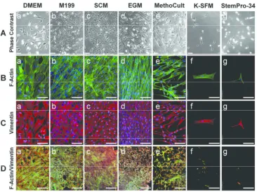
Effects of the seven initial media types were compared by imaging CSSC morphology at P0,
after 5 days of culture in the respective media (figure 2). Phase contrast imaging showed that
there were subjectively lower numbers of cells in DMEM (a) and MethoCult (e) than M199
(b), SCM (c) and EGM (d). There were very few cells in K-SFM (f) and in StemPro-34 (g),
the cells had begun to become rounded and detach from the plate, a process commonly seen
with loss of viability. F-actin (figure 2B) and vimentin (figure 2C) staining demonstrated
differences in morphology between the cell cultures. Cells in M199 and EGM began to align
spontaneously with each other. Cells cultured in DMEM were much larger and spread over a
greater area. Cells cultured in M199 had strong vimentin staining with filaments circling the
nucleus and actin filaments at the cell periphery and stretched across the cell. Cells in SCM 3
For Peer Review Only
were smaller than M199, and appeared to contain a higher proportion of actin fibres. Cells in
EGM were the smallest cells, had aligned with each other and did not have vimentin rings
around the nucleus. When cultured in MethoCult, CSSC had a more heterogenous appearance
with cells of many sizes. CSSC in both K-SFM and StemPro-34 were sparse, individual cells
displaying a vimentin and actin cytoskeleton. However, there were too few cells in these
media to determine differences in morphology. Low magnification images of actin/vimentin
indicated the density of the cell monolayer, with M199 and SCM having the densest
monolayers at day 7.
Effect of different media on protein expression of CSSC
CSSC at P2 were seeded in glass chamber slides and immunocytochemistry was performed to
identify expression of key markers associated with MSC, keratocytes and pluripotent stem
cells. Images were then quantified to estimate the purity of the cell populations in each
media. The medium StemPro-34 was not included due to the previously demonstrated rapid
loss of cell viability. Figure 3 shows the expression of cluster of differentiation (CD) markers
positively and negatively associated with MSC. Figure 4 shows the quantification of these
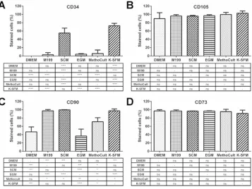
markers. CD34 expression is typically considered a marker absent on MSC; however, it is
also a keratocyte marker. CD34 expression can clearly be seen in individual cells in EGM
and K-SFM, and in a number of cells in SCM.CD34 staining was not observed in DMEM,
M199 and MethoCult. CD105, CD90 and CD73 are markers of MSC. The percentage of cells
expressing CD34 was significantly higher in SCM and K-SFM than all other media and when quantified, CD34 was stained in approximately 55.2% cells in SCM and 72.7% cells in
K-SFM (figure 4A). CD105 was observed on all cells in all media and the merged image with
CD34 shows the individual cells that are expressing CD34 also express CD105. There were
For Peer Review Only
no significant differences between percentages of cells expressing CD105 in different media,
with nearly 100% of cells expressing CD105 in each media (figure 4B). CD90 staining was
strong in M199, SCM and K-SFM. However, it was not present in EGM and only appeared in
some cells in DMEM and MethoCult. Quantification of the percentage of cells stained for CD90 revealed that there were significantly higher numbers in M199 (97.4%), SCM (99.8%)
and K-SFM (97.4%), than DMEM (46.8%), EGM (36.8%) and MethoCult (71.1%). CD73
was seen in all cells in every medium and all media had approximately 100% of cells staining
for CD73
Immunocytochemistry was performed for a range of other markers associated with
keratocytes and stem cells (figure 5) and subsequently quantified (figure 6). ABCG2 was
detected in over 80% of cells in SCM (90.2%), EGM (98.5%), MethoCult (87.1%) and
K-SFM (83.3%), and to a lesser degree in the FBS-containing media DMEM (62.9%) and M199
(11.6%). Keratocan, a proteoglycan secreted by keratocytes, was detected in all cultures;
however higher levels of staining were seen in cell cultured in DMEM (93.3%), EGM
(81.0%), MethoCult (62.8%)and K-SFM (90.3%), than in SCM (36.8%) and M199 (10.6%).
SSEA-4, a cell surface marker found on pluripotent stem cells was seen to a high degree in
SCM (72.4%) and EGM (75.0%) but to a lesser degree in DMEM (37.5%), M199 (13.3%), MethoCult (9.0%)and K-SFM (12.0%). Oct-4A, a transcription factor associated with
pluripotent stem cells was seen in almost all the nuclei of cells cultured in M199 (93.8%),
SCM (94.1%), EGM (92.3%)and MethoCult (96.1%), but in only half of DMEM (51.6%)
and very few in K-SFM (5.6%). PAX6 was seen predominantly in SCM (86.8%), EGM
(98.5%), MethoCult (68.2%) and K-SFM (76.7%).
Effect of different media on mRNA expression of CSSC
For Peer Review Only
RT-qPCR was performed on CSSC at P2 to determine relative differences in mRNA level of
CD34, THY1 (gene name for CD90), POU5F1 (gene name for Oct-4) and PAX6 (figure 7).
CD34, THY1 and PAX6 mRNA level was significantly higher in SCM than in any other
medium. For CD34 and THY1 there were no other significant differences in mRNA levels
between DMEM, M199, EGM and MethoCult. POU5F1 was downregulated in MethoCult
compared to DMEM, but no differences were seen between the other media. PAX6 was
downregulated in M199, EGM and MethoCult, when compared to DMEM.
Effect of differentiation on keratocyte differentiation
CSSC that had been cultured in different media up to P2 were changed to a serum-free
keratocyte differentiation media for 21 days. Immunocytochemistry and RT-qPCR were
performed to assess levels of differentiation compared to non-differentiated controls (figure
8). Immunocytochemistry showed increased levels of ALDH3A1 in all media (fig. 8A vi-x).
However, CSSC, in SCM, EGM and MethoCult appeared to contain higher levels of
ALDH3A1 in the non-differentiated controls (fig. 8A i-v). Myofibroblast marker α-SMA was
present in both the control and differentiated samples in DMEM (fig. 8B i/vi) and M199 (fig.
8B ii/vii). Non-differentiated controls in SCM contained some cells expressing α-SMA (fig.
8Biii) but these were not present in the differentiated sample (fig. 8Bviii). CSSC cultured in
EGM media, showed the reverse, with no staining for α-SMA in control samples (fig. 8Biv) but increased staining in the differentiated samples (fig. 8Bix). No α-SMA was seen in
MethoCult samples. Staining for CD34 was increased in the differentiation samples for all media (fig. 8C vi-x). However, SCM controls also contained some cells that stained for
CD34. RT-qPCR showed that ALDH3A1 was significantly increased in differentiated CSSC from DMEM, M199, and SCM culture compared to controls, but not in EGM or MethoCult
For Peer Review Only
(fig. 8D). ACTA2 (gene name for α-SMA) was significantly increased in both DMEM and EGM cells when differentiated, and was not significantly decreased in differentiated cells
from any media (fig. 8E). CD34 expression was increased in differentiated cells in all media, but only significantly in SCM and MethoCult (fig. 8F). Increased mRNA levels of KERA (gene name for keratocan, fig. 8G) and COL1A1 (collagen-I, fig. 8H) were seen in differentiated cells from all media, but were increased significantly in cells propagated in
SCM, EGM and MethoCult, indicating increased ECM production.
Discussion
Corneal reconstruction with amniotic membrane [40, 41] has demonstrated that the acellular
amniotic stroma is repopulated by corneal keratocytes that migrate from the stroma through
breaks in the Bowman’s membrane. These cells are initially CD34 negative but α-SMA and
vimentin positive, indicating that they are an “activated” phenotype. When corneal buttons
were obtained from corneal grafts performed around 9 to 12 months subsequent to the
amniotic membrane graft, several cells in the amnion stroma were positive for CD34,
indicating that immunophenotypical transitions occur over time during corneal regeneration
in vivo [40]. This holds promise for use of in vitro expanded CSSC in cell therapies for
corneal regeneration.
As the potential for the application of regenerative cell therapies for the cornea is realised and
research gathers momentum, it is essential that common denominators are established in
relation to methodology, so that outcomes are comparable and understood by all. Culture
media constituents and respective concentrations can influence cell behaviour and
differentiation. Hence, even if we start with an identical cell population, different media and
culture environments will result in different cell populations after extended culture, although 3
For Peer Review Only
most or all are collectively referred to as ‘progenitor or stem cells’. This study, wherein a
comprehensive comparison of CSSC media was undertaken concerning cell morphology,
proliferation, protein and gene expression, and keratocyte differentiation ability addresses this
issue by identifying an optimal medium and an optimal in vitro progenitor cell phenotype,
with the potential to generate keratocytes. For the purposes of this study, we considered an
optimal culture medium to have the following properties: the ability to maintain viability and
proliferation of the CSSC in a moderate manner; the promotion of stem cell characteristics
including typical markers for progenitor and pluripotent stem cells; and the ability to allow
differentiation back to a keratocyte phenotype. If the cell were to become part of a cell therapy, the
optimal medium would also need to allow easy transition to a clinical manufacturing process. For this
reason, media containing FBS would be unsuitable due to its animal origin and variable non-defined
composition.
Our results demonstrate that when an extracted cell population is divided into different
media, the resulting cultured cell populations show very different characteristics. Although,
some markers such as vimentin, ALDH3A1, CD73 and CD105 were conserved across all
media, these markers are associated with fibroblasts and MSC and may potentially be
induced by culture on two-dimensional plastic surfaces. This also suggests that the cells
fundamentally express a range of baseline markers in vitro, but changing the medium or
supplements can cause additional markers to be either maintained or expressed, such as
ABCG2, CD34 and PAX6.
Previous media comparison studies have focused solely on the production and maintenance
of an MSC phenotype in CSSC [23, 24], as defined by the ISCT [42]. However, the current
minimal criteria for MSC are not ideal for assessing the phenotype of CSSC, as one criterion
states that MSC should not express CD34. Nevertheless, our group has showed CD34 is a
well-established marker for quiescent keratocytes in vivo [14, 15], and CD34 has been linked 3
For Peer Review Only
to the stem-cell properties of CSSC and other potentially related cell types [23, 43]. The
function of CD34 expression in keratocytes has not yet been elucidated, although it has been
speculated that CD34 plays roles in regulation of differentiation, adhesion and quiescence
[14, 43]. It has been suggested that CD34+ keratocytes are of hematopoietic origin [27], however, these cells do not form hematopoietic colonies when cultured in semi-solid
medium, as shown by culture in MethoCult in this study, and in reality display a plastic
adherent, fibroblastic morphology [18]. For these reasons, a medium that satisfies the
minimum criteria for an MSC phenotype, such as M199, may not be as desirable for culture
of CSSC as, for example SCM, a medium that promotes markers associated with an MSC
phenotype (CD73, CD90, CD105), alongside other progenitor/stem cell markers, such as
CD34, ABCG2, PAX6, Oct-4, and SSEA-4.
ABCG2 is a molecular determinant of the side population phenotype that is characteristic of
stem cells, and has been used previously to identify CSSC [26, 44]. ABCG2 is expressed by a
wide variety of stem cells including haematopoietic stem cells [45], embryonic stem cells
[46], neural stem cells [47], and perhaps most relevantly limbal epithelial stem cells [48].
ABCG2 is often downregulated during stem cell differentiation [44], suggesting that it plays
a regulatory role in maintaining stem cells in an undifferentiated state. PAX6, although not
conventionally a stem or progenitor marker, is strongly associated with eye development.
PAX6 has also been associated with keratocyte progenitors [49]. SSEA-4 and OCT-4 are
markers that are used to identify human pluripotent stem cells [50]. However, these markers
have also been found on adult stem cells including MSC [51]. The presence of all these
markers on the cells in SCM suggests a level of plasticity, which more differentiated cells
may not possess. This plasticity will allow the exploitation of CSSC as cellular therapies,
either to repopulate the corneal stroma as keratocytes, or in broader regenerative medicine
areas, as they are a readily available and easy to isolate stem cell source. 3
For Peer Review Only
Many researchers are aiming to use CSSC as part of regenerative cellular therapies. For this
purpose, CSSC would need to be expanded in large quantities whilst maintaining the desired
phenotype. This would rule out the use of any medium similar to K-SFM or StemPro-34.
DMEM and MethoCult also had slow proliferation rates, so it would be unlikely that media
similar to either of these would be used. The use of DMEM in particular, seemed to favour a
characteristic fibroblastic phenotype, with very large cells and low expression of CD34,
CD90, ABCG2 and SSEA-4. Cells in MethoCult had a more heterogeneous appearance that
may have signified different populations of differentiating cells potentially caused by the
inclusion of factors such as interleukin-3 and erythropoietin in the medium. Growth factors
such as bFGF, PDGF, and isoforms of TGF-β have all been associated with regulation of
wound healing and a keratocyte phenotype in the corneal stroma [52, 53].
Ingredients within a basal medium can have a large effect on the final cell phenotype.
Comparisons between the DMEM and M199 formulations reveals that M199 contains a
number of constituents not found in DMEM including numerous pyrimidines and purines
including ATP, ribose, cholesterol and vitamins which likely drive proliferation [54]. M199
contains the same amount of D-glucose as DMEM ‘low glucose’ formulations (1000 mg/L)
whereas the ‘high glucose’ DMEM used in this study contains significantly higher concentrations (4500 mg/L). This indicates that the much faster proliferation rates seen in
M199 compared to DMEM was not down to glucose concentration.
The success of SCM in promoting markers associated with progenitor cells and pluripotency
is most likely because it is a medium predominantly used in the culture of human embryonic
stem cells (hESC) and induced pluripotent stem cells (iPSC). SCM does not contain FBS but
instead uses knockout serum replacement (KSR). In cultures of hESC and iPSC, FBS is not
suitable as it is an undefined biological product, which varies from batch-to-batch, and can
trigger spontaneous differentiation of cells. KSR is chemically defined and the composition 3
For Peer Review Only
can be found in its patent [55] and in this publication by Garcia-Gonzalo and Belmonte [56].
KSR predominantly contains a mix of amino acids, vitamins, antioxidants, trace elements,
insulin, transferrin and lipid-rich albumin, which has been shown to be the ingredient most
involved in regulation of pluripotency [56]. SCM also contains the recombinant proteins
bFGF and LIF. bFGF is associated with differentiation to the keratocyte phenotype [57] and
is essential for the maintenance of pluripotency of hESC [58]. LIF is a factor that is essential
in the maintenance of pluripotency in mouse ESC [59] but has also been identified as
important in naïve hESC [60]. The combination of these ingredients may allow SCM to
induce or maintain a naïve or progenitor phenotype within the CSSC population. SCM was
the most successful propagation medium in studies assessing the ability to differentiate back
to a keratocyte in vitro. This may be because SCM retained markers associated with the
keratocyte phenotype more successfully than the other media types.
From those tested, we have identified SCM as being the medium with the most potential for
cell therapy applications; however in its current form it would still not be ideal for the clinical
production of CSSC as it is not completely free of animal derived products (xeno-free).
However, all the ingredients have available xeno-free alternatives, which are currently
expensive, and would add to the cost of research and clinical translation. It is also worth
noting that although the culture medium is important in deriving a phenotype of choice; other
aspects of in vitro cell culture can have an effect. Culturing in a three-dimensional
environment rather than two-dimensional can help to dedifferentiate cells back to their
original form [61], thus changing to a scaffold based culture method may have an additional
effect on top of culture medium. Other changes to the environment can include the use of
hypoxia, which has been shown to induce faster proliferation whilst maintaining stem cell
potential and delaying the cellular ageing process [62]. However, culture in a
three-dimensional environment can be complicated and have many unpredictable effects on the
For Peer Review Only
cells, and a hypoxic environment has been shown to promote chondrogenic differentiation,
which may be an issue as the cells are of mesenchymal origin [63, 64].
This study has shown that there can be considerable variation in the properties of CSSC when
cultured in different media. This indicates that in many cases, studies between groups may
not be comparable if performed in different media, due to induced variations in final
phenotype. For the eventual aim of producing a cell therapy from CSSC, it will be imperative
to find a medium that supports a stem cell phenotype and in which the cells can be stably
expanded and maintained as stocks in tissue banks, for release when required for clinical use.
Acknowledgements
This work was funded by grants from the Royal College of Surgeons of Edinburgh and Fight
for Sight. This study was supported by COST Action BM1302 Joining Forces in Corneal
Regeneration Research.
Disclosure of Interests
The authors report no conflicts of interest.
References
[1] Whitcher JP, Srinivasan M,Upadhyay MP, Corneal blindness: a global perspective. Bull
World Health Organ 2001; 79: 214-21.
[2] Tabbara KF, Blindness in the eastern Mediterranean countries. Br J Ophthalmol 2001; 85:
771-5.
[3] Dua HS, Miri A,Said DG, Contemporary limbal stem cell transplantation - a review. Clin
Experiment Ophthalmol 2010; 38: 104-17.
For Peer Review Only
[4] Jester JV, Petroll WM,Cavanagh HD, Corneal stromal wound healing in refractive surgery:
the role of myofibroblasts. Prog Retin Eye Res 1999; 18: 311-56.
[5] Funderburgh JL, Mann MM,Funderburgh ML, Keratocyte phenotype mediates proteoglycan
structure: a role for fibroblasts in corneal fibrosis. J Biol Chem 2003; 278: 45629-37.
[6] Hassell JR,Birk DE, The molecular basis of corneal transparency. Exp Eye Res 2010; 91:
326-35.
[7] Hoar RM, Embryology of the eye. Environ Health Perspect 1982; 44: 31-4.
[8] West-Mays JA,Dwivedi DJ, The keratocyte: corneal stromal cell with variable repair
phenotypes. Int J Biochem Cell Biol 2006; 38: 1625-31.
[9] Poole CA, Brookes NH,Clover GM, Keratocyte networks visualised in the living cornea using
vital dyes. J Cell Sci 1993; 106 ( Pt 2): 685-91.
[10] Meek KM,Boote C, The organization of collagen in the corneal stroma. Exp Eye Res 2004;
78: 503-12.
[11] Scott JE,Thomlinson AM, The structure of interfibrillar proteoglycan bridges (shape
modules') in extracellular matrix of fibrous connective tissues and their stability in various chemical environments. J Anat 1998; 192 ( Pt 3): 391-405.
[12] Michelacci YM, Collagens and proteoglycans of the corneal extracellular matrix. Braz J Med Biol Res 2003; 36: 1037-46.
[13] Jester JV, Moller-Pedersen T, Huang J, Sax CM, Kays WT, Cavangh HD, et al., The cellular
basis of corneal transparency: evidence for 'corneal crystallins'. J Cell Sci 1999; 112 ( Pt 5): 613-22.
[14] Joseph A, Hossain P, Jham S, Jones RE, Tighe P, McIntosh RS, et al., Expression of CD34
and L-selectin on human corneal keratocytes. Invest Ophthalmol Vis Sci 2003; 44: 4689-92.
[15] Perrella G, Brusini P, Spelat R, Hossain P, Hopkinson A,Dua HS, Expression of
haematopoietic stem cell markers, CD133 and CD34 on human corneal keratocytes. Br J
Ophthalmol 2007; 91: 94-9.
[16] Beales MP, Funderburgh JL, Jester JV,Hassell JR, Proteoglycan synthesis by bovine
keratocytes and corneal fibroblasts: maintenance of the keratocyte phenotype in culture. Invest Ophthalmol Vis Sci 1999; 40: 1658-63.
[17] Masur SK, Dewal HS, Dinh TT, Erenburg I,Petridou S, Myofibroblasts differentiate from
fibroblasts when plated at low density. Proc Natl Acad Sci U S A 1996; 93: 4219-23.
[18] Branch MJ, Hashmani K, Dhillon P, Jones DR, Dua HS,Hopkinson A, Mesenchymal stem
cells in the human corneal limbal stroma. Invest Ophthalmol Vis Sci 2012; 53: 5109-16.
[19] Du Y, Carlson EC, Funderburgh ML, Birk DE, Pearlman E, Guo N, et al., Stem cell therapy
restores transparency to defective murine corneas. Stem Cells 2009; 27: 1635-42.
[20] Choong PF, Mok PL, Cheong SK,Then KY, Mesenchymal stromal cell-like characteristics of
corneal keratocytes. Cytotherapy 2007; 9: 252-8.
[21] Helary C, Ovtracht L, Coulomb B, Godeau G,Giraud-Guille MM, Dense fibrillar collagen
matrices: a model to study myofibroblast behaviour during wound healing. Biomaterials 2006; 27: 4443-52.
[22] Polisetty N, Fatima A, Madhira SL, Sangwan VS,Vemuganti GK, Mesenchymal cells from
limbal stroma of human eye. Mol Vis 2008; 14: 431-42.
[23] Hashmani K, Branch MJ, Sidney LE, Dhillon PS, Verma M, McIntosh OD, et al.,
Characterisation of corneal stromal stem cells with the potential for epithelial transdifferentiation. Stem Cell Res Ther 2013; 4: 75.
[24] Bray LJ, Heazlewood CF, Atkinson K, Hutmacher DW,Harkin DG, Evaluation of methods
for cultivating limbal mesenchymal stromal cells. Cytotherapy 2012; 14: 936-47.
For Peer Review Only
[25] Li GG, Zhu YT, Xie HT, Chen SY,Tseng SC, Mesenchymal stem cells derived from human
limbal niche cells. Invest Ophthalmol Vis Sci 2012; 53: 5686-97.
[26] Pinnamaneni N,Funderburgh JL, Concise review: Stem cells in the corneal stroma. Stem Cells
2012; 30: 1059-63.
[27] Sosnova M, Bradl M,Forrester JV, CD34+ corneal stromal cells are bone marrow-derived and
express hemopoietic stem cell markers. Stem Cells 2005; 23: 507-15.
[28] Ainscough SL, Linn ML, Barnard Z, Schwab IR,Harkin DG, Effects of fibroblast origin and
phenotype on the proliferative potential of limbal epithelial progenitor cells. Exp Eye Res 2011; 92: 10-9.
[29] Jester JV, Barry-Lane PA, Cavanagh HD,Petroll WM, Induction of alpha-smooth muscle
actin expression and myofibroblast transformation in cultured corneal keratocytes. Cornea 1996; 15: 505-16.
[30] Park SH, Kim KW, Chun YS,Kim JC, Human mesenchymal stem cells differentiate into
keratocyte-like cells in keratocyte-conditioned medium. Exp Eye Res 2012; 101: 16-26.
[31] Pei Y, Sherry DM,McDermott AM, Thy-1 distinguishes human corneal fibroblasts and
myofibroblasts from keratocytes. Exp Eye Res 2004; 79: 705-12.
[32] Basu S, Hertsenberg AJ, Funderburgh ML, Burrow MK, Mann MM, Du Y, et al., Human
limbal biopsy-derived stromal stem cells prevent corneal scarring. Sci Transl Med 2014; 6: 266ra172.
[33] Du Y, Funderburgh ML, Mann MM, SundarRaj N,Funderburgh JL, Multipotent stem cells in
human corneal stroma. Stem Cells 2005; 23: 1266-75.
[34] Kawakita T, Espana EM, He H, Smiddy R, Parel JM, Liu CY, et al., Preservation and
expansion of the primate keratocyte phenotype by downregulating TGF-beta signaling in a low-calcium, serum-free medium. Invest Ophthalmol Vis Sci 2006; 47: 1918-27.
[35] Garfias Y, Nieves-Hernandez J, Garcia-Mejia M, Estrada-Reyes C,Jimenez-Martinez MC,
Stem cells isolated from the human stromal limbus possess immunosuppressant properties. Mol Vis 2012; 18: 2087-95.
[36] Li GG, Chen SY, Xie HT, Zhu YT,Tseng SC, Angiogenesis potential of human limbal
stromal niche cells. Invest Ophthalmol Vis Sci 2012; 53: 3357-67.
[37] Katikireddy KR, Dana R,Jurkunas UV, Differentiation potential of limbal fibroblasts and
bone marrow mesenchymal stem cells to corneal epithelial cells. Stem Cells 2014; 32: 717-29.
[38] Kopher RA, Penchev VR, Islam MS, Hill KL, Khosla S,Kaufman DS, Human embryonic
stem cell-derived CD34+ cells function as MSC progenitor cells. Bone 2010; 47: 718-28.
[39] Zhao S,Fernald RD, Comprehensive algorithm for quantitative real-time polymerase chain
reaction. J Comput Biol 2005; 12: 1047-64.
[40] Said DG, Nubile M, Alomar T, Hopkinson A, Gray T, Lowe J, et al., Histologic features of
transplanted amniotic membrane: implications for corneal wound healing. Ophthalmology 2009; 116: 1287-95.
[41] Nubile M, Dua HS, Lanzini M, Ciancaglini M, Calienno R, Said DG, et al., In vivo analysis
of stromal integration of multilayer amniotic membrane transplantation in corneal ulcers. Am J Ophthalmol 2011; 151: 809-22 e1.
[42] Dominici M, Le Blanc K, Mueller I, Slaper-Cortenbach I, Marini F, Krause D, et al., Minimal
criteria for defining multipotent mesenchymal stromal cells. The International Society for Cellular Therapy position statement. Cytotherapy 2006; 8: 315-7.
[43] Sidney LE, Branch MJ, Dunphy SE, Dua HS,Hopkinson A, Concise review: evidence for
CD34 as a common marker for diverse progenitors. Stem Cells 2014; 32: 1380-9.
For Peer Review Only
[44] Ding XW, Wu JH,Jiang CP, ABCG2: a potential marker of stem cells and novel target in
stem cell and cancer therapy. Life Sci 2010; 86: 631-7.
[45] Scharenberg CW, Harkey MA,Torok-Storb B, The ABCG2 transporter is an efficient Hoechst
33342 efflux pump and is preferentially expressed by immature human hematopoietic progenitors. Blood 2002; 99: 507-12.
[46] Apati A, Orban TI, Varga N, Nemeth A, Schamberger A, Krizsik V, et al., High level
functional expression of the ABCG2 multidrug transporter in undifferentiated human embryonic stem cells. Biochim Biophys Acta 2008; 1778: 2700-9.
[47] Islam MO, Kanemura Y, Tajria J, Mori H, Kobayashi S, Hara M, et al., Functional expression
of ABCG2 transporter in human neural stem/progenitor cells. Neurosci Res 2005; 52: 75-82.
[48] Watanabe K, Nishida K, Yamato M, Umemoto T, Sumide T, Yamamoto K, et al., Human
limbal epithelium contains side population cells expressing the ATP-binding cassette transporter ABCG2. FEBS Lett 2004; 565: 6-10.
[49] Funderburgh ML, Du Y, Mann MM, SundarRaj N,Funderburgh JL, PAX6 expression
identifies progenitor cells for corneal keratocytes. Faseb J 2005; 19: 1371-3.
[50] Adewumi O, Aflatoonian B, Ahrlund-Richter L, Amit M, Andrews PW, Beighton G, et al.,
Characterization of human embryonic stem cell lines by the International Stem Cell Initiative. Nat Biotechnol 2007; 25: 803-16.
[51] Riekstina U, Cakstina I, Parfejevs V, Hoogduijn M, Jankovskis G, Muiznieks I, et al., Embryonic stem cell marker expression pattern in human mesenchymal stem cells derived from bone marrow, adipose tissue, heart and dermis. Stem Cell Rev 2009; 5: 378-86.
[52] Lim M, Goldstein MH, Tuli S,Schultz GS, Growth factor, cytokine and protease interactions
during corneal wound healing. Ocul Surf 2003; 1: 53-65.
[53] Kim A, Lakshman N, Karamichos D,Petroll WM, Growth factor regulation of corneal
keratocyte differentiation and migration in compressed collagen matrices. Invest Ophthalmol Vis Sci 2010; 51: 864-75.
[54] Kondo M, Yamaoka T, Honda S, Miwa Y, Katashima R, Moritani M, et al., The rate of cell
growth is regulated by purine biosynthesis via ATP production and G(1) to S phase transition. J Biochem 2000; 128: 57-64.
[55] Price PJ, Goldsborough MD,Tilkins ML, Embryonic stem cell serum replacement. I.P.
Application, 1998, WO/1998/030679
[56] Garcia-Gonzalo FR,Izpisua Belmonte JC, Albumin-associated lipids regulate human
embryonic stem cell self-renewal. PLoS One 2008; 3: e1384.
[57] Wu J, Du Y, Mann MM, Yang E, Funderburgh JL,Wagner WR, Bioengineering organized,
multilamellar human corneal stromal tissue by growth factor supplementation on highly aligned synthetic substrates. Tissue Eng Part A 2013; 19: 2063-75.
[58] Xu C, Inokuma MS, Denham J, Golds K, Kundu P, Gold JD, et al., Feeder-free growth of
undifferentiated human embryonic stem cells. Nat Biotechnol 2001; 19: 971-4.
[59] Williams RL, Hilton DJ, Pease S, Willson TA, Stewart CL, Gearing DP, et al.,
Myeloid-leukemia inhibitory factor maintains the developmental potential of embryonic stem cells. Nature 1988; 336: 684-87.
[60] Gafni O, Weinberger L, Mansour AA, Manor YS, Chomsky E, Ben-Yosef D, et al.,
Derivation of novel human ground state naive pluripotent stem cells. Nature 2013; 504: 282-6.
[61] Baker BM,Chen CS, Deconstructing the third dimension: how 3D culture microenvironments
alter cellular cues. J Cell Sci 2012; 125: 3015-24.
[62] Haque N, Kasim NH,Rahman MT, Optimization of pre-transplantation conditions to enhance
the efficacy of mesenchymal stem cells. Int J Biol Sci 2015; 11: 324-34.
For Peer Review Only
[63] Muller J, Benz K, Ahlers M, Gaissmaier C,Mollenhauer J, Hypoxic conditions during
expansion culture prime human mesenchymal stromal precursor cells for chondrogenic differentiation in three-dimensional cultures. Cell Transplant 2011; 20: 1589-602.
[64] Markway BD, Tan GK, Brooke G, Hudson JE, Cooper-White JJ,Doran MR, Enhanced
chondrogenic differentiation of human bone marrow-derived mesenchymal stem cells in low oxygen environment micropellet cultures. Cell Transplant 2010; 19: 29-42.
[65] De Francesco F, Tirino V, Desiderio V, Ferraro G, D'Andrea F, Giuliano M, et al., Human
CD34/CD90 ASCs are capable of growing as sphere clusters, producing high levels of VEGF and forming capillaries. PLoS One 2009; 4: e6537.
[66] Carotta S, Pilat S, Mairhofer A, Schmidt U, Dolznig H, Steinlein P, et al., Directed
differentiation and mass cultivation of pure erythroid progenitors from mouse embryonic stem cells. Blood 2004; 104: 1873-80.
For Peer Review Only
Table 1. Details of experimental culture media. Medium Basal
Medium
Supplements Reference
DMEM Dulbecco’s
Modified Eagle’s Medium (Gibco, Life Technologies)
10% (v/v) foetal bovine serum (Sigma Aldrich)
2 mM L-glutamine (Sigma Aldrich) 20 ng/mL gentamicin, 0.5 ng/mL amphotericin B (Life Technologies)
[20, 27, 28]
M199 Medium 199
(Sigma Aldrich)
20% (v/v) foetal bovine serum 2 mM L-glutamine
20 ng/mL gentamicin, 0.5 ng/mL amphotericin B [18, 23] Stem Cell Medium (SCM) DMEM/F12 (Gibco, Life Technologies)
20% (v/v) knock-out serum replacement (KSR, Life Technologies)
1% (v/v) non-essential amino acids (NEAA, Life Technologies)
4 ng/mL bFGF (Life Technologies)
5 ng/mL hLIF (New England Biolabs, UK) 20 ng/mL gentamicin, 0.5 ng/mL
amphotericin B (Life Technologies)
[36, 37] Endothelial Growth Media (EGM) EBM-2 Basal Medium (Lonza, Belgium)
EGM-2 SingleQuot Kit (CC-4147, Lonza) [38]
MethoCult™ MethoCult® H4034 Optimum (STEMCELL Technologies, UK)
20 ng/mL gentamicin, 0.5 ng/mL amphotericin B (Life Technologies)
[65] Keratinocyte Serum Free Medium (K-SFM) Keratinocyte-SFM (Life Technologies)
Keratinocyte Supplement (Bovine Pituitary Extract, Epidermal Growth Factor (EGF), Life Technologies)
20 ng/mL gentamicin, 0.5 ng/mL amphotericin B (Life Technologies)
[24, 34] StemPro®-34 StemPro®-34 Serum Free Medium (Life Technologies
StemPro®-34 Nutrient Supplement 20 ng/mL gentamicin, 0.5 ng/mL amphotericin B (Life Technologies)
For Peer Review Only
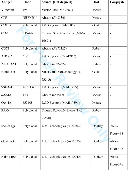
Table 2. Details of antibodies used in immunocytochemistry
Antigen Clone Source (Catalogue #) Host Conjugate
Vimentin V9 Vector Labs (VPV684) Mouse -
CD34 QBEND10 Abcam (Ab8536) Mouse -
CD105 Polyclonal R&D Systems (AF1097) Goat -
CD90 F15-42-1 Thermo Scientific Pierce
(MA5-16671)
Mouse -
CD73 Polyclonal Abcam (Ab71322) Rabbit -
ABCG2 5D3 R&D Systems (MAB995) Mouse -
ALDH3A1 Polyclonal Abcam (ab76976) Rabbit -
Keratocan Polyclonal Santa Cruz Biotechnology
(sc-33243)
Goat -
SSEA-4 MC813-70 R&D Systems (MAB1435) Mouse -
α-SMA 1A4 Abcam (ab7817) Mouse -
Oct-4A 653108 R&D Systems (MAB17591) Mouse -
PAX6 Polyclonal Thermo Scientific Pierce
(PA5-25970)
Rabbit
Mouse IgG Polyclonal Life Technologies (A-21202) Donkey Alexa
Fluor-488
Goat IgG Polyclonal Life Technologies (A-11056) Donkey Alexa
Fluor-546
Rabbit IgG Polyclonal Life Technologies (A-10040) Donkey Alexa
Fluor-546 3
For Peer Review Only
Figure Legends
Figure 1. Effect of culture medium on proliferation of CSSC. Cells were cultured in (a) DMEM (b) M199 (c) SCM (d) EGM (e) MethoCult (f) K-SFM (g) StemPro-34. Graph shows
number of P0 cells cultured in different media at day 1, 3, 7 and 10. Data shown as mean
±SEM of 3 independent experiments each with 3 replicates (n=3).
Figure 2. Effect of culture medium on morphology and cytoskeletal arrangements of CSSC. Cells were cultured in (a) DMEM (b) M199 (c) SCM (d) EGM (e) MethoCult (f) K-SFM (g) StemPro-34. (A) Phase contrast images at day 5 (scale bar=90 µm). (B) F-Actin
(green) staining counterstained with DAPI (blue) (scale bar=100 µm). (C) Vimentin (red)
staining counterstained with DAPI (blue) (scale bar=100 µm). (D) Merged F-Actin, vimentin,
DAPI staining taken at low magnification (x40, scale bar=500 µm).
Figure 3. Effect of culture medium on mesenchymal stem cell markers. Cells were cultured in (A) DMEM (B) M199 (C) SCM (D) EGM (E) MethoCult (F) K-SFM.
Immunocytochemistry was performed for (a) CD34, green; (b) CD105, red; (c) CD90, green;
and (d) CD73, red. Negative control images (secondary antibody only) of (e) donkey
anti-mouse IgG Alexa Fluor-488, (f) donkey anti-goat IgG Alexa Fluor-546 (g) donkey anti-rabbit
IgG Alexa Fluor-546. All images shown with DAPI counterstain (blue), scale bar=100 µm.
Figure 4. Quantification of number of cells expressing mesenchymal stem cells markers.
Immunocytochemistry images were quantified to calculate the number of cells in each media
expressing (A) CD34, (B) CD105, (C) CD90 and (D) CD73. Data shown as mean±SD of 3
independent experiments (n=3). Statistical significances comparing each media to each other are shown in the table below each graph: ns, non-significant, *p≤0.05, **p≤0.01,
***p≤0.001, ****p≤0.0001 (one-way ANOVA).
For Peer Review Only
Figure 5. Effect of culture medium on pluripotency and keratocyte markers. Cells were cultured in (A) DMEM (B) M199 (C) SCM (D) EGM (E) MethoCult (F) K-SFM.
Immunocytochemistry was performed for (a) ABCG2, green; (b) Keratocan, green; (c)
SSEA-4, green; (d) Oct-4A, red and (e) Pax6, red. Negative controls (secondary antibody only) can be seen in figure 3. All images shown with DAPI counterstain (blue), scale bar=100
µm.
Figure 6. Quantification of cell number expressing pluripotency and keratocyte
markers. Immunocytochemistry images were quantified to calculate the number of cells in
each media expressing (A) ABCG2, (B) Keratocan, (C) SSEA-4, (D) Oct-4A and (E) Pax6.
Data shown as mean±SD of 3 independent experiments (n=3). Statistical significances
comparing each media to each other are shown in the table below each graph: ns,
non-significant, *p≤0.05, **p≤0.01, ***p≤0.001, ****p≤0.0001 (one-way ANOVA).
Figure 7. Comparative effect of culture medium on gene expression of CSSC. CSSC were continually cultured in DMEM, M199, SCM, EGM or MethoCult to P2. Relative levels
of mRNA were determined by RT-qPCR for the following genes (a) CD34 (b) THY1, (c)
POU5F1 and (d) PAX6. Expression of each target gene is normalised to GAPDH and
represented relative to mRNA expression in DMEM. Data shown as mean±SEM of 3
independent experiments (n=3), each with 3 replicates. Statistical significance vs. DMEM
*p≤0.05, **p≤0.01, ***p≤0.001, ****p≤0.0001 (one-way ANOVA).
Figure 8. Effect of culture medium on differentiation back to a keratocyte phenotype.
CSSC at P3 were differentiated back to a keratocyte phenotype using a serum-free medium containing bFGF, for 21 days. Immunocytochemistry was performed for (A) ALDH3A1, (B)
α-SMA, (C) CD34 on non-differentiated control samples (i-v) and differentiated samples
(vi-x). CSSC had ben propagated in DMEM, M199, SCM, EGM or MethoCult prior to
For Peer Review Only
differentiation. All images shown with DAPI counterstain (blue), scale bar=100 µm. Relative
levels of mRNA were determined by RT-qPCR for the following genes (D) ALDH3A1 (E) ACTA2 (F) CD34 (G) KERA and (H) COL1A1. Expression of each target gene is normalised to GAPDH and represented relative to mRNA expression in the non-differentiated control of that media. Data shown as mean±SEM of 3 independent experiments (n=3), each with 2
replicates. Statistical significance vs. control *p≤0.05, **p≤0.01, ***p≤0.001, ****p≤0.0001
(two-way ANOVA).
For Peer Review Only
Figure 1. Effect of culture medium on proliferation of CSSC. Cells were cultured in (a) DMEM (b) M199 (c) SCM (d) EGM (e) MethoCult (f) K-SFM (g) StemPro-34. Graph shows number of P0 cells cultured in different
media at day 1, 3, 7 and 10. Data shown as mean ±SEM of 3 independent experiments each with 3 replicates (n=3).
119x144mm (300 x 300 DPI) 3
For Peer Review Only
Figure 2. Effect of culture medium on morphology and cytoskeletal arrangements of CSSC. Cells were cultured in (a) DMEM (b) M199 (c) SCM (d) EGM (e) MethoCult (f) K-SFM (g) StemPro-34. (A) Phase contrast images at day 5 (scale bar=90 µm). (B) F-Actin (green) staining counterstained with DAPI (blue) (scale bar=100 µm). (C) Vimentin (red) staining counterstained with DAPI (blue) (scale bar=100 µm). (D)
Merged F-Actin, vimentin, DAPI staining taken at low magnification (x40, scale bar=500 µm). 140x104mm (300 x 300 DPI)
For Peer Review Only
Figure 3. Effect of culture medium on mesenchymal stem cell markers. Cells were cultured in (A) DMEM (B) M199 (C) SCM (D) EGM (E) MethoCult (F) K-SFM. Immunocytochemistry was performed for (a) CD34, green; (b) CD105, red; (c) CD90, green; and (d) CD73, red. Negative control images (secondary antibody
only) of (e) donkey anti-mouse IgG Alexa Fluor-488, (f) donkey anti-goat IgG Alexa Fluor-546 (g) donkey anti-rabbit IgG Alexa Fluor-546. All images shown with DAPI counterstain (blue), scale bar=100 µm.
119x195mm (300 x 300 DPI)
For Peer Review Only
Figure 4. Quantification of number of cells expressing mesenchymal stem cells markers.
Immunocytochemistry images were quantified to calculate the number of cells in each media expressing (A) CD34, (B) CD105, (C) CD90 and (D) CD73. Data shown as mean±SD of 3 independent experiments (n=3).
Statistical significances comparing each media to each other are shown in the table below each graph: ns, non-significant, *p≤0.05, **p≤0.01, ***p≤0.001, ****p≤0.0001 (one-way ANOVA).
80x59mm (300 x 300 DPI)
For Peer Review Only
Figure 5. Effect of culture medium on pluripotency and keratocyte markers. Cells were cultured in (A) DMEM (B) M199 (C) SCM (D) EGM (E) MethoCult (F) K-SFM. Immunocytochemistry was performed for (a) ABCG2,
green; (b) Keratocan, green; (c) SSEA-4, green; (d) Oct-4A, red and (e) Pax6, red. Negative controls (secondary antibody only) can be seen in figure 3. All images shown with DAPI counterstain (blue), scale
bar=100 µm.
119x179mm (300 x 300 DPI) 3
For Peer Review Only
Figure 6. Quantification of cell number expressing pluripotency and keratocyte markers.
Immunocytochemistry images were quantified to calculate the number of cells in each media expressing (A) ABCG2, (B) Keratocan, (C) SSEA-4, (D) Oct-4A and (E) Pax6. Data shown as mean±SD of 3 independent
experiments (n=3). Statistical significances comparing each media to each other are shown in the table below each graph: ns, non-significant, *p≤0.05, **p≤0.01, ***p≤0.001, ****p≤0.0001 (one-way ANOVA).
99x109mm (300 x 300 DPI)
For Peer Review Only
[image:40.612.132.470.119.398.2]Figure 7. Comparative effect of culture medium on gene expression of CSSC. CSSC were continually cultured in DMEM, M199, SCM, EGM or MethoCult to P2. Relative levels of mRNA were determined by RT-qPCR for
the following genes (a) CD34 (b) THY1, (c) POU5F1 and (d) PAX6. Expression of each target gene is normalised to GAPDH and represented relative to mRNA expression in DMEM. Data shown as mean±SEM of
3 independent experiments (n=3), each with 3 replicates. Statistical significance vs. DMEM *p≤0.05, **p≤0.01, ***p≤0.001, ****p≤0.0001 (one-way ANOVA).
62x48mm (300 x 300 DPI)
For Peer Review Only
Figure 8. Effect of culture medium on differentiation back to a keratocyte phenotype. CSSC at P3 were differentiated back to a keratocyte phenotype using a serum-free medium containing bFGF, for 21 days. Immunocytochemistry was performed for (A) ALDH3A1, (B) α-SMA, (C) CD34 on non-differentiated control
samples (i-v) and differentiated samples (vi-x). CSSC had ben propagated in DMEM, M199, SCM, EGM or MethoCult prior to differentiation. All images shown with DAPI counterstain (blue), scale bar=100 µm. Relative levels of mRNA were determined by RT-qPCR for the following genes (D) ALDH3A1 (E) ACTA2 (F) CD34 (G) KERA and (H) COL1A1. Expression of each target gene is normalised to GAPDH and represented relative to mRNA expression in the non-differentiated control of that media. Data shown as mean±SEM of 3
independent experiments (n=3), each with 2 replicates. Statistical significance vs. control *p≤0.05, **p≤0.01, ***p≤0.001, ****p≤0.0001 (two-way ANOVA).
95x150mm (300 x 300 DPI)