Original Article
Devazepide, a nonpeptide antagonist of CCK receptor,
induces cell apoptosis and inhibits contraction in
human prostatic stromal myofibroblasts
Bingbing Wei1*, Jun Ruan1*, Jiabei Liang2*, Yuanyuan Mi3*, Jian Zhang1, Zhirong Wang1, Qiang Hu1
1Department of Urology, Affiliated Wuxi People’s Hospital, Nanjing Medical University, Wuxi 214023, China; 2Department of Pathology, Affiliated Wuxi People’s Hospital, Nanjing Medical University, Wuxi 214023, China; 3
De-partment of Urology, Huashan Hospital, Fudan University, 12 Wulumuqi Middle Road, Shanghai 200040, China. *Equal contributors.
Received January 13, 2017; Accepted March 18, 2017; Epub April 15, 2017; Published April 30, 2017
Abstract: Human prostatic stromal myofibroblasts have been shown to be related to lower urinary tract symptoms
(LUTS) in aging men. Devazepide, also known as a nonpeptide antagonist of CCK receptor, has been found to inhibit the proliferation in tumor cells. Here, we aim to investigate the effects of Devazepide on growth and contraction
ability in human prostatic stromal WPMY-1 myofibroblasts. Human prostatic stromal WPMY-1 myofibroblasts were
treated with various concentrations of Devazepide. The cell viability was determined by WST-8 assay. TUNEL assay and Annexin-V/Propidium iodide (PI) staining were performed to determine cell apoptosis. Expression levels of cleaved Caspase-3 and cleaved Caspase-9 were examined by Western blot. Contraction ability in WPMY-1 cells was
evaluated by collagen gel contraction assay. We found that cell viability of WPMY-1 cells was significantly inhibited
in the presence of Devazepide. Devazepide inhibited growth of WPMY-1 cells in a dose and time-dependent manner.
In addition, Devazepide could significantly induce G2/M cell cycle arrest and apoptosis in WPMY-1 cells. Increased
expression levels of cleaved Caspase-3 and Caspase-9 proteins were found in Devazepide-treated WPMY-1 cells. Collagen gel contraction assay further showed that Devazepide could inhibit contraction ability of WPMY-1 cells, which might be associated with growth inhibition and decreased expression levels of Col1A1 and Col1A2 in WPMY-1 cells. Taken together, Devazepide could inhibit in vitro growth and contraction ability in human prostatic stromal
myofibroblasts, which might be a novel therapeutic target of LUTS in aging men.
Keywords: Devazepide, CCK receptor, myofibroblast, prostate
Introduction
Lower urinary tract symptoms (LUTS) have been shown to be an important medical challenge in aging men. The main symptoms include noctu-ria, intermittent or hesitant urination, weak stream, and sensation of incomplete emptying [1]. If LUTS were not treated effectively, multi-ple complications eventually occurred, such as urinary retention, bladder calculi, and renal impairment [2]. It has been reported that many diseases could lead to LUTS, such as benign prostatic hyperplasia (BPH) [3]. However, effec-tive treatment of LUTS is still a medical chal-lenge due to less understanding of etiology of LUTS.
Fibrosis is characterized by myofibroblast
ac-cumulation, collagen, and extracellular matrix
(ECM) deposition [4, 5]. Multiple cell types co-
uld differentiate into myofibroblasts including fibrocytes, pericytes, and fibroblasts [6]. Recent
years, multiple evidences showed that
periure-thral ECM deposition and fibrosis were closely
associated with LUTS in aging men [7, 8]. In the study [9], it was reported that periurethral
calci-fication (PUC) was independently associated with peak urinary flow rate (Qmax) and
urin-ary symptoms, which provided strong evidence
that periurethral fibrosis and stiffness could
lead to LUTS-BPH in aging men. It was reported that prostate tissues from men with LUTS were
significantly stiffer than those without LUTS [8].
Further study showed that it was associated
with significantly higher collagen content and
significantly increased in BPH nodules, which suggested that large fibers might be involved in
BPH/LUTS [10].
Currently, although anti-androgens, smooth-muscle relaxers, and surgical operation could be used to treat LUTS in aging men, they were
not effective to all patients. Several findings showed that anti-fibrotic therapeutics might be
effective for the treatment of LUTS [7, 11, 12].
In addition, it has been reported that myofibro -blast accumulation has been found in the
peri-urethral area of prostate. Myofibroblast accu -mulation was thought to be associated with ECM deposition, tissue stiffness, and urinary voiding dysfunction [8, 12]. Considering the
important roles of myofibroblasts in LUTS, myo
-fibroblasts may be a novel target for LUTS treat -ment in aging men.
Devazepide is known as a CCK-1 receptor antagonist, which has been shown to inhibit Ewing tumor growth and HT-29 cell prolifera- tion [13, 14]. In this study, we investigated the effects of Devazepide on proliferation and con-traction ability in human prostatic stromal
WPMY-1 myofibroblasts. We found that
Deva-zepide could induce apoptosis and inhibit in vitro contraction ability in human WPMY-1 cells. Materials and methods
Cell culture, reagents
Human prostatic stromal myofibroblast cell line
WPMY-1 was obtained from American Type Culture Collection (ATCC). WPMY-1 cells were
cultured in Dulbecco’s modified Eagle’s medi -um (DMEM) supplemented with 10% fetal
bovine serum (FBS) at 37°C in a humidified 5%
CO2 atmosphere. The CCK-1 receptor antago-nist Devazepide and CCK-2 receptor antagoni- st LY225910 were purchased from Tocris Biosciences (Bristol, UK). Non-selective CCK antagonist Proglumide was purchased from Sigma Aldrich (MO, USA).
WST-8 cell viability assay
Cell viability was evaluated by Cell Counting Kit-8 (CCK-8) (Dojindo, Japan). The WPMY-1 cells were seeded at a density of 0.8 × 10⁴ cells per well in 96-well plates. After 12 h incu- bation, the medium was replaced with fresh medium containing various concentrations of
Devazepide, LY225910, and Proglumide. At dif-ferent time points, WST-8 was used to detect cell viability. The experiments were repeated four times, and cell growth rate was computed
using the following formula: Growth rate = (OD
test/OD control) × 100%. Immunofluorescence assay
Human WPMY-1 cells were grown on glass
cov-erslips and fixed in 4% paraformaldehyde for
15 min. After washing with PBS, WPMY-1 cells were permeabilized with 0.2% Triton X-100 for 15 min at room temperature. Then, 5% goat
serum was used to block nonspecific binding.
Cells were incubated with rabbit polyclonal KI-67 antibody at a dilution of 1:200 overnight at 4°C, and subsequently were exposed to goat
anti-rabbit IgG Rhodamine (TRITC) (1:500 dilu -tion) for one hour at 37°C. DAPI was used to stain nuclei of cells.
RNA extraction and quantitative real-time RT-PCR
Total RNA in WPMY-1 cells was extracted us- ing the RNAiso Plus reagent (TaKaRa) accord-ing to the manufacturer’s instruction. Com- plementary DNA (cDNA) was obtained from RNA using the PrimeScriptTM RT reagent Kit with gDNA Eraser kit (TaKaRa). Expression
lev-els of KI-67, COL1A1, COL1A2, and GAPDH
mRNA were examined by quantitative real-time RT-PCR (qRT-PCR). The qRT-PCR was performed using the SYBR® Premix Ex TaqTM II (Tli RNaseH Plus) (TaKaRa). The primer sequences for qRT-PCR were shown as follows: for KI-67, 5’-CCC-
AAGACACCTTTGGAGAA-3’ (forward) and 5’-AT-CCTCACCTCCTGGTACTT-3’ (reverse); for COL-1A1, 5’-CCCTGGAAAGAATGGAGATG-3’ (forwa-rd) and 5’-CCACTGAAACCTCTGTGTCC-3’ (reve-rse); for COL1A2, 5’-CAGAACATCACCTACCAC-TGCAA-3’ (forward) and 5’-TTCAACATCGTTGG-AACCCTG-3’ (reverse); for GAPDH, 5’-TGCACC-ACCAACTGCTTAGC-3’ (forward) and reverse primer 5’-GGCATGGACTGTGGTCATGAG-3’
(rev-erse).
Analysis of cell cycle
Human WPMY-1 cells were seeded in 60-mm dishes and treated with Devazepide. Cells were subsequently collected and analyzed for DNA content at different time points. To analyze
cold ethanol. Then, cells were treated with RNase and propidium iodide for 10 min at room
temperature, and further analyzed by flow
cytometry. Western blot
Human WPMY-1 cells were treated with De- vazepide. Then, WPMY-1 cells were treated with RIPA lysis buffer on ice for 15 min. Subsequently, proteins were separated by
SDS-PAGE and transferred to polyvinylidene difluo -ride membranes (PVDF). Non-fat milk (5%) in TBS-T (Tris-buffered saline containing 0.1%
Tween-20) was prepared to block nonspecific
binding. The membranes were immunoblotted with the primary antibodies overnight at 4°C, and subsequently incubated with secondary
antibody goat anti-rabbit IgG (H+L) HRP (1:5000
dilution, Bioworld Technology, Minneapolis, USA) for one hour at room temperature. The pri-mary antibodies in this study were shown as
fol-lows: Polyclonal rabbit anti-GAPDH (1:5000
dilution, Bioworld Technology, Minneapolis, USA), p-Chk1 (Ser280) (1:1000 dilution, Bio- world Technology, Minneapolis, USA), p-Chk2 (Thr68) (1:500 dilution, Bioworld Technology, Minneapolis, USA), Cyclin B1 (1:1000, Bioworld Technology, Minneapolis, USA), p21 (1:1000, Bioworld Technology, Minneapolis, USA), Cas- pase-3 (1:1000 dilution, Abcam, Cambridge, UK), Caspase-9 (1:1000 dilution, Abcam, Cambridge, UK), cleaved Caspase-3 (1:1000 dilution, Abcam, Cambridge, UK), and cleaved Caspase-9 (1:1000 dilution, Abcam, Cambridge, UK).
TUNEL assay
TUNEL assay was conducted with the one-step
in situ TUNEL fluorescent kit (Beyotime, China). Briefly, WPMY-1 cells were treated with Devazepide and were fixed with 4% paraformal -dehyde for 15 min at room temperature. Subsequently, cells were permeabilized with 0.2% Triton X-100/PBS for 15 min, followed with Cyanine 3 (Cy3)-labelled TUNEL reaction mixture for one hour at 37°C. The Cy3-labeled
cells were photographed using fluorescent
microscope.
Flow cytometric analysis of cell apoptosis
Apoptosis was analyzed using Annexin V-FITC/ Propidium iodide Apoptosis Detection kit acc-
ording to the manufacturer’s instruction (Do-
jindo Molecular Technologies, Japan). Briefly,
after Devazepide treatment, cells were
harvest-ed and suspendharvest-ed in 200 μL binding buffer containing 10 μL Annexin V-FITC and 5 μL PI.
Cells were incubated at room temperature
for 15 min, and then analyzed using flow
cytometry.
Collagen gel contraction assay
Collagen gel contraction assay was performed
to evaluate contraction ability of myofibroblasts using modifications of a previously described method [12]. Briefly, collagen gel was prepared
on ice by mixing 3 ml of collagen solution (Stem Cell Technologies, Vancouver, BC) with 5 ml DMEM. The mixture was adjusted to pH 7.4. Then, 600 µl of the mixture was added to each well of 24-well plate and allowed to solidify at
37°C. WPMY-1 cells were seeded on solidified
collagen gels, and allowed to attach for 24 h.
Solidified collagen gel was detached from the
wall of 24-well plate and cells were treated with Devazepide at 37°C in a 5% CO2 incubator. The gels were photographed at different time
points. Gel surface areas were quantified
us-ing Image-Pro Plus 6.0 software (Media Cybernetics, Inc., MD, USA).
Statistical analysis
All data were calculated based on at least three independent experiments and expressed as means ± standard derivations (SD). Statistical differences were analyzed by Student’s t test for intergroup comparison using SPSS 19.0 software (SPSS Inc., USA). A P value < 0.05 was
statistically considered significant.
Results
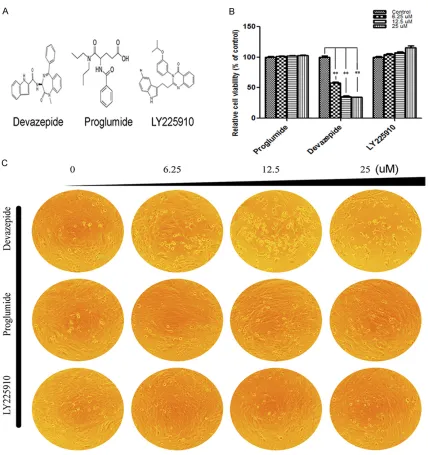
Devazepide inhibited proliferation in human prostatic stromal WPMY-1 myofibroblasts
investigated the effects of CCK-1 receptor antagonist Devazepide on human prostatic
stromal WPMY-1 myofibroblasts. WPMY-1 cells
were treated with indicated concentrations of Devazepide and cell viability was evaluated by WST-8 assay. After treatment for 72 h, it was found that Devazepide could inhibit the prolif-eration of WPMY-1 cells by about 41.7%
[image:4.612.92.520.68.523.2]com-pared with the control group. In addition, we found that Devazepide could inhibit the growth of WPMY-1 cells in a time and dose-dependent manner (Figure 1). Furthermore, for non-selec-tive CCK receptor antagonist Proglumide and selective CCK-2 receptor antagonist LY225910, no inhibitory effects were found on the prolifer-ation of WPMY-1 cells (Figure 1).
We also analyzed the changes in cell morp- hology in the presence of Devazepide. After Devazepide treatment, WPMY-1 cells became
shrunken and floating. Hoechst 33258 staining
was also used to detect nuclear change under Devazepide treatment (data not shown). We found that Devazepide-treated WPMY-1 cells showed morphological changes including nuclear chromatin condensation, which were consistent with cell apoptosis. In Proglumide- and LY225910-treated groups, no apoptotic
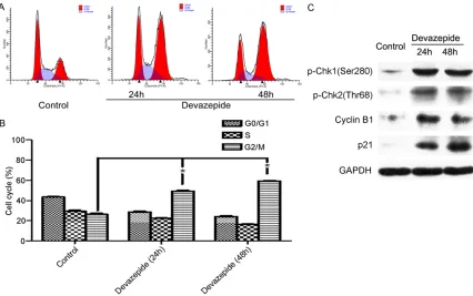
cell cycle phase. To determine this speculation, we treated human WPMY-1 cells with indicated concentrations of Devazepide. Then, cell cycle
was analyzed using flow cytometry at different time points. Representative cell cycle profiles
and histograms of Devazepide-untreated and -treated cells were shown in Figure 3. It was
found that Devazepide significantly decreased
the percentage of WPMY-1 cells in S phase and increased the percentage of WPMY-1 cells in
[image:5.612.91.373.70.470.2]G2/M phase. After Devazepide treatment, the
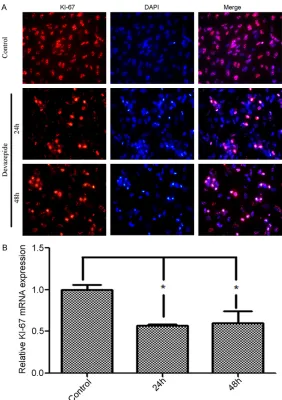
Figure 2. Selective CCK-1 receptor antagonist Devazepide downregulated KI-67 expression in WPMY-1 cells. A. Representative images of low expres-sion of KI-67 in Devazepide-treated WPMY-1 cells by immunofluorescence staining. Cells were stained using 6-diamidino-2-phenylindole (DAPI) to vi-sualize nuclei. B. KI-67 mRNA expression was decreased in Devazepide-treated WPMY-1 cells by quantitative real-time RT-PCR. Data are presented as means ± SD. Asterisks indicate statistically significant difference (*P < 0.05).
morphological changes were found (Figure 1).
Devazepide downregulated KI-67 expression
KI-67 is a nuclear protein which is closely associated with cell proliferation. Several studies showed that KI-67 was also related to ribosomal RNA transcription [15, 16]. It was well known that KI-67 was a valuable cellular mark-er for prolifmark-eration, which was associated with cell prolifera-tion [17]. We found that De- vazepide could inhibit the growth of WPMY-1 cells. This led us to investigate the effe- ct of Devazepide on KI-67
expression. Using immunoflu -orescence assay, we found that KI-67 was mainly local-ized in nuclei of WPMY-1 cells. Furthermore, weak staining of KI-67 protein was found in Devazepide-treated WPMY-1 cells (Figure 2). In addition, we found that Devazepide also inhibited KI-67 mRNA expression in WPMY-1 cells (Figure 2).
Devazepide induced G2/M cell cycle arrest in human WPMY-1 cells
percentage of WPMY-1 cells in G0/G1 phase significantly decreased to 25.45±2.77% and
21.88±2.49%, respectively. Consistently, the percentage of Devazepide-treated WPMY-1
cells in G2/M phase significantly increased to
45.90±4.26% and 50.56±7.78%, respectively. Devazepide upregulated phosphorylation of Chk1 and Chk2 in human WPMY-1 cells
It has been reported that phosphorylation of
Chk1 and Chk2 may be involved in G2/M cell
cycle arrest in human cervical carcinoma HeLa cells when treated with 8-ADEQ [18]. Because
Devazepide could induce G2/M cell cycle
arr-est in human WPMY-1 cells, we examined the effects of Devazepide on phosphorylation lev-els of Chk1 and Chk2. We found that Devaze-
pide significantly upregulated phosphorylation
levels of Chk1 and Chk2 at Ser280 and Thr68 sites (Figure 3), respectively. It suggests that
Cantharidin increased cell population in G2/M
phase and Cyclin B1 expression in
hepatocel-lular carcinoma stem cells [19]. In this study, increased expression level of Cyclin B1 was detected in WPMY-1 cells in the presence of Devazepide (Figure 3). In addition, Devazepide could also upregulate p21 expression in WPMY-1 cells (Figure 3).
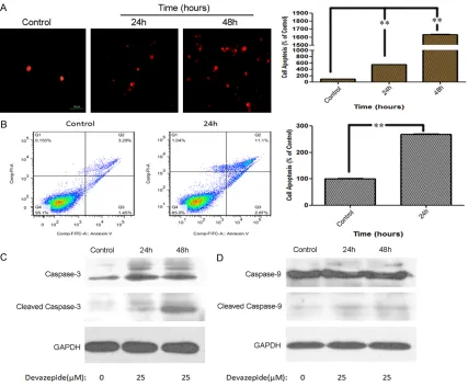
Devazepide induced cell apoptosis in human WPMY-1 cells via intrinsic apoptosis pathway
To investigate the effects of Devazepide on apoptosis, we treated human WPMY-1 cells with indicated concentrations of Devazepide. Using TUNEL assay, apoptosis in WPMY-1 cells was detected (Figure 4). Furthermore, Annexin V-FITC apoptosis detection kit was used to eval-uate apoptosis in WPMY-1 cells in the presence of Devazepide. It was found that Devazepide
could significantly induce apoptosis in
[image:6.612.94.520.71.338.2]apoptosis signaling. In this study, we found
that Devazepide could significantly upregulate
cleaved Caspase-9 and cleaved Caspase-3 by Western blot (Figure 4).
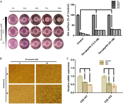
Devazepide inhibited in vitro contraction ability of human WPMY-1 cells
It suggests that myofibroblasts could form con
-tractile stress fibers on stiff collagen gels [20].
The contraction ability of WPMY-1 cells may be associated with LUTS in aging men. Therefore, collagen gel contraction assay was performed to evaluate the effect of Devazepide on in vitro contraction ability of WPMY-1 cells. We found
that Devazepide could significantly inhibit the
contraction of WPMY-1 cells at different time
points (Figure 5). Devazepide also induced cell death in WPMY-1 cells (Figure 5), which might
influence gel contraction. In addition, we found
that Devazepide could downregulate the expression levels of COL1A1, and COL1A2 (Figure 5).
Discussion
Devazepide, as a benzodiazepine drug, could block CCK-mediated activation of CCK-1 recep-tor. It suggests that CCK could affect the con-traction of gall bladder and relaxation of Sphincter of Oddi(Glisson’s sphincter), which is
involved in the delivery of bile into the small
intestine. Here, we reported a novel finding that
[image:7.612.93.520.72.425.2]WPMY-1 cells. In other studies, the effects of Devazepide on cell growth have been reported. It suggests that Devazepide could induce apop-tosis in HT-29 cells [14]. It was also found that Devazepide could inhibit the growth of Ewing
tumor cells [13]. Our studies firstly showed that
Devazepide could induce death of human WPMY-1 cells.
KI-67 has been extensively studied in past sev-eral years and was reported to be closely asso-ciated with cell proliferation. Furthermore, it was found that KI-67 could regulate ribosomal RNA transcription and influence cell prolifera -tion. KI-67 has been known as a marker for cell
proliferation in scientific research of glioma
[21], breast cancer [22], and prostate cancer [23]. In our study, it was observed that KI-67
expression was significantly decreased in
Devazepide-treated WPMY-1 cells, which was a valuable marker for cell proliferation in the presence of Devazepide.
Cell cycle is series of events occurring in cell growth, which could cause division and replica-tion of DNA to produce daughter cells. In mam-mals, cell cycle consists of three periods: inter-phase, the mitotic (M) inter-phase, and cytokinesis.
The G2/M checkpoint, as an important cell
[image:8.612.93.520.74.436.2]Devazepide could arrest Ewing tumor cells in
G2/M cell cycle phase. In our study, we found that WPMY-1 cells in G2/M cell cycle phase were significantly increased when treated with
Devazepide. This suggested Devazepide could
induce G2/M cell cycle arrest not only in tumor
cells, but also in normal human prostate my-
ofibroblasts. Therefore, Devazepide might in-duce cell death via affecting G2/M cell cycle
checkpoint.
The underlying molecular mechanisms
regulat-ing G2/M cell cycle in cells are still not well
understood. It reports that Chk1, as a Serine/
threonine-specific protein kinase in humans, regulates the G2/M phase transition [24]. And
Chk1 plays an important role in the initiation of cell cycle checkpoints, and cell cycle arrest. Chk2 is another important checkpoint kinase, which could phosphorylate the cell-cycle tran-scription factor E2F1 and promyelocytic leuke-mia protein [25]. The phosphorylation of Chk1
and Chk2 has been shown to influence G2/M
phase transition. Cyclin B1 is also a vital
mole-cule regulating G2/M phase transition, and found to be upregulated in cells of G2/M cell
cycle arrest. In this study, we found that phos-phorylation levels of Chk1 and Chk2 were
sig-nificantly increased in human WPMY-1 cells in
the presence of Devazepide. Meanwhile, incre- ased expression of Cyclin B1 was also detected in WPMY-1 cells when treated with Devazepide.
These findings suggested that Devazepide could induce G2/M cell cycle arrest in WPMY-1
cells, which was involved in activation of Chk1, and Chk2.
It has been found that apoptosis is a process of programmed cell death. Apoptosis could be activated through intrinsic and extrinsic path-ways. In intrinsic apoptotic pathway, Caspase-9 is activated and the activated Caspase-9 could further cleave and activate other executioner Caspases, such as Caspase-3. In this study, we found that Devazepide could activate Cas- pase-9 and Caspase-3 in WPMY-1 cells. These
findings suggested that Devazepide induced
apoptosis in human WPMY-1 cells via activating intrinsic apoptotic pathway.
Type I collagen is found to be the most abun-dantcollagenof human body, which is present in scar tissue and involved in fibrosis [26]. Myofibroblasts could express type I collagen
and play a vital role in fibrosis [27]. It suggests
that COL1A1 gene encodes a component of type I collagen, also known as pro-alpha1(I) chain, which combines with another a pro-alpha2(I) chain (encoded by COL1A2 gene) to produce a molecule of type I procollagen. Hence, both COL1A1 and COL1A2 genes could affect the production of type I collagen. In the study, we found that Devazepide could inhibit COL1A1 and COL1A2 expression in human
WPMY-1 myofibroblasts. In addition, we also
found that Devazepide inhibited in vitro con-traction ability of human WPMY-1 cells. We
speculated that Devazepide could inhibit myofi
-broblasts-mediated fibrosis via suppressing
the production oftype I collagen.
On the whole, we found that Devazepide could induce cell death and inhibit contraction ability
of human prostate WPMY-1 myofibroblasts. Considering the important roles of myofibro
-blasts in fibrosis, Devazepide may be a novel
target for LUTS treatment in aging men. Acknowledgements
The work was partially supported by grants from the Key Program of Hospital Manage-
ment Center, China (YGZX1112); the
Founda-tion of Nanjing Medical University, China (2013NJMU157); the Research program of Wuxi Health and Family Planning Commission (Q201612).
Disclosure of conflict of interest None.
Address correspondence to: Qiang Hu and Zhirong Wang, Department of Urology, Affiliated Wuxi People’s Hospital, Nanjing Medical University, No. 299 Qingyang Road, Wuxi 214023, China. Tel: +86-510-85350150; Fax: +86-510-85350150; E-mail: qianghu123@outlook.com (QH); Tel: +86-510-85351150; Fax: +86-+86-510-85351150; E-mail: wangzr656@163.com (ZRW)
References
[1] Laborde EE and McVary KT. Medical manage-ment of lower urinary tract symptoms. Rev Urol 2009; 11: S19-25.
[3] Egan KB. The pidemiology of benign prostatic hyperplasia associated with lower urinary tract symptoms: prevalence and incident rates. Urol Clin North Am 2016; 43: 289-297.
[4] Wynn TA. Cellular and molecular mechanisms
of fibrosis. J Pathol 2008; 214: 199-210.
[5] Hinz B. Formation and function of the myofibro -blast during tissue repair. J Invest Dermatol 2007; 127: 526-537.
[6] Hinz B, Phan SH, Thannickal VJ, Galli A, Bocha
-ton-Piallat ML and Gabbiani G. The myofibro -blast: one function, multiple origins. Am J Pathol 2007; 170: 1807-1816.
[7] Rodriguez-Nieves JA and Macoska JA. Prostatic
fibrosis, lower urinary tract symptoms, and
BPH. Nat Rev Urol 2013; 10: 546-550. [8] Ma J, Gharaee-Kermani M, Kunju L, Holling
-sworth JM, Adler J, Arruda EM and Macoska
JA. Prostatic fibrosis is associated with lower
urinary tract symptoms. J Urol 2012; 188: 1375-1381.
[9] Han JH, Kwon JK, Lee JY, Kang DH, Choi HC,
Lee JS and Cho KS. Is periurethral calcification associated with urinary flow rate and symptom
severity in men with lower urinary tract symp-toms-benign prostatic hyperplasia? A retro-spective review. Urology 2015; 85: 1156-1161. [10] Bauman TM, Nicholson TM, Abler LL, Eliceiri
KW, Huang W, Vezina CM and Ricke WA.
Char-acterization of fibrillar collagens and
extra-cellular matrix of glandular benign prostatic hyperplasia nodules. PLoS One 2014; 9: e109102.
[11] Gharaee-Kermani M, Moore BB and Macoska
JA. Resveratrol-mediated repression and
rever-sion of prostatic myofibroblast phenoconver -sion. PLoS One 2016; 11: e0158357.
[12] Gharaee-Kermani M, Kasina S, Moore BB,
Thomas D, Mehra R and Macoska JA. CXC-type
chemokines promote myofibroblast phenocon
-version and prostatic fibrosis. PLoS One 2012;
7: e49278.
[13] Carrillo J, Agra N, Fernandez N, Pestana A and Alonso J. Devazepide, a nonpeptide an-tagonist of CCK receptors, induces apoptosis and inhibits Ewing tumor growth. Anticancer Drugs 2009; 20: 527-533.
[14] Gonzalez-Puga C, Garcia-Navarro A, Escames G, Leon J, Lopez-Cantarero M, Ros E and Acu -na-Castroviejo D. Selective A but not CCK-B receptor antagonists inhibit HT-29 cell prolif-eration: synergism with pharmacological levels of melatonin. J Pineal Res 2005; 39: 243-250. [15] Bullwinkel J, Baron-Luhr B, Ludemann A,
Wohlenberg C, Gerdes J and Scholzen T. Ki-67
protein is associated with ribosomal RNA tran-scription in quiescent and proliferating cells. J Cell Physiol 2006; 206: 624-635.
[16] Rahmanzadeh R, Huttmann G, Gerdes J and
Scholzen T. Chromophore-assisted light
inacti-vation of pKi-67 leads to inhibition of ribosom-al RNA synthesis. Cell Prolif 2007; 40: 422-430.
[17] Scholzen T and Gerdes J. The Ki-67 protein:
from the known and the unknown. J Cell Physi-ol 2000; 182: 311-322.
[18] Kim JY, Choi HE, Lee HH, Shin JS, Shin DH, Choi JH, Lee YS and Lee KT. Resveratrol ana-logue (E)-8-acetoxy-2-[2-(3,4-diacetoxyphenyl)
ethenyl]-quinazoline induces G(2)/M cell cycle
arrest through the activation of ATM/ATR in hu-man cervical carcinoma HeLa cells. Oncol Rep 2015; 33: 2639-2647.
[19] Le AP, Zhang LL, Liu W and Shi YF. Cantharidin inhibits cell proliferation and induces
apopto-sis through G2/M phase cell cycle arrest in he -patocellular carcinoma stem cells. Oncol Rep 2016; 35: 2970-2976.
[20] Tuxhorn JA, Ayala GE, Smith MJ, Smith VC,
Dang TD and Rowley DR. Reactive stroma in
human prostate cancer: induction of myofibro -blast phenotype and extracellular matrix re-modeling. Clin Cancer Res 2002; 8: 2912-2923.
[21] Chen WJ, He DS, Tang RX, Ren FH and Chen G.
Ki-67 is a valuable prognostic factor in glio-mas: evidence from a systematic review and meta-analysis. Asian Pac J Cancer Prev 2015; 16: 411-420.
[22] Pathmanathan N and Balleine RL. Ki67 and proliferation in breast cancer. J Clin Pathol 2013; 66: 512-516.
[23] Pascale M, Aversa C, Barbazza R, Marongiu B, Siracusano S, Stoffel F, Sulfaro S, Roggero E,
Bonin S and Stanta G. The proliferation marker
Ki67, but not neuroendocrine expression, is an independent factor in the prediction of prognosis of primary prostate cancer patients. Radiol Oncol 2016; 50: 313-320.
[24] Zhang Y and Hunter T. Roles of Chk1 in cell bi-ology and cancer therapy. Int J Cancer 2014; 134: 1013-1023.
[25] Cai Z, Chehab NH and Pavletich NP. Structure and activation mechanism of the CHK2 DNA damage checkpoint kinase. Mol Cell 2009; 35: 818-829.
[26] Bartis D, Crowley LE, D’Souza VK, Borthwick L, Fisher AJ, Croft AP, Pongracz JE, Thompson
R, Langman G, Buckley CD and Thickett DR.
Role of CD248 as a potential severity marker in
idiopathic pulmonary fibrosis. BMC Pulm Med
2016; 16: 51.
[27] Mattyasovszky SG, Wollstadter J, Martin A, Ritz
U, Baranowski A, Ossendorf C, Rommens PM and Hofmann A. Inhibition of contractile
func-tion in human joint capsule myofibroblasts by targeting the TGF-beta1 and PDGF pathways.