A
NOS1
Variant Implicated in Cognitive
Performance Influences Evoked Neural Responses
During A High Density EEG Study of Early Visual
Perception
Therese O’Donoghue,
1,2Derek W. Morris,
1Ciara Fahey,
1Andreia Da Costa,
1John J. Foxe,
2,3,4Doreen Hoerold,
2Daniela Tropea,
1Michael Gill,
1,2Aiden Corvin,
1,2and Gary Donohoe
1,2*
1
Neuropsychiatric Genetics Group and Department of Psychiatry, Institute of Molecular Medicine,
Trinity College Dublin, St. James Hospital, Dublin 8, Ireland
2
Trinity College Institute of Neuroscience, Trinity College, Dublin 2, Ireland
3
The Cognitive Neurophysiology Lab, Children’s Evaluation and Rehabilitation Center (CERC),
Department of Pediatrics, Albert Einstein College of Medicine, Bronx, New York
4
Cognitive Neurophysiology Laboratory, Program in Cognitive Neuroscience and Schizophrenia,
Nathan Kline Institute for Psychiatry Research, 140, Old Orangeburg Road,
Orangeburg, NY 10962, USA
r r
Abstract: Background: The nitric oxide synthasase-1 gene (NOS1) has been implicated in mental disor-ders including schizophrenia and variation in cognition. TheNOS1variant rs6490121 identified in a ge-nome wide association study of schizophrenia has recently been associated with variation in general intelligence and working memory in both patients and healthy participants. Whether this variant is also associated with variation in early sensory processing remains unclear.Methods:We investigated differen-ces in the P1 visual evoked potential in a high density EEG study of 54 healthy participants. Given both
NOS1’s association with cognition and recent evidence that cognitive performance and P1 response are correlated, we investigated whetherNOS1’s effect on P1 response was independent of its effects on cog-nition using CANTAB’s spatial working memory (SWM) task.Results:We found that carriers of the pre-viously identified risk ‘‘G’’ allele showed significantly lower P1 responses than non-carriers. We also found that while P1 response and SWM performance were correlated,NOS1continued to explain a sig-nificant proportion of variation in P1 response even when its effects on cognition were accounted for.
Conclusion:The schizophrenia implicatedNOS1variants rs6490121 influences visual sensory processing as measured by the P1 response, either as part of the gene’s pleiotropic effects on multiple aspects of brain function, or because of a primary influence on sensory processing that mediates the effects already seen in higher cognitive processes.Hum Brain Mapp 00:000–000, 2011. VC2011Wiley-Liss,Inc.
Key words: visual evoked potentials; working memory; NOS1 gene; schizophrenia
r r
Contract grant sponsor: Science Foundation Ireland (SFI); Contract grant numbers: G22128, G20339; Contract grant sponsor: NARSAD. *Correspondence to: Dr. Gary Donohoe, Department of Psychia-try, The Trinity Center, St. James’s Hospital, Dublin 8, Ireland. E-mail: [email protected]
Received for publication 7 September 2010; Revised 6 January 2011; Accepted 13 January 2011
DOI: 10.1002/hbm.21281
INTRODUCTION
Nitric oxide (NO) is a highly reactive messenger mole-cule, which diffuses freely across membranes stimulating guanylyl cyclase and modifying protein structure with multiple roles in immune, cardiac, and neurological func-tion. NO stimulates synthesis of cGMP and strongly influ-ences glutamate neurotransmission via N-methyl-D -aspartate (NMDA) receptor interaction [Akyol et al., 2004; Brenman and Bredt, 1997]. NO is also involved in uptake, release and storage of other CNS neurotransmitters includ-ing acetylcholine, dopamine, noradrenaline, and GABA [Boehning and Snyder, 2003; Pepicelli et al., 2004]. Abnor-mal distribution of nitrinergic neurons in frontal and tem-poral lobes in schizophrenia (SZ) [Akbarian et al., 1996], increased NO metabolites in the serum of patients with SZ [Das et al., 1995; Taneli et al., 2004; Yilmaz et al., 2007], and postmortem increasedNOS1 messenger RNA in pre-frontal cortex of patients [Baba et al., 2004] collectively suggest a functional role for NO in abnormal signaling. NO is produced by different nitric oxidase synthetase (NOS) enzymes including neuronal NOS and transported to different cellular compartments by adaptor proteins to minimize non-specific interactions. Neuronal nitric oxide synthase (nNOS) accounts for 90% of nitric oxide (NO) in the central nervous system, production of which is dynam-ically controlled both during development and in response to brain injury.
The nitric oxide synthasase-1 gene (NOS1; OMIM 163731), encoding nNOS and mapping to 12q24, shows some evidence of association with risk for psychiatric disor-ders. In schizophrenia,NOS1falls within a region showing modest evidence of linkage to schizophrenia [Abkevich et al., 2003; Bailer et al., 2000, 2002; DeLisi et al., 2002]. Four of five publishedNOS1candidate gene association studies in schizophrenia suggest evidence of association [DeLisi et al., 2002; Fallin et al., 2005; Reif et al., 2006; Shinkai et al., 2002; Tang et al., 2008], the exception being Liou et al. [2003]. Molecular pathway analysis of structural variants implicated in SZ by Walsh et al. [2008], identified a signifi-cant excess of disrupted genes involving the NO signaling pathway. In their SZ genome-wide association study (GWAS), O’Donovan et al. [2008] identified a single-nucleo-tide polymorphism (SNP) at theNOS1locus (rs6490121) as being 1 of 12 SNPs with strong initial statistical evidence for association (P ¼ 9.82 106). The same allele at this
SNP was significantly associated in a replication sample of 1,664 cases and 3,541 controls of European ancestry but not in a sample of mixed European and Asian ancestry and not in subsequent schizophrenia GWAS. Three further replica-tion studies have been reported for rs6490121, one report-ing a positive association in an Asian sample [Cui et al., 2010] and two reporting negative associations in European and Asian samples, respectively [Okumura et al., 2009; Riley et al., 2009].
Although the role of NOS1in schizophrenia susceptibil-ity is uncertain, more consistent evidence of association
with variation in cognitive function in both animal and human studies has been reported. In mouse models,NOS1
knockouts have repeatedly been associated with variance in cognition [Kirchner et al., 2004; Weitzdoerfer et al., 2004]. Notably, phencyclidine hydrochloride-induced cog-nitive and behavioral deficits that model SZ symptoms (including pre-pulse inhibition, habituation of acoustic startle, latent inhibition, spatial learning, spatial reference memory, and working memory) can all be prevented by interfering with the production of NO [Johansson et al., 1997, 1998; Klamer et al., 2001, 2004a,b, 2005; Pa˚lsson et al., 2007; Wass et al., 2006]. In patients with SZ, Reif et al. [2006] reported that two of four genetic markers tested at theNOS1 locus were associated with variance in performance on measures of prefrontal function (the Con-tinuous Performance Task, P300 peak amplitude, and response latency). We recently found that the risk ‘‘G’’ al-lele at the NOS1 SNP rs6490121 identified by O’Donovan et al., is associated with significantly poorer performance in measures of both verbal intelligence and working mem-ory in both patients with schizophrenia and healthy con-trols; findings which we replicated in independent samples of German patients and controls [Donohoe et al., 2009]. Based on this evidence, we concluded that NOS1’s association with SZ may reflect this gene’s broader role in cognition.
A critical question for cognitive neuroscience regards how individual genes contribute to variation in cognitive function. Among several possibilities (e.g., impact on grey matter volume, white matter structure, white matter integ-rity), one hypothesis relevant to SZ is that genetic variants impact on cognitive ability via an influence on sensory level processing. In schizophrenia, observed deficits in sen-sory level processing [Butler et al., 2007; Foxe et al., 2001] are predicted to lower signal-to-noise ratio and increase the cognitive demands and errors made during cognitive task performance [Butler et al., 2007]. Javitt [2009] has sug-gested that deficits in encoding both auditory and visual information, as measured by sensory evoked potentials such as the P50, N1, P1, and the MMN may contribute to a variety of higher-level difficulties in SZ, including pho-netic processing and facial recognition. Supporting this theory there is already evidence that at least one SZ candi-date gene (DTNBP1) is associated both deficits in higher cognitive functions and deficits in early visual processing [Donohoe et al., 2007, 2008]. Whether this represents the ‘‘bottom up’’ effects of DTNBP1 on cognition, or multiple pleiotropic effects on sensory and cognitive processing [Donohoe et al., 2009] remains unclear.
In the present study we examined whether the NOS1
whether this response in turn predicted variation in cogni-tive ability, based on the SWM task employed in our pre-vious neuropsychological study of NOS1. Empirical evidence that the amplitude of the visual P1 response can partially predict SWM response has recently been reported [Haenschel et al., 2009]. Finally, we sought to determine if the P1 response mediated the relationship between NOS1
and SWM (a ‘‘bottom up’’ effect) or, alternatively, if the effects ofNOS1 on SWM had a ‘‘top down’’ effect on the P1 such that the relationship betweenNOS1and either the P1 or SWM disappeared after the effects of the other had been accounted for.
MATERIALS AND METHODS
Participants
Informed consent was obtained from 54 participants, Aged 18–60 who satisfied the criteria of having (a) no tory of psychosis (based on clinical interview), (b) no his-tory of head injury or loss of consciousness; (c) no hishis-tory of drug or alcohol abuse, (d) Irish descent (Irish parents and grandparents on both sides), (e) no first degree rela-tive with an Axis I Diagnosis (DSM-IV); and (f) no current cannabis abuse or history of drugs or alcohol abuse. None of the controls were on psychotropic medication at the time of testing. All participants had been included in our original neuropsychological study of NOS1 [Donohoe et al., 2008] and represented those who, when re-con-tacted, were consenting and available to participate in an EEG assessment.
Presentation
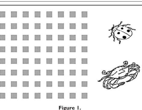
Participants were seated in a comfortable chair in a dimly lit room, 110 cm from the computer screen. Stimuli were presented with ‘‘Presentation’’ (version 14.2 Neurobe-havioral Systems). For the P1 paradigm, which was di-vided into a series of 3-min blocks to allow resting periods, participants were presented with isolated-check images containing an 8 8 matrix of checks (7.3wide by 7.3tall at 64% contrast, 100 per block), and line drawings of two kinds of animals (5.2 wide by 3.6 tall; 40 per block) on a white background [Yeap et al., 2006]. The 64% contrast condition was chosen to stimulate both the mag-nocellular and parvocellular systems. Each image appeared for 60 ms with a variable inter-stimulus-interval (ISI) between 740 and 1,540 ms (randomly in steps of 200 ms) during which there was a blank white screen. The purpose of the target animal stimuli was to encourage ticipants to attend to the screen. Each block required par-ticipants to press the key-pad when they identified a target animal they were shown at the start of each block. Participants were directed to only respond to the target animal and refrain from responding to the non-target ani-mal. Target and non-target animals were presented
ran-domly intermixed with the isolated-check stimuli, with both target and non-target animals appearing with equal probability. Each block contained a different animal pair (see Fig. 1) with each animal-pairing being somewhat simi-lar to ensure the task was sufficiently challenging and to promote alertness. On average participants completed 9.62 blocks (SD 0.86).
Electrophysiological Data Acquisition
Continuous electroencephalographic (EEG) data were recorded to computer with the Biosemi Acquisition pro-gramme: ActiView/www.biosemi.com/. EEG was recorded using 128 scalp electrodes. Horizontal and verti-cal electro-oculograms were also recorded by means of electrodes placed at the left and right external canthi and an electrode below the left eye. Data were recorded contin-uously at a digitization rate of 512 Hz with an open pass-band. The Biosemi amplification system replaces the ‘‘ground’’ electrodes with two separate electrodes: com-mon mode sense (CMS) active electrode and driven right leg (DRL) passive electrode (for more on the function of the CMS and DRL electrodes, see www.biosemi.com/faq/ cms&drl.htm). For analysis and display purposes, data were subsequently filtered with a 0-phase-shift 40-Hz low-pass filter (48 dB/octave) after acquisition. No high low-pass filter was used. Only sweeps related to the isolated-check stimuli were included in the analysis.
Spatial Working Memory Assessment
[image:3.612.316.552.68.252.2]All participants completed the SWM test from the Cam-bridge automated test battery (CANTAB Eclipse version, Cambridge Cognition, 2004) The touch screen computer task involves searching for ‘‘hidden’’ tokens in boxes whose number increases from trial to trial. Participants are
Figure 1.
instructed to remember which box they visit as a token will never be hidden in the same box twice. An error is committed when a participant returns to a box location from which a token has already been recovered. The de-pendent variable was the total numbers of errors made. Participants also completed subtests from the Wechsler adult intelligence test (WAIS, 3rd edition) and the Wechs-ler Memory Scale (WMS, 3rd edition) to ensure that all participants’ scored at or above the average range for IQ [see Donohoe et al., 2009].
Genetic Analysis
The SNP rs6490121 was genotyped using a TaqmanVR
SNP genotyping assay on a 7900HT sequence detection system (Applied Biosystems). The call rate for the Taqman genotyping was 100% and samples were in Hardy-Wein-berg equilibrium (P > 0.05). Along with these samples, a number of HapMap CEU DNA samples (www.hapma-p.org) were genotyped for rs6490121 for quality control purposes and were all found to be concordant with avail-able online HapMap data for this SNP. Only five partici-pants were identified as GG genotype carriers (9.25% of sample). For statistical analyses, therefore, participants were grouped as GG carriers and AG genotype carriers (n ¼ 29) versus AA genotype carriers (n ¼ 24). Mean scores and standard deviations are also reported for each geno-type group separately to provide evidence of allele dosage effects.
ERP Analyses
ERP analyses were performed using BESA Software Ver-sion 5.2. Any EEG channels which were noisy or which were not connected properly during recording were identi-fied and switched off for further analysis. Across partici-pants, the average number of channels excluded in this manner from analysis was 9.92 5.35. The surrogate model [Berg and Scherg, 1994] was then used for further artifact correction. Artifact correction in the current study was based on a model [Berg and Scherg, 1994; Lins et al., 1993] of artifact topography (the averaged artifact) and a set of brain topographies (multiple dipoles). The result was an estimation of artifact activation based on the linear combination of brain and artifact activities. Corrected-epoched data were also inspected for other artifacts using the BESA artifact rejection interface [Berg and Scherg, 1994]. Grand averages were generated for each participant from the isolated-check stimuli only. Approximately 654 241 sweeps per individual were averaged for the AA group and 669 262 for the GG þ AG group with an epoch of -200 to 1,000 msec. The average number of bad channels for the AA group was 8.41 and 11.15 for the GG
þ AG group. The P1 was defined as the area under the curve (versus the 0-lV baseline) generated by the 64% con-trast isolated-checkerboard stimuli within the
post-stimu-lus window of 70–110 msec spanning the P1 component. For the baseline correction, a baseline between200 and 0 msec was set. A set of six symmetrical pairs of scalp sites were chosen over occipital scalp sites from which P1 amplitudes were extracted (Left hemisphere: P1/P3/P03; Right hemisphere: P4/P6/P04). These sites were chosen based on topographical analysis of the grand-average group data which revealed lateral-occipital topographies consistent with those previously reported in the literature [e.g., Foxe and Simpson, 2002], for left and right hemi-spheres, respectively.
For statistical analyses, P1 measures were submitted to analysis-of-covariance (ANCOVA) using SPSS Software (SPSS, Chicago, IL Version 16.0) with theNOS1 genotypes (AA versus GG þ AG) as the between subject factor and P1 response (both averaged across all six target electrodes and for left lateral occipital and right lateral occipital regions taken separately) as the within-subjects factor, with age and gender entered as covariates of no interest in the analysis. To further investigate possible relationships between P1 and SWM in relation to NOS1, multiple regression analysis was performed, first to examine whether the P1 response predicted SWM performance, and second to examine whether any relationship observed between NOS1 and P1 performance was independent of variance in the P1 due to variance in SWM.
RESULTS
Demographic and Behavioral Differences
Associated With
NOS1
As this study was based on an opportunistic sample of consenting individuals who were still available following our original neuropsychological study, NOS1 genotype groups were not matched in advance for age, years in edu-cation or gender. No differences in age, eduedu-cation, or handedness were observed (age: AA¼ 24.8 [SD ¼ 12.45], AG/GG ¼ 29.1 [SD ¼ 12.45], t ¼ 0.69; P ¼ 0.49; years in education: AA ¼ 16.0 [SD ¼ 2.1], AG/GG ¼ 16.3 [SD ¼ 2.2]; t ¼ 0.46; P ¼ 0.64; handedness: AA: 21/22 right handed; AG/GG: 28/30 right handed;v2¼0.15;P¼0.69). Differences in gender were observed (AA: 10/24 male, AG/GG: 21/29 male;v2 ¼7.41;P ¼0.006). Consequently, gender was used as a covariate in all subsequent analysis of ERP components; age was also included as a covariate of no interest in the analyses.
AG group was 192.24 (12.95). The mean rate of incorrect responses to non-targets was 12.53 (6.66) for the AA group and 8.52 (4.24) for the GGþ AG group. Between group differences on each of these metrics of performance were all non-significant (all P-values >0.05). Collectively these data suggested that participants in both groups were equally engaged in the task, and given the high hit-rates, clearly focused their attention centrally toward the screen throughout each block presentation.
Differences in P1 VEP According to
NOS1
Genotype
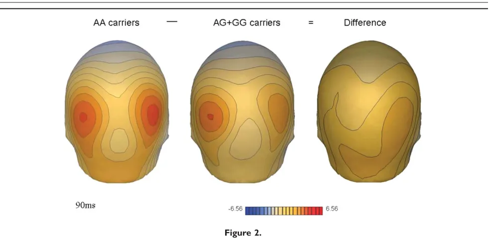
Figure 2 shows the bilateral occipital distribution of the P1 inNOS1risk ‘‘GGþ AG’’ and non-risk ‘‘AA’’ genotype groups. The map of the difference topography between these genotype groups (captured at maximal amplitude at 90ms) illustrates the reduction in P1 amplitude in the ‘‘GG
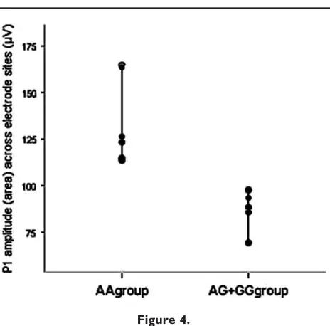
þAA’’ group relative to the ‘‘non-risk’’ AA group. Figure 3 illustrates the individual P1 morphology for electrode sites included in the statistical analysis. At each site the ‘‘risk’’ GG þ AG genotype group shows a reduced P1 response compared to the ‘‘non-risk’’ AA genotype group. Over the right lateral occipital region, where the P1 amplitude differ-ence was maximal, the mean P1 amplitude was 147.25 75.25 for the AA genotype group and 86.84 52.42 for the GG þ AG group. Figure 4 presents a scatterplot of P1 amplitudes measured at electrode sites included in the anal-yses (10/20 equivalents of: P1/P3/P03 and P4/P6/P04).
Reflecting these differences, a significant main effect of ge-notype group was observed, showing reduced P1 (measured as area under the curve) in the ‘‘risk’’ GGþ AG genotypes
group compared to the ‘‘non-risk’’ AA genotypes group (F(2,52) ¼ 13.85; P ¼ 0.001). Differences associated with
NOS1were found to be more robust over the right than the left hemiscalp [right: F(3,52) ¼ 16.73, P ¼ 0.00016]; [left:
F(3,52)¼3.14,P¼0.083]. As mentioned the low frequency of GG carriers (n ¼ 5) prevented a statistical analysis of GGvAGvAA groups separately. However, inspection of means and standard deviations across these groups sug-gested a gene dosage effect such that GG genotype individu-als showed a less robust P1 evoked response than the AG group, who in turn showed a less robust P1 evoked response than the AA group for both hemiscalps (see Table I).
Group differences were also calculated for the N1 (97– 185 msec) and P2 (160–300 msec). No significant differen-ces were observed for either right or left hemisphere elec-trodes for these ERPs. Latency measures were also examined. The mean latency for AA carriers was 85.59 12.32 and was 93.31 12.92 for AG þGG carriers. These differences were not found to be significant [F(1,50) ¼ 3.30,P¼0.07].
P1 VEP, NOS1
Genotype, and SWM Performance
Given previous evidence of association between P1 and SWM performance, and evidence of association between [image:5.612.62.553.68.308.2]NOS1 and SWM in our previous study, we investigated whether P1 performance predicted SWM performance in the present study using regression analyses. For this analy-sis SWM task performance was entered as the dependent variable. Age and gender were entered on the first step of the equation as covariates of no interest and P1 perform-ance (electrode sites for left and right hemiscalps averaged
Figure 2.
together) was entered on the second step as the independ-ent variable of interest. After the effects of age and gender were accounted for, P1 response explained a further 12.9% of variance in SWM performance (F change (1,40) ¼ 7.74,
P¼0.009).
NOS1
Effects on Sensory and Cognitive
Processing: Top Down Versus Bottom Up
Influences
We next determined, using a multiple regression analy-sis, whetherNOS1’s observed influence on the P1 response might be accounted for by the previously observed influ-ence of NOS1 on SWM performance. To do this the P1 response was entered as the dependent variable, SWM performance as the independent variable in the first step of the analysis, followed byNOS1as the independent
vari-able in the second step. P1 response was again measured in terms of the area under the curve, based on the elec-trode site in which differences betweenNOS1risk carriers and non-carriers were maximal (i.e., right occipital electro-des P4/P6/P04). We reasoned that if the effects of NOS1
on the P1 response were being mediated by SWM,NOS1’s effects on the P1 response would become non-significant once the variance attributable to SWM was accounted for. Instead, we found that even after accounting for the effects of SWM performance on the P1 response (which accounted for 26% of the variance in P1 response), NOS1
independently explained a further 9% of variation in P1 response. (R2 ¼
0.35; F(1,40)¼ 5.04, P ¼ 0.03). This sug-gested that at least some of the effects ofNOS1on P1 per-formance are independent of NOS1’s previously reported influence on SWM. We also intended to examine whether
[image:6.612.66.553.66.456.2]NOS1’s influence on SWM performance was mediated by P1 response. Unfortunately, we were prevented from
Figure 3.
doing so due to insufficient power to detect association between NOS1 and SWM performance in the restricted EEG sample (n¼54 versus the overall neuropsychological sample of n ¼ 160) and so this analysis could not be undertaken.
DISCUSSION
We have previously reported evidence that the risk ‘‘G’’ allele at the SZ GWAS identified NOS1 variant rs6490121 was associated with poorer performance in SWM and verbal IQ in independent samples of both SZ patients and healthy controls. Following up these findings, the present study investigated whether the same NOS1 variant was also associated with poorer performance in sensory level processing as measured by the P1 visual evoked potential in a sample of healthy participants. Consistent with our hypothesis, we observed that the associated risk allele at rs6490121 was associated with a significantly reduced P1 response bilaterally. No differences in N1 or P2 response associated withNOS1were observed.
As an endophenotypic measure, the P1 has the major advantage of being relatively easy to measure quickly and accurately. The large differences between healthy controls and both patients and their first-degree relatives suggest this component is heritable [Donohoe et al., 2007, 2008; Haenschel et al., 2009; Yeap et al., 2006; Walters and Owen]. As a largely automatic response, it is not as sus-ceptible to the same motivational factors or fluctuations in clinical state as later cognitive components such as the P300. However, this is not to say that the P1 is not cogni-tively penetrable. Although early stages of perceptual processing (from as early as 50100 msec post-stimulus)
serve an important role in ‘‘spotlighting’’ of relevant infor-mation for later processing, these early processing stages (from 70 msec onward) appear to be reciprocally modu-lated by higher processing areas [Martinez et al., 1999].
It is interesting to speculate about the twin effects of
NOS1on (in our larger sample) SWM, and (in the present study), the P1 response. These associations may reflect the reciprocal relationship between early sensory and higher cognitive function, particularly for visual information. On one hand, deficits in ‘‘capturing’’ visual information are likely to increase difficulties in efficiently maintaining and updating that information ‘‘online’’ during SWM tasks. Conversely, an inability to maintain context during later stages of processing leads to difficulties focusing on rele-vant information during earlier stages of visual processing. A relationship between the P1 response and SWM per-formance has been empirically demonstrated previously [Haenschel et al., 2007] and, in the present study, we were able to replicate this evidence: the P1 response signifi-cantly predicted SWM task performance in our participants.
For the first time (to our knowledge) we were able to partly test whether the genetic effects on either of these stages of processing (early visual sensory versus SWM) were being mediated by the other. Although insufficient power prevented us from determining whether NOS1’s effects on SWM were mediated by P1 performance, we were able to reject the hypothesis thatNOS1’s effect on the P1 was being mediated in a ‘‘top-down’’ fashion by
NOS1’s influence on SWM performance. In a multiple regression analysis, while SWM significantly predicted variance in the P1 response,NOS1 continued to explain a significant amount of variance in the P1 response even af-ter the variance associated with SWM was accounted for. We interpret these data as suggesting that NOS1 has a direct influence on visual sensory processing as measured by the P1 response, either because of pleiotropic effects of this gene on multiple aspects of brain function, or because of a primary influence on sensory processing that mediate the effects already seen in higher cognitive processes. This evidence supports the increasingly popular theory that some deficits in cognitive processing may result at least in part from sensory level processing deficits [Javitt, 2009], but require testing in a larger sample to confirm the effect of the P1 response as mediating the influence ofNOS1 on
Figure 4.
[image:7.612.65.297.71.300.2]Scatterplot of P1 amplitudes (area under the curve) across elec-trodes used in statistical analysis.
TABLE I. Differences in P1 response according to genotype group (measured as the area under the curve)
for left hemisphere electrode sites, right hemisphere electrode sites, and averaged across electrode sites
GG (n¼5) AG (n¼25) AA (n¼22)
P1 Left hemisphere 65.77 (55.46) 97.70 (60.98) 118.64 (59.56) P1 Right hemisphere 78.53 (51.24) 88.47 (52.45) 150.04 (76.02) P1 Both hemispheres
combined
cognition. A limitation of our findings concerned the observed gender and sex differences between genotype groups. Although these differences was co-varied for in the analysis and did not appear to influence the signifi-cance of our results, replication of these findings in more gender and sex matched genotype groups will enable a better assessment of the contribution of these variables. In the current study, the GG groups were grouped together as the frequency of the GG genotype group was too low. Future replication studies could also include a sample where AG and GG groups are better individually represented.
NOS1
: Molecular Mechanism and Functional
Implications
The implicated SNP (rs6490121) has no obvious func-tional effect and may reflect a proxy association with 1 or more other causal genetic variants in SZ. Based on Hap-Map CEU data, rs6490121 is not in high linkage disequili-brium (LD;r2>
0.80) with any other common SNP at this locus. NOS1 is characterized by complex transcriptional regulation. We previously investigated whether the cogni-tive effects of thisNOS1variant could be explained by the dinucleotide variable-number tandem repeat located in the core promoter region of Exon 1f, the short arm of which is associated with electrophysiological measures of atten-tional control [Reif et al., 2009] and which is in partial LD with this SNP [D’¼ 0.70,r2¼0.26; Donohoe et al., 2009].
However, we failed to find evidence that this variant explained variation in cognition in our samples. Similarly, to explore potential mechanisms by which NO could exert an effect on cognitive processes, we previously screened experimentally validated protein–protein interactions of
NOS1 using the protein-protein interaction databases and identified 19 confirmed human binary interactions, includ-ing SZ relevant susceptibility genes involved in presynap-tic synaptogenesis (NOS1AP and syntrophin [SNTA1] [OMIM 601017]) and postsynaptically through the Postsy-naptic Density 95 (PSD95). Elements of PSD95 signaling cascades have been targeted in SZ genetic association stud-ies including erbB4/neuregulin signaling and the N -methyl-D-aspartate receptor complex, which is involved in long-term potentiation, memory, and learning. Of these, we have investigated the neuropsychological effects of the
NOS1AP SNP implicated in SZ risk (rs12742393) and found no evidence of association with variation in cogni-tion (data available on request). We have as yet to explore the influence of this or other interacting genes on the vis-ual evoked potentials reported here.
Since its original identification as a common genetic var-iant associated with SZ risk by [O’Donovan et al., 2008] none of the subsequent genome wide association studies of SZ have identified NOS1 rs6490121 as achieving ge-nome wide level significance [Stefansson et al., 2008; The International Schizophrenia Consortium, 2009; Walsh
et al., 2008]. We have previously suggested that NOS1
may be a modifier gene that influences cognitive ability without having a direct influence on disease risk. The present data suggest an even broader role forNOS1 in in-formation processing, impacting early sensory as well as later cognitive function. This broad influence on informa-tion processing is consistent with the known biology of
NOS1, including negative feedback on N-methyl-D -aspar-tate (NMDA) receptor function and inhibition of synaptic reuptake of dopamine. This position at the crossroads of two mutually regulating messenger systems, and its ubiq-uitous expression throughout the brain, together make a discrete influence on only one level of information proc-essing unlikely. A wider role for genetic variants influenc-ing NMDA at the levels of both SWM and P1 response has already been reported in the case of Dysbindin-1 [DTNBP1; Donohoe et al., 2007, 2008].
CONCLUSION
As originally conceived, the use of cognitive and EEG measures as ‘‘intermediate’’ or ‘‘endo’’-phenotypes was proposed as a strategy for reducing the genetic complexity of broader clinical phenotypes that would allow greater power for identifying genes of small effects [Gottesman and Gould, 2003]. Since then, several EEG studies have focused on confirming the effects of variants already asso-ciated with increased disease risk on individual brain sys-tems for the purposes of characterizing the effects on these variants on individual aspects of brain function. Such an approach may be helpful in elucidating gene-disease path-ways [Walters and Owen] and, eventually, therapeutic tar-gets. However, there is currently little evidence that the genetic architecture of cognition is much less complex than that of disease phenotypes. Thus, cognitive neuro-science studies of psychiatric disease associated variants, in which information processing is disrupted, is likely to have an equally valuable role in elucidating the molecular biology of information processing in the general popula-tion. Evidence of NOS1’s role in early visual processing presented here is therefore likely to be relevant not just to schizophrenia pathophysiology, but to understanding the molecular basis of visual processing more generally.
ACKNOWLEDGMENTS
The authors sincerely thank all patients who contributed to this study and all staff who facilitated their involvement.
REFERENCES
Akbarian S, Sucher NJ, Bradley D, Tafazzoli A, Trinh D, Hetrick WP, Potkin SG, Sandman CA, Bunney WE Jnr, Jones EG (1996): Selective alterations in gene expression for NMDA re-ceptor subunits in prefrontal cortex of schizophrenics. J Neuro-sci 16:19–30.
Akyol O, Zoroglu SS, Armutcu F, Sahin S, Gurel A (2004): Nitric oxide as a physiopathological factor in neuropsychiatric disor-ders. In Vivo 18:377–390.
Baba HCA, Suzuki T, Arai H, Emson PC (2004): Expression of nNOS and soluble guanylate cyclase in schizophrenic brain. Neuroreport 15:677–680.
Bailer U, Leisch F, Meszaros K, Lenzinger E, Willinger U, Strobl R, Gerhardt C, Gerhard E, Fuchs K, Sieghart W, Kasper S, Hornik K, Aschauer HN (2000): Genome scan for susceptibility loci for schizophrenia. Neuropsychobiology 42:175–182. Bailer U, Leisch F, Meszaros K, Lenzinger E, Willinger U, Strobl
R, Heiden A, Gebhardt C, Doge E, Fuchs K, Sieghart W, Kaspe S, Hornik K, Aschauer HN (2002): Genome scan for suscepti-bility loci for schizo-phrenia and bipolar disorder. Biol Psychi-atry 52:40–52.
Berg P, Scherg M (1994): A multiple source approach to the cor-rection of eye artifacts. Electroencephalogr Clin Neurophysiol 90:229–241.
Boehning D, Snyder SH (2003): Novel neural modulators. Annu Rev Neurosci 26:105–131.
Brenman JE, Bredt DS (1997): Synaptic signaling by nitric oxide. Curr Opin Neurobiol 7:374–378.
Butler PD, Martinez A, Foxe JJ, Kim D, Zemon V, Silipo G, Maho-ney J, Shapaner M, Jalbrzikowski M, Javitt DC (2007): Subcorti-cal visual dysfunction in schizophrenia drives secondary cortical impairments. Brain 130:417.
Cui H, Nishiguchi N, Yanagi M, Fukutake M, Mouri K, Kitamura N, Hashimoto T, Shirakawa O, Hishimoto A (2010): A putative cis-acting polymorphism in the NOS1 gene is associated with schizophrenia and NOS1 immunoreactivity in the postmortem brain. Schizophr Res 121:172–178.
Das I, Khan NS, Puri BK, Sooranna SR, Debelleroche J, Hirsch SR (1995): Elevated platelet calcium mobilization and nitric oxide synthase activity may reflect abnormalities in schizophrenic brain. Biochem Biophys Res Commun 212:375–380.
DeLisi LE, Shaw SH, Crow TJ, Shields G, Smith AB, Larach VW, Wellman N, Loftus J, Nanthakumar B, Razi K, Stewart J, Comazzi M, Vita A, Heffner T, Sherrington R (2002): A ge-nome-wide scan for linkage to chromosomal regions in 382 sibling pairs with schizophrenia or schizoaffective disorder. Am J Psychiatry 159:803–812.
Donohoe G, Morris DW, Clarke S, McGhee KA, Schwaiger S, Nan-gle JM, Garavan H, Robertson IH, Gill M, Corvin A (2007): Variance in neurocognitive performance is associated with dysbindin-1 in schizophrenia: A preliminary study. Neuropsy-chologia 45:454–458.
Donohoe G, Morris DW, De Sanctis P, Magno E, Montesi JL, Garavan HP, Robertson IH, Javitt DC, Gill M, Corvin AP, Foxe JJ (2008): Early visual processing deficits in dysbindin-associ-ated schizophrenia. Biol Psychiatry 63:484–489.
Donohoe G, Walters J, Morris DW, Quinn EM, Judge R, Norton N, Giegling AM, Hartmann HJ, Moller P, Muglia H, Williams V, Moskvina R, Peel R, O’ Donoghue T, Owen MJ, O’ Donovan MC, Gill M, Rujescu D, Corvin AP (2009): Influence of NOS1 on verbal intelligence and working memory in both patients with schizophrenia and healthy control subjects. Arch Gen Psychiatry 66:1045–1054.
Fallin MD, Lasseter VK, Avramopoulos D, Nicodemus KK, Woly-niec PS, McGrath JA, Nestadt G, Valle D, Liang KY, Pulver AE (2005): Bipolar I disorder and schizophrenia: A 440-single-nu-cleotide polymorphism screen of 64 candidate genes among Ashkenazi Jewish case-parent trios. Am J Hum Genet 77:918– 936.
Foxe JJ, Doniger GM, Javitt DC (2001): Early visual processing def-icits in schizophrenia: Impaired P1 generation revealed by high-density electrical mapping. Neuroreport 12:3815–3820. Foxe JJ, Simpson GV (2002): Flow of activation from V1 to frontal
cortex in humans. Experimental Brain Res 142:139–150. Gottesman II, Gould TD (2003): The endophenotype concept in
psychiatry: Etymology and strategic intentions. Am J Psychia-try 160:636–645.
Haenschel C, Bittner RA, Haertling F, Rotarska-Jagiela A, Maurer K, Singer W, Linden DEJ (2007): Contribution of impaired early-stage visual processing to working memory dysfunction in adoles-cents with schizophrenia. Arch Gen Psychiatry 64:1229–40.
Haenschel C, Bittner RA, Waltz J, Haertling F, Wibral M, Singer W, Linden DEJ, Rodriguez E (2009): Cortical oscillatory activity is critical for working memory as revealed by deficits in early-onset schizophre-nia. J Neurosci 29:9481–9489.
Javitt DC (2009): When doors of perception close: Bottom-up mod-els of disrupted cognition in schizophrenia. Annu Rev Clin Psychol 5:249–275.
Johansson C, Jackson DM, Svensson L (1997): Nitric oxide syn-thase inhibition blocks phencyclidine-induced behavioral effects on prepulse inhibition and locomotor activity in the rat. Psychopharmacology 131:167–173.
Johansson C, Magnusson O, Deveney AM, Jackson DM, Zhang J, Engel JA, Svensson L (1998): The nitric oxide synthase inhibi-tor. L-NAME, blocks certain phencyclidine-induced but not amphetamine-induced effects on behavior and brain biochem-istry in the rat. Prog Neuro-Psychopharmacol Biol Psychiatry 22:1341–1360.
Kirchner L, Weitzdoerfer R, Hoeger H, Url A, Schmidt P, Engel-mann M, Villa SR, Fountoulakis M, Lubec G, Lubec B (2004): Impaired cognitive performance in neuronal nitric oxide syn-thase knockout mice is associated with hippocampal protein derangements. Nitric Oxide 11:316–330.
Klamer D, Engel JA, Svensson L (2001): The nitric oxide synthase inhibitor, L-NAME, blocks phencyclidine-induced disruption of prepulse inhibition in mice. Psychopharmacologia 156:182– 186.
Klamer D, Engel JA, Svensson L (2004a): The neuronal selective nitric oxide synthase inhibitor, Nomega-propyl-L-arginine, blocks the effects of phencyclidine on prepulse inhibition and locomotor activity in mice. Eur J Pharmacol 503:103–107. Klamer D, Pa˚lsson E, Revesz A, Engel JA, Svensson L (2004b):
Habituation of acoustic startle is disrupted by psychotomi-metic drugs: Differential dependence on dopaminergic and ni-tric oxide modulatory mechanisms. Psychopharmacology 176:440–450.
Klamer D, Engel JA, Svensson L (2005): Effects of phencyclidine on acoustic startle and prepulse inhibition in neuronal nitric oxide synthase deficient mice. Eur Neuropsychopharmacol 15:587–590.
Liou Y-J, Tsai S-J, Hong C-J, Liao D-L (2003): Association analysis for the CA repeat polymorphism of the neuronal nitric oxide synthase (NOS1) gene and schizophrenia. Schizophrenia Res 65:57–59.
Martinez A, Anllo-Vento L, Sereno MI, Frank LR, Buxton RB, Dubowitz DJ, Wong EC, Hinrichs H, Heinze HJ, Hillyard SA (1999): Involvement of striate and extrastriate visual cortical areas in spatial attention. Nat Neuroscience 2:364–369. O’Donovan MC, Craddock N, Norton N, Williams H, Peirce T,
Moskvina V, Nikolov I, Hamshere M, Carroll L, Georgieva L, Divyer S, Holmans P, Marchini JL, Spencer CCA, Howie B, Leung H-T, Hartmann AM, Moller DW, Shi Y, Feng G, Hoff-mann P, Propping P, Vasilescu C, Maier W, Rietschel M, Zam-mit S, Schumacher J, Quinn EM, Schulze TG, Williams NM, Giegling I, Iwata N, Ikeda M, Darvasi A, Shifman S, He L, Duan J, Sanders AR, Levinson DF, Gejman PV, Molecular Genetics of Schizophrenia Collaboration, Cichon S, Nothen MM, Gill M, Corvin A, Rujescu D, Kirov G, Owen MJ (2008): Identification of loci associated with schizophrenia by genome-wide association and follow-up. Nat Genet 40:1053–1055. Okumura T, Okochi T, Kishi T, Ikeda M, Kitajima T, Yamanouchi
Y, Kinoshita Y, Kawashima K, Tsunoka T, Ujike H, Inada T, Ozaki N, Iwata N (2009): No association between polymor-phisms of neuronal oxide synthase 1 gene (NOS1) and schizo-phrenia in a Japanese population. Neuromol Med 11:123–127. Pa˚lsson E, Fejgin K, Wass C, Engel J, Svensson L, Klamer D
(2007): The amino acid L-lysine blocks the disruptive effect of phencyclidine on prepulse inhibition in mice. Psychopharma-cology 192:9–15.
Pepicelli O, Raiteri M, Fedele E (2004): The NOS/sGC pathway in the rat central nervous system: A microdialysis overview. Neu-rochem Int 45:787–797.
Reif A, Herterich S, Strobel A, Ehlis AC, Saur D, Jacob CP, Wienker T, To¨pner T, Fritzen S, Walter U, Schmitt A, Fallgatter AJ, Lesch KP (2006): A neuronal nitric oxide synthase (NOS-1) haplotype associated with schizophrenia modifies prefrontal cortex function. Molecular Psychiatry 11:286–300.
Reif A, Jacob CP, Rujescu D, Herterich S, Lang S, Gutknecht L, Bahne C, Strobel A, Freitag CM, Giegling I, Romanus M, Hart-mann A, Rosler M, Renner TJ, Fallgatter AJ, Retz W, Ehlis A-C, Lesch K-P (2009): Influence of functional variant of neuro-nal nitric oxide synthase on impulsive behaviors in humans. Archives General Psychiatry 66:41–50.
Riley B, Thiselton D, Maher BS, Bigdeli T, Wormley B, McMichael GO, Fanous AH, Vladimirov V, O’ Neill FA, Walsh D, Kendler KS (2009): Replication of association between schizophrenia and ZNF804A in the Irish case-control study of schizophrenia sample. Molecular Psychiatry 15:29–37.
Shinkai T, Ohmori O, Hori H, Nakumura J (2002): Allelic associa-tion of the neuronal nitric oxide synthase (NOS1) gene with schizophrenia. Mol Psychiatry 7:560–563.
Stefansson H, Rujescu D, Cichon S, Pietilainen OPH, Ingason A, Steinberg S, Fossdal R, Sigurdsson E, Sigmundsson T,
Buizer-Voskamp JE, Hansen T, Jakobsen KD, Muglia P, Francks C, Mathews PM, Gylfason A, Halldorsson BV, Gudbjartsson D, Thorgeirsson TE, Sigurdsson A, Jonasdottir A, Bjornsson A, Mattiasdottir S, Blondal T, Haradlsson M, Magnusdottir BB, Giegling I, Moller HJ, Hartmann A, Shianna KV, Ge D, Need AC, Crombie C, Fraser G, Walker N, Lonnqvist J, Suvisaari J, Tuulio-Henriksson A, Paumo T, Toulopoulou T, Bramon E, DiForti M, Murray R, Ruggeri M, Vassos E, Tosato S, Walshe M, Li T, Vasilescu C, Muhleisen TW, Wang AG, Ullum H, Djurovic S, Melle I, Olesen J, Kiemeney LA, Franke B, GROUP, Sabatti C, Freimer NB, Gulcher JR, Thorsteinsdottir U, Kong A, Andreassen OA, Ophoff RA, Georgia A, Rietschel M, Werge T, Petursson H, Goldstein DB, Nothen MM, Peltonen L, Collier DA, St. Clair D, Stefansson K (2008): Large recurrent microde-letions associated with schizophrenia. Nature 455:232–236. Taneli F, Pırıldar S, Akdeniz F, Uyanık BS, Arı Z (2004): Serum
nitric oxide metabolite levels and the effect of antipsychotic therapy in schizophrenia. Arch Med Research 35:401–405. Tang HK, Tang R, Zhou G, Fang C, Zhang J, Du L, Feng G, He L,
Shi Y (2008): Evidence for association between the 5’ flank of the NOS1 gene and schizophrenia in the Chinese population. Int J Neuropsychopharmacology 11:1063–1071.
The International Schizophrenia Consortium (2009): Common polygenic variation contributes to risk of schizophrenia and bipolar disorder. Nature 460:748–752.
Walsh T, McClellan JM, McCarthy SE, Addington AM, Pierce SB, Cooper GM, Nord AS, Kusenda M, Malhorta D, Bhandari A, Stray SM, Rippey CF, Roccanova P, Makarov V, Lakshmi B, Findling RL, Sikich L, Stromberg T, Merriman B, Gogtay N, Butler P, Eckstrand K, Noory L, Gochman P, Long T, Chen Z, Davis S, Baker C, Eichler EE, Meltzer PS, Nelson SF, Singleton AB, Lee MK, Rapoport JL, King M-C, Sebat J (2008): Rare structural variants disrupt multiple genes in neurodevelop-mental pathways in schizophrenia. Science 320:539–543. Walters JTR, Owen MJ (2007): Endophenotypes in psychiatric
genetics. Mol Psychiatry 12:886–890.
Wass C, Archer T, Pa˚lsson E, Fejgin K, Alexandersson A˚ , Klamer D, Engel D, Engel JA, Svensson L (2006): Phencyclidine affects memory in a nitric oxide dependent manner: Working and ref-erence memory. Beh Brain Research 174:49–55.
Weitzdoerfer R, Hoeger H, Engidawork E, Engelmann M, Singe-wald N, Lubec G, Lubec B (2004): Neuronal nitric oxide syn-thase knock-out mice show impaired cognitive performance. Nitric Oxide 10:130–140.
Yeap S, Kelly SP, Sehatpour P, Magno E, Javitt DC, Garavan H, Thakore JH, Foxe JJ (2006): Early visual sensory deficits as endophenotypes for schizophrenia: High density electrical mapping in clinically unaffected first-degree relatives. Arch Gen Psychiatry 63:1180–1188.