Acta Cryst.(2001). E57, o1113±o1115 DOI: 10.1107/S1600536801017834 Erika Kaiser-Morriset al. C6H8N2
o1113
organic papers
Acta Crystallographica Section E Structure Reports Online
ISSN 1600-5368
2,6-Dimethylpyrazine at 20 K: a neutron-diffraction
study
Erika Kaiser-Morris,a* Alain Cousson,aWerner Paulusband Francois Fillauxc
aLaboratoire LeÂon Brillouin, CEA Saclay, 91191
Gif-sur-Yvette CEDEX, France,bUniversite de
Rennes 1, LCSIM/UMR 6511, Campus de Beaulieu, Avenue du GeÂneÂral Leclerc, 35042 Rennes CEDEX, France, andcLADIR, 2 rue
Henry Dunant, 94320 Thiais, France
Key indicators
Single-crystal neutron study
T= 20 K
Mean(C±C) = 0.002 AÊ
Rfactor = 0.031
wRfactor = 0.019 Data-to-parameter ratio = 9.2
For details of how these key indicators were automatically derived from the article, see http://journals.iucr.org/e.
#2001 International Union of Crystallography Printed in Great Britain ± all rights reserved
Single crystal neutron diffraction techniques are used to determine the crystal structure of 2,6-dimethylpyrazine (DMP), C6H8N2, at 20 K. The space group isP21/awithZ=
4, as at room temperature. The methyl groups are ordered. There are two crystallographically inequivalent methyl groups in the unit cell.
Comment
Light particles experiencing potential functions with topolo-gical degeneracy manifest their quantum natureviatunnelling. The magnitude of the tunnel splitting depends on the particle mass and potential shape (distances between identical sites and barrier height). For methyl groups the threefold symmetry ensures strict topological degeneracy and rotational tunnelling has been observed with inelastic neutron-scattering techni-ques (INS) in many crystals (Press, 1981; Prager & Heide-mann, 1995). The INS spectrum of DMP at 2 K exhibits tunnelling lines at 20 and 29meV (NicolaõÈ, Kaiseret al., 1998). The presence of more than one tunnelling transition in the same system is rather rare. It reveals either inequivalent methyl groups or dynamical coupling. A precise knowledge of the crystalline structure at the same temperature of the tunnelling measurements is necessary to interpret the INS spectra (Johnsonet al., 1996; Johnsonet al., 1997; Neumann & Johnson, 1997; NicolaõÈ, Kearleyet al., 1998). We present in this paper the structure determination at 20 K. The structure at the temperature of the tunnelling experiment, 5 K, is presented in the following article (Kaiser-Morriset al., 2001).
The space groupP21/a(monoclinic) with four formula units
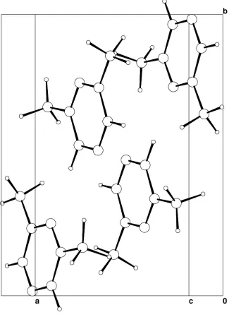
per unit cell was obtained from a preliminary study at 253 K with X-rays (Morriset al., 1998) and a recent X-ray diffraction study at 180 K with a better resolution (Thalladiet al., 2000). A preliminary neutron-diffraction experiment at 260 K, close to the melting point of the title compound, and neutron-diffraction experiments on a single crystal at 20 and 5 K con®rmed the space groupP21/a(Fig. 1). There is no evidence
for any phase transition between 260 and 20 K, but there are signi®cant changes of the lattice parameters below 260 K: at
organic papers
o1114
Erika Kaiser-Morriset al. C6H8N2 Acta Cryst.(2001). E57, o1113±o1115 260 K, the cell parameters are:a= 7.467 (8),b= 10.859 (7),c=7.558 (7) AÊ,= 90.8 (4).
Experimental
2,6±Dimethylpyrazine (DMP) is hygroscopic and melts at 311 K. We performed neutron-diffraction experiments with a single crystal at 260, 20 and 5 K on the four-circle neutron diffractometer 5-C2 at the LLB (Saclay, France). A large single crystal (1 1 5 cm) was obtained at low temperature. A small single crystal (555 mm) was cut, glued on a goniometer head and oriented on 5-C2. The measurements were performed with the!scan mode and an incident wavelength close to 0.83 AÊ selected with the Cu (220) mono-chromator.
Crystal data
C6H8N2
Mr= 108.14 Monoclinic,P21=a
a= 7.288 (5) AÊ
b= 10.73 (1) AÊ
c= 7.444 (7) AÊ = 90.10 (8)
V= 582.4 AÊ3
Z= 4
Dx= 1.23 Mg mÿ3
Neutron radiation = 0.8308 AÊ
Cell parameters from 16 re¯ections
= 9.8±21.5
= 0.08 mmÿ1
T= 20 K Prism, white 5.05.05.0 mm
Data collection
OrpheÂe reactor (Saclay, France): 5-C2 four-circle
!scans
Absorption correction: none 2230 measured re¯ections 1918 independent re¯ections 1337 re¯ections withI> 3(I)
Rint= 0.074
max= 37.5
h=ÿ10!2
k= 0!15
l=ÿ10!10 2 standard re¯ections
frequency: 450 min intensity decay: none
Re®nement
Re®nement onF R= 0.031
wR= 0.019
S= 1.03 1337 re¯ections 146 parameters
All H-atom parameters re®ned Weighting scheme: Chebychev
polynomial with 5 parameters: 1.04,ÿ3.06,ÿ0.108,ÿ0.644,
ÿ0.800 (Carruthers & Watkin, 1979)
(/)max= 0.007 max= 0.83 e AÊÿ3 min=ÿ0.89 e AÊÿ3 Extinction correction: Larson
(1970)
Extinction coef®cient: 1.52 (13) Atomic scattering factors from
Sears (1992)
Table 1
Selected geometric parameters (AÊ,).
N1ÐC1 1.3354 (9)
N1ÐC4 1.341 (1)
N2ÐC2 1.3399 (9)
N2ÐC3 1.340 (1)
C1ÐC2 1.4040 (11)
C2ÐC5 1.5005 (11)
C3ÐC4 1.3960 (11)
C3ÐC6 1.5019 (11)
C1ÐN1ÐC4 116.19 (6)
C2ÐN2ÐC3 117.47 (6)
N1ÐC1ÐC2 122.06 (7)
N2ÐC2ÐC1 120.99 (7)
N2ÐC2ÐC5 118.56 (7)
C1ÐC2ÐC5 120.45 (7)
N2ÐC3ÐC4 120.78 (7)
N2ÐC3ÐC6 117.72 (7)
C4ÐC3ÐC6 121.50 (7)
N1ÐC4ÐC3 122.51 (7)
Data collection:DIF4N(modi®ed Linux version ofDIF4; Stoe & Cie, 2000); cell re®nement: DIF4N; data reduction: PRON (modi®ed version of REDU4; Stoe & Cie, 2000); program(s) used to re®ne structure: CRYSTALS (Watkin et al., 1996); molecular graphics:
CAMERON(Watkinet al., 1996); software used to prepare material for publication:CRYSTALS.
We thank J. Godard from the Parc d'Orsay, France for providing the single crystals.
References
Carruthers, J. R. & Watkin, D. J. (1979).Acta Cryst.A35, 698±699.
Johnson, M. R., Frick, B. & Rommsdorff, H. P. (1996).Chem. Phys. Lett.258, 187±193.
Johnson, M. R., Neumann, M., NicolaõÈ, B., Smith, P. & Kearley, G. J. (1997).J. Chem. Phys.215, 343±353.
Kaiser-Morris, E., NicolaõÈ, B., Cousson, A., Paulus, W. & Fillaux, F. (2001).
Acta Cryst.E57, o1115±o1116.
Larson A. (1970).Crystallographic Computing, edited by F. R. Ahmed, pp. 291±294. Copenhagen: Munksgaard.
Morris, E., Cousson, A. & Paulus, W. (1998).Z. Kristallogr. New Cryst. Struct. 213, 79.
Neumann, M. & Johnson, M. R. (1997).J. Chem. Phys.107, 1725±1731. NicolaõÈ, B., Kaiser, E., Fillaux, F., Kearley, G. J., Cousson, A. & Paulus, W.
(1998).Chem. Phys.26, 1±13.
Figure 1
NicolaõÈ, B., Kearley, G. K., Johnson, M. R., Fillaux, F. & Suard, E. (1998).J. Chem. Phys.109, 9062±9074.
Prager, M. & Heidemann, A. (1995).Chem. Rev.97, 2933±2966. Press, W. (1981).Springer Tracts in Modern Phys.92, 1. Sears, V. F. (1992).Neutron News,3, 26±37.
Stoe & Cie (2000). DIF4 and PRON Software. Stoe & Cie, Darmstadt, Germany.
Thalladi, V. R., Gherke, A. & Boese, R. (2000).New J. Chem.24, 463±470. Watkin, D. J., Prout, C. K. & Pearce, L. J. (1996).CAMERON. Chemical
Crystallography Laboratory, Oxford, England.
Watkin, D. J., Prout, C. K., Carruthers, J. R. & Betteridge, P. W. (1996).
CRYSTALS Issue 10. Chemical Crystallography Laboratory, Oxford, England.
supporting information
sup-1
Acta Cryst. (2001). E57, o1113–o1115
supporting information
Acta Cryst. (2001). E57, o1113–o1115 [doi:10.1107/S1600536801017834]
2,6-Dimethylpyrazine at 20
K: a neutron-diffraction study
Erika Kaiser-Morris, Beatrice Nicola
ï
, Alain Cousson, Werner Paulus and Francois Fillaux
S1. Comment
Light particles experiencing potential functions with topological degeneracy manifest their quantum nature via tunnelling. The magnitude of the tunnel splitting depends on the particle mass and potential shape (distances between identical sites
and barrier height). For methyl groups the threefold symmetry ensures strict topological degeneracy and rotational
tunnelling has been observed with inelastic neutron-scattering techniques (INS) in many crystals (Press, 1981; Prager &
Heidemann, 1995). The INS spectrum of DMP at 2 K exhibits tunnelling lines at 20 and 29 µeV (Nicolaï, Kaiser et al., 1998). The presence of more than one tunnelling transition in the same system is rather rare. It reveals either inequivalent
methyl groups or dynamical coupling. A precise knowledge of the crystalline structure at the same temperature of the
tunnelling measurements is necessary to interpret the INS spectra (Johnson et al., 1996; Johnson et al., 1997; Neumann & Johnson, 1997; Nicolaï, Kearley et al., 1998). We present in this paper the structure determination at 20 K. The structure at the temperature of the tunnelling experiment, 5 K, is presented in the following article (Kaiser-Morris et al., 2001).
The space group P21/a (monoclinic) with four formula units per unit cell was obtained from a preliminary study at 253
K with X-rays (Morris et al., 1998) and a recent X-ray diffraction study at 180 K with a better resolution (Thalladi et al., 2000). A preliminary neutron-diffraction experiment at 260 K, close to the melting point of the title compound, and
neutron diffraction experiments on a single-crystal at 20 and 5 K confirmed the space group P21/a (Fig. 1). There is no
evidence for any phase transition between 260 and 20 K, but there are significant changes of the lattice parameters below
260 K: at 260 K, the cell parameters are: a = 7.467 (8), b = 10.859 (7), c = 7.558 (7) Å, β = 90.8 (4)°.
S2. Experimental
2,6-Dimethylpyrazine (DMP) is hygroscopic and melts at 311 K. We performed neutron diffraction experiments with a
single-crystal at 260, 20 and 5 K on the four-circle neutron diffractometer 5 C2 at the LLB (Saclay, France). A large
single-crystal (1 × 1 × 5 cm) was obtained at low temperature. A small single-crystal (5 × 5 × 5 mm) was cut, glued on a
supporting information
sup-2
[image:5.610.144.469.74.523.2]Acta Cryst. (2001). E57, o1113–o1115
Figure 1
The crystal structure of (I) at 20 K. For a single molecule representation, see the following paper (Kaiser-Morris et al., 2001).
2,6-dimethylpyrazine
Crystal data C6H8N2
Mr = 108.14
Monoclinic, P21/a
a = 7.288 (5) Å b = 10.73 (1) Å c = 7.444 (7) Å β = 90.10 (8)° V = 582.4 Å3
Z = 4
F(000) = 114.73 Dx = 1.23 Mg m−3
Melting point: not measured K Neutron radiation, λ = 0.8308 Å Cell parameters from 16 reflections θ = 9.8–21.5°
supporting information
sup-3
Acta Cryst. (2001). E57, o1113–o1115
T = 20 K Prism, white
5.0 × 5.0 × 5.0 mm
Data collection
Orphée reactor (Saclay, France): 5C2 four-circle diffractometer
Radiation source: Orphée reactor Saclay France Cu (220) monochromator
ω scans
2230 measured reflections 1918 independent reflections 1337 reflections with I > 3σ(I)
Rint = 0.074
θmax = 37.5°, θmin = 1°
h = −10→2 k = 0→15 l = −10→10
2 standard reflections every 450 min intensity decay: none
Refinement Refinement on F
Least-squares matrix: full R[F2 > 2σ(F2)] = 0.031
wR(F2) = 0.019
S = 1.03 1337 reflections 146 parameters
All H-atom parameters refined
Chebychev polynomial with 5 parameters: 1.04, -3.06, -0.108, -0.644, -0.800 (Carruthers & Watkin, 1979)
(Δ/σ)max = 0.007
Δρmax = 0.83 e Å−3
Δρmin = −0.89 e Å−3
Extinction correction: Larson, 1970 Extinction coefficient: 1.52 (13)
Fractional atomic coordinates and isotropic or equivalent isotropic displacement parameters (Å2)
x y z Uiso*/Ueq
N1 0.12257 (8) 0.98276 (5) 0.23638 (8) 0.0070
N2 0.19670 (7) 0.74325 (5) 0.36768 (7) 0.0057
C1 0.19500 (11) 0.96465 (7) 0.3992 (1) 0.0062
C2 0.23295 (11) 0.84500 (7) 0.4658 (1) 0.0052
C3 0.1245 (1) 0.76032 (7) 0.20383 (11) 0.0049
C4 0.08847 (11) 0.88000 (7) 0.13941 (11) 0.0060
C5 0.31567 (12) 0.82858 (8) 0.64895 (11) 0.0080
C6 0.08306 (11) 0.64661 (8) 0.09342 (11) 0.0079
H11 0.2239 (3) 1.04718 (17) 0.4806 (3) 0.0215
H41 0.0295 (3) 0.8933 (2) 0.0052 (3) 0.0205
H51 0.3174 (6) 0.7323 (2) 0.6885 (4) 0.0437
H52 0.4564 (4) 0.8622 (4) 0.6503 (4) 0.0447
H53 0.2403 (5) 0.8820 (3) 0.7480 (3) 0.0439
H61 0.0101 (6) 0.6703 (3) −0.0285 (4) 0.0422
H62 0.2068 (3) 0.5988 (3) 0.0548 (5) 0.0441
H63 0.0000 (5) 0.5815 (2) 0.1698 (4) 0.0374
Atomic displacement parameters (Å2)
U11 U22 U33 U12 U13 U23
N1 0.0092 (2) 0.0050 (2) 0.0067 (2) 0.00117 (18) −0.00155 (17) 0.00074 (17)
N2 0.0075 (2) 0.0043 (2) 0.0052 (2) 0.00022 (17) −0.00173 (19) 0.00042 (16)
C1 0.0090 (3) 0.0033 (3) 0.0062 (3) 0.0001 (2) −0.0013 (2) −0.0004 (2)
C2 0.0071 (3) 0.0046 (3) 0.0041 (3) −0.0003 (2) −0.0014 (2) 0.0002 (2)
supporting information
sup-4
Acta Cryst. (2001). E57, o1113–o1115
C4 0.0073 (3) 0.0052 (3) 0.0053 (3) 0.0002 (2) −0.0017 (2) 0.0010 (2)
C5 0.0111 (3) 0.0075 (3) 0.0054 (3) −0.0002 (3) −0.0035 (3) −0.0002 (2)
C6 0.0087 (3) 0.0067 (3) 0.0083 (3) −0.0007 (3) −0.0021 (3) −0.0020 (3)
H11 0.032 (1) 0.0122 (7) 0.0198 (8) 0.0016 (7) −0.0060 (7) −0.0047 (6)
H41 0.0271 (9) 0.0196 (8) 0.0147 (7) 0.0013 (7) −0.0088 (7) 0.0026 (6)
H51 0.081 (2) 0.0180 (9) 0.0319 (13) −0.0042 (12) −0.0274 (14) 0.0070 (8)
H52 0.025 (1) 0.078 (2) 0.0317 (11) −0.0182 (13) −0.0105 (9) 0.0152 (13)
H53 0.0547 (16) 0.0587 (18) 0.0184 (9) 0.0308 (15) −0.001 (1) −0.011 (1)
H61 0.070 (2) 0.0286 (12) 0.0280 (11) 0.0020 (12) −0.0292 (13) −0.0041 (9)
H62 0.021 (1) 0.0407 (14) 0.071 (2) 0.0046 (9) 0.0017 (11) −0.0339 (14)
H63 0.0537 (16) 0.0260 (11) 0.0325 (11) −0.0220 (11) 0.0127 (11) −0.0061 (8)
Geometric parameters (Å, º)
N1—C1 1.3354 (9) C3—C6 1.5019 (11)
N1—C4 1.341 (1) C4—H41 1.0963 (19)
N2—C2 1.3399 (9) C5—H51 1.074 (2)
N2—C3 1.340 (1) C5—H52 1.087 (3)
C1—C2 1.4040 (11) C5—H53 1.084 (3)
C1—H11 1.0937 (19) C6—H61 1.081 (3)
C2—C5 1.5005 (11) C6—H62 1.078 (2)
C3—C4 1.3960 (11) C6—H63 1.086 (3)
C1—N1—C4 116.19 (6) C3—C4—H41 120.43 (13)
C2—N2—C3 117.47 (6) C2—C5—H51 111.49 (16)
N1—C1—C2 122.06 (7) C2—C5—H52 110.32 (15)
N1—C1—H11 117.42 (13) H51—C5—H52 107.8 (3)
C2—C1—H11 120.51 (13) C2—C5—H53 110.67 (16)
N2—C2—C1 120.99 (7) H51—C5—H53 109.1 (3)
N2—C2—C5 118.56 (7) H52—C5—H53 107.3 (3)
C1—C2—C5 120.45 (7) C3—C6—H61 111.48 (16)
N2—C3—C4 120.78 (7) C3—C6—H62 111.47 (15)
N2—C3—C6 117.72 (7) H61—C6—H62 107.4 (3)
C4—C3—C6 121.50 (7) C3—C6—H63 110.40 (15)
N1—C4—C3 122.51 (7) H61—C6—H63 108.5 (3)