Acta Cryst.(2003). E59, m67±m68 DOI: 10.1107/S1600536803001077 Alexander D. Vasilievet al. K+C2H2N5O2ÿ
m67
metal-organic papers
Acta Crystallographica Section E
Structure Reports Online
ISSN 1600-5368
Potassium 4-nitramino-1,2,4-triazolate
Alexander D. Vasiliev,a* Alexander M. Astachov,b Ludmila A. Kruglyakovaband Rudolf S. Stepanovb
aInstitute of Physics, Krasnoyarsk 660036,
Russia, andbSiberian State Technological
University, pr. Mira 82, Krasnoyarsk 660049, Russia
Correspondence e-mail: [email protected]
Key indicators
Single-crystal X-ray study
T= 293 K
Mean(N±C) = 0.003 AÊ
Rfactor = 0.048
wRfactor = 0.124
Data-to-parameter ratio = 12.4
For details of how these key indicators were automatically derived from the article, see http://journals.iucr.org/e.
#2003 International Union of Crystallography Printed in Great Britain ± all rights reserved
The title compound, K+C
2H2N5O2ÿ, has an anion
conforma-tion similar to the reported conformaconforma-tion of 4-nitramino-1,2,4-triazole. Seven O and N atoms of six ligand molecules
coordinate the potassium cation with short values of K O
and K N interatomic distances.
Comment
The structure of 4-nitramino-1,2,4-triazole, (II), has a zwit-terionic conformation and has been solved previously (Vasil-ievet al., 1999). To determine the in¯uence of the transition from (II) to its anion on the molecular conformation and on its nitrimine fragment, the structure of the potassium salt of (II),
viz. the title compound, (I), was investigated. The project
arose from a search of structure±property relationships in a series of energetic nitramines. This knowledge is needed for an estimation of the properties of new hypothetical energetic molecules (Astachov, 1999).
The structural formula for (I) (see Scheme above) does not precisely re¯ect the true location of a negative charge in the anion of (I). However, as will be shown, the representation of (I) using the conventional principles of chemical structural formulae, which takes into consideration atom valence and experimental geometry parameters, was a dif®cult task.
As a whole, the transition from (II) to its anion does not affect the molecular geometry parameters. Like (II), the molecule in (I) is not planar, but consists of two planar frag-ments: a 1,2,4-triazole ring (all deviations < 0.001 AÊ) and an
NÐNÐNO2nitrimide group [r.m.s. deviation 0.012 (1) AÊ and
maximum deviation 0.019 (1) AÊ]. The N4ÐN6ÐN7ÐO2 torsion angle is 3.0 (3)[ÿ2.6for (II)]. Maximum changes of
valence angles are near 5for the triazole ring and less than 2
for the nitrimide fragment.
The bond lengths of the nitrimide fragment of (I) are practically the same as in (II) [in brackets]: N4ÐN6 1.408 (2) AÊ [1.407 (2) AÊ], N6ÐN7 1.322 (3) AÊ [1.319 (2) AÊ], N7ÐO1 1.261 (2) AÊ [1.259 (2) AÊ] and N7ÐO2 1.243 (3) AÊ [1.235 (2) AÊ]. Some interatomic distances in the triazole ring changed rather more: N1ÐN2 1.393 (3) AÊ [1.362 (2) AÊ], N2Ð
C3 1.295 (3) AÊ [1.300 (2) AÊ], C3ÐN4 1.351 (3) AÊ
[1.358 (2) AÊ], N4ÐC5 1.352 (3) AÊ [1.341 (2) AÊ] and C5ÐN1 1.299 (3) AÊ [1.308 (2) AÊ]. These data allow the assignment of single and double bonds more de®nitely than in the molecule of (II); this is re¯ected in theScheme. The valence angles are given in Table 1.
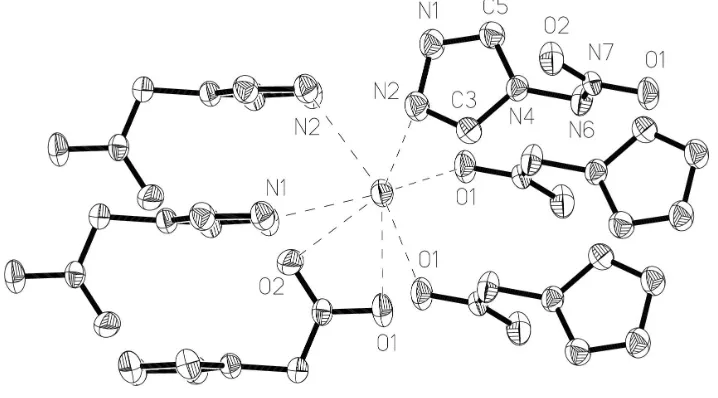
In the crystal structure of (I), the seven atoms O1, O2, N1 and N2 of six anions coordinate the potassium cation (Fig. 1). It is worth noting that atom O1 has a tetrahedral environment, consisting of atom N7 and three potassium ions. Short values
of K O and K N interatomic distances [2.788±2.934 (2) AÊ]
(Table 1) do not allow exact location of a negative charge on some of the anion atoms. Taking account of atom valency and the non-variability of the nitrimide fragment geometrical parameters, atom N6 was assigned the negative charge in the structural formula of (I), but it does not coordinate the potassium ion.
The X-ray analysis, in particular, con®rms the earlier proposal for the ®rst step of thermolysis; namely, as in (II), it involves breaking the N4ÐN6 bond (Astachov, 1999). The established bond lengths show that the N4ÐN6 bond, which connects the nitrimine group and the triazole ring, is the weakest bond in the ligand of (I) and in the molecule of (II) (Vasilievet al., 1999).
Experimental
Several drops of water were added to a boiling suspension of 4-nitramino-1,2,4-triazole (1 g) in caustic alcohol (0.8 g potassium hydroxide in 20 ml of 95% ethanol) for solution homogenization. To the transparent solution was added diethyl ether (20 ml) and the mixture was cooled slowly to room temperature. A precipitae of ®ne needle-shaped crystals was separated by ®ltration, washed with ethanol and dried. The yield of (I) was 1.0 g (77%).
Crystal data
K+C 2H2N5O2ÿ Mr= 167.19 Monoclinic,P21=c a= 7.9671 (6) AÊ b= 4.8182 (4) AÊ c= 16.812 (1) AÊ = 110.165 (7) V= 605.81 (8) AÊ3 Z= 4
Dx= 1.833 Mg mÿ3 Cu Kradiation Cell parameters from 25
re¯ections = 22±28 = 7.30 mmÿ1 T= 293 (2) K Lump, colourless 0.270.250.22 mm
Data collection
Kuma KM-4 diffractometer /2scans
Absorption correction: none 1230 measured re¯ections 1154 independent re¯ections 1039 re¯ections withI> 2(I) Rint= 0.022
max= 70.0 h=ÿ9!0 k= 0!5 l=ÿ18!20 2 standard re¯ections
every 50 re¯ections intensity variation: 0.4%
Re®nement
Re®nement onF2 R[F2> 2(F2)] = 0.048 wR(F2) = 0.124 S= 1.01 1154 re¯ections 93 parameters
Only H-atomU's re®ned
w= 1/[2(F
o2) + (0.1004P)2 + 0.2012P]
whereP= (Fo2+ 2Fc2)/3 (/)max= 0.003
max= 0.80 e AÊÿ3
min=ÿ0.55 e AÊÿ3
Extinction correction:SHELXL97 Extinction coef®cient: 0.0085 (17)
Table 1
Selected geometric parameters (AÊ,).
K O1i 2.7886 (18)
K N1ii 2.862 (2)
K O1iii 2.9001 (18)
K N2iv 2.914 (2)
K O1 2.923 (2) K O2 2.9257 (18) K N2v 2.934 (2)
C5ÐN1ÐN2 107.3 (2) C3ÐN2ÐN1 106.4 (2) N2ÐC3ÐN4 111.2 (2) C3ÐN4ÐC5 104.9 (2) C3ÐN4ÐN6 126.7 (2) C5ÐN4ÐN6 128.1 (2)
N1ÐC5ÐN4 110.3 (2) N7ÐN6ÐN4 109.2 (2) O2ÐN7ÐO1 121.0 (2) O2ÐN7ÐN6 124.3 (2) O1ÐN7ÐN6 114.7 (2)
Symmetry codes: (i) ÿx;yÿ1
2;12ÿz; (ii) ÿx;1ÿy;1ÿz; (iii) ÿx;12y;12ÿz; (iv)
ÿx;ÿy;1ÿz; (v)xÿ1;1 2ÿy;zÿ12.
Two H atoms were found in a difference Fourier map and were re®ned as riding atoms with a common isotropic displacement parameter.
Data collection: KM-4 Software (Kuma, 1991); cell re®nement:
KM-4 Software; data reduction: DATARED in KM-4 Software; program(s) used to solve structure: SHELXS97 (Sheldrick, 1997); program(s) used to re®ne structure:SHELXL97 (Sheldrick, 1997); molecular graphics:SHELXTL (Sheldrick, 1995); software used to prepare material for publication:SHELXL97.
We are grateful to the Russian Foundation of Fundamental Investigations for partial ®nancial support of the work (grant No. 00-15-96790).
References
Astachov, A. M. (1999). PhD thesis, Siberian State Technological University, Krasnoyarsk, Russia.
Kuma (1991).KM-4Software. Kuma Diffraction, Wrocøaw, Poland. Sheldrick, G. M. (1995).SHELXTL.Version 5. Siemens Analytical X-ray
Instruments Inc., Madison, Wisconsin, USA.
Sheldrick, G. M. (1997). SHELXS97 and SHELXL97. University of GoÈttingen, Germany.
Vasiliev, A. D., Astachov, A. M., Stepanov, R. S. & Kirik, S. D. (1999).Acta Cryst.C55, 830±832.
Figure 1
The arrangement of anions around the potassium ion in the crystal structure of (I), with the atomic numbering scheme (H atoms not shown).
supporting information
sup-1
Acta Cryst. (2003). E59, m67–m68
supporting information
Acta Cryst. (2003). E59, m67–m68 [doi:10.1107/S1600536803001077]
Potassium 4-nitramino-1,2,4-triazolate
Alexander D. Vasiliev, Alexander M. Astachov, Ludmila A. Kruglyakova and Rudolf S. Stepanov
S1. Comment
The structure of 4-nitramino-1,2,4-triazole, (II), has a zwitterionic conformation and has been solved previously (Vasiliev
et al., 1999). To determine the influence of the transition from (II) to its anion on the molecular conformation and on its nitrimine fragment, the structure of the potassium salt of (II), viz. the title compound, (I), was investigated. The task arose
out of a search of relationship `structure–property′ in a series of energetic nitramines. This knowledge is needed for an
estimation of properties of new hypothetic energetic molecules (Astachov, 1999).
The structural formula for (I) (see Scheme above) does not precisely reflect the true location of a negative charge in the
anion of (I). However, as will be shown, the representation of (I) using conventional principles of chemical structural
formula spelling, which takes into consideration the atom valence, and experimental geometry parameters, was a difficult
task.
As a whole, the transition from (II) to its anion does not affect the molecular geomerty parameters. Like (II), the
molecule of (I) is not planar, but consists of two planar fragments: 1,2,4-triazole cycle [maximum deviation 0.000 (1) Å]
and nitrimide group N—N—NO2 [r.m.s. deviation 0.012 (1) Å and maximum deviation 0.019 (1) Å]. The N4—N6—N7
—O2 torsion angle is 3.0 (3)° [−2.6° for (II)]. Maximum changes of valence angles are near 5° for the triazole cycle and
less than 2° for the molecular nitrimide fragment.
The bond lengths of a nitrimide fragment of (I) are practically the same as in (II) [in brackets]: N4—N6 1.408 (2) Å
[1.407 (2) Å], N6—N7 1.322 (3) Å [1.319 (2) Å], N7—O1 1.261 (2) Å [1.259 (2) Å] and N7—O2 1.243 (3) Å [1.235 (2)
Å]. Some interatomic distances in a triazole cycle changed rather more: N1—N2 1.393 (3) Å [1.362 (2) Å], N2—C3
1.295 (3) Å [1.300 (2) Å], C3—N4 1.351 (3) Å [1.358 (2) Å], N4—C5 1.352 (3) Å [1.341 (2) Å] and C5—N1 1.299 (3)
Å [1.308 (2) Å]. The facts allow the separation of single and double bonds more definitely than in the molecule of (II).
The separation is reflected in the cited above structural formula. The valence angle data is collected in Table 1.
In the crystal of (I), the seven atoms O1, O2, N1 and N2 of six anion molecules coordinate the potassium cation (Fig.
1). It is worth noting that atom O1 has a tetrahedral environment coordinated by atom N7 and three potassium ions. Close
values of K···O and K···N interatomic distances [2.788–2.934 (2) Å] (Table 1) do not allow exact location of a negative
charge on some of the anion atoms. Beginning with atom valency and invariability of nitrimide fragment geometry
parameters, atom N6 was supplied by negative anion charge in the structural formula of (I), but the atom does not
coordinate the potassium ion.
The X-ray analysis allows, in particular, the confirmation of the suggested before supposition about first step of
thermolysis; namely, like (II), it is a break of the N4—N6 bond (Astachov, 1999). The established bond lengths show that
the N4—N6 bond, which connects the nitrimine group and the triazole cycle is the weakest bond in the molecule of (I)
S2. Experimental
Several drops of water were added to a boiling suspension of 4-nitramino-1,2,4-triazole (1 g) in caustic alcohol (0.8 g
potassium hydroxide in 20 ml of 95% ethanol) for solution homogenization. To the transparent solution was added
di-ethyl ether (20 ml) and the mixture was cooled slowly to room temperature. A fine needle-shaped precipitate was
separated by filtration, washed with ethanol and dried. The yield of (I) was 1.0 g (77%).
S3. Refinement
Two H atoms were found in a difference Fourier map and were refined as riding atoms with a common isotropic
[image:4.610.127.486.210.409.2]displacement parameter.
Figure 1
The arrangement of anion molecules near the potassium ion in the crystal of (I), with the atomic numbering scheme (H
atoms not shown). Dash lines are K···O and K···N contacts.
Potassium 4-nitramino-1,2,4-triazolate
Crystal data K+·C2H2N5O2− Mr = 167.19
Monoclinic, P21/c Hall symbol: -P 2ybc a = 7.9671 (6) Å b = 4.8182 (4) Å c = 16.812 (1) Å β = 110.165 (7)° V = 605.81 (8) Å3 Z = 4
F(000) = 336 Dx = 1.833 Mg m−3
Cu Kα radiation, λ = 1.5418 Å Cell parameters from 25 reflections θ = 22–28°
µ = 7.30 mm−1 T = 293 K
Transparent lump, colourless 0.27 × 0.25 × 0.22 mm
Data collection Kuma KM-4
diffractometer
Radiation source: fine-focus sealed tube Graphite monochromator
θ/2θ scans
1230 measured reflections
1154 independent reflections 1039 reflections with I > 2σ(I) Rint = 0.022
θmax = 70.0°, θmin = 5.6° h = −9→0
supporting information
sup-3
Acta Cryst. (2003). E59, m67–m68 l = −18→20
2 standard reflections every 50 reflections
intensity decay: variation 0.4%
Refinement Refinement on F2 Least-squares matrix: full R[F2 > 2σ(F2)] = 0.048 wR(F2) = 0.124 S = 1.01 1154 reflections 93 parameters 0 restraints
Primary atom site location: structure-invariant direct methods
Secondary atom site location: difference Fourier map
Hydrogen site location: difference Fourier map Only H-atom displacement parameters refined w = 1/[σ2(F
o2) + (0.1004P)2 + 0.2012P] where P = (Fo2 + 2Fc2)/3
(Δ/σ)max = 0.003 Δρmax = 0.80 e Å−3 Δρmin = −0.55 e Å−3
Extinction correction: SHELXL97, Fc*=kFc[1+0.001xFc2λ3/sin(2θ)]-1/4 Extinction coefficient: 0.0085 (17)
Special details
Geometry. All e.s.d.'s (except the e.s.d. in the dihedral angle between two l.s. planes) are estimated using the full covariance matrix. The cell e.s.d.'s are taken into account individually in the estimation of e.s.d.'s in distances, angles and torsion angles; correlations between e.s.d.'s in cell parameters are only used when they are defined by crystal symmetry. An approximate (isotropic) treatment of cell e.s.d.'s is used for estimating e.s.d.'s involving l.s. planes.
Refinement. Refinement of F2 against ALL reflections. The weighted R-factor wR and goodness of fit S are based on F2, conventional R-factors R are based on F, with F set to zero for negative F2. The threshold expression of F2 > σ(F2) is used only for calculating R-factors(gt) etc. and is not relevant to the choice of reflections for refinement. R-factors based on F2 are statistically about twice as large as those based on F, and R- factors based on ALL data will be even larger.
Fractional atomic coordinates and isotropic or equivalent isotropic displacement parameters (Å2)
x y z Uiso*/Ueq
K −0.24012 (6) 0.08386 (11) 0.25514 (3) 0.0309 (3)
N1 0.2758 (3) 0.5141 (5) 0.62639 (13) 0.0367 (5)
N2 0.3902 (3) 0.2865 (5) 0.64968 (12) 0.0354 (5)
C3 0.4030 (3) 0.1883 (5) 0.58028 (15) 0.0334 (5)
H1 0.4716 0.0348 0.5775 0.057 (7)*
N4 0.3037 (2) 0.3378 (4) 0.51240 (12) 0.0282 (5)
C5 0.2270 (3) 0.5384 (5) 0.54458 (16) 0.0337 (6)
H2 0.1503 0.6745 0.5127 0.057 (7)*
N6 0.2994 (3) 0.3078 (5) 0.42839 (12) 0.0336 (5)
N7 0.1644 (3) 0.1445 (4) 0.38656 (11) 0.0260 (5)
O1 0.1502 (3) 0.0975 (4) 0.31070 (10) 0.0382 (5)
O2 0.0554 (2) 0.0459 (4) 0.41692 (11) 0.0394 (5)
Atomic displacement parameters (Å2)
U11 U22 U33 U12 U13 U23
K 0.0307 (4) 0.0384 (4) 0.0224 (4) 0.00076 (18) 0.0076 (2) −0.00247 (17)
N1 0.0466 (13) 0.0368 (11) 0.0257 (10) 0.0019 (10) 0.0110 (9) −0.0042 (9)
N2 0.0355 (11) 0.0435 (12) 0.0244 (10) −0.0006 (9) 0.0066 (8) −0.0001 (8)
C3 0.0309 (12) 0.0390 (13) 0.0277 (12) 0.0025 (10) 0.0069 (9) 0.0014 (10)
C5 0.0372 (13) 0.0343 (12) 0.0273 (11) 0.0033 (10) 0.0084 (10) −0.0019 (10)
N6 0.0353 (10) 0.0448 (13) 0.0227 (10) −0.0089 (9) 0.0125 (8) −0.0043 (8)
N7 0.0289 (10) 0.0303 (9) 0.0175 (9) 0.0039 (8) 0.0062 (7) 0.0032 (7)
O1 0.0528 (11) 0.0414 (10) 0.0194 (9) 0.0014 (8) 0.0112 (8) −0.0018 (7)
O2 0.0342 (9) 0.0548 (11) 0.0276 (9) −0.0106 (8) 0.0088 (7) 0.0041 (8)
Geometric parameters (Å, º)
K—O1i 2.7886 (18) K—O2 2.9257 (18)
K—N1ii 2.862 (2) K—N2v 2.934 (2)
K—O1iii 2.9001 (18) C3—H1 0.9300
K—N2iv 2.914 (2) C5—H2 0.9300
K—O1 2.923 (2)
C5—N1—N2 107.3 (2) O2—N7—O1 121.0 (2)
C3—N2—N1 106.4 (2) O2—N7—N6 124.3 (2)
N2—C3—N4 111.2 (2) O1—N7—N6 114.7 (2)
C3—N4—C5 104.9 (2) N2—C3—H1 124.4
C3—N4—N6 126.7 (2) N4—C3—H1 124.4
C5—N4—N6 128.1 (2) N1—C5—H2 124.8
N1—C5—N4 110.3 (2) N4—C5—H2 124.8
N7—N6—N4 109.2 (2)
C5—N1—N2—C3 0.0 (3) N6—N4—C5—N1 −173.2 (2)
N1—N2—C3—N4 0.0 (3) C3—N4—N6—N7 95.2 (3)
N2—C3—N4—C5 0.0 (3) C5—N4—N6—N7 −93.0 (3)
N2—C3—N4—N6 173.3 (2) N4—N6—N7—O2 3.0 (3)
N2—N1—C5—N4 0.0 (3) N4—N6—N7—O1 −178.4 (2)
C3—N4—C5—N1 0.0 (3)