www.wjpr.net Vol 7, Issue 01, 2018. 1562
DEVELOPMENT OF HERBAL FORMULATION WITH
AJWA
SEED
(
PHOENIX DACTYLIFERA
L.) EXTRACT AND ZAM-ZAM WATER
FOR
INVITRO
-ANTICANCER ACTIVITY
Ibrahim Afsal V. T.1* and B. Arul2
1
Department of Pharmaceutics, Research Scholar, Institute of Pharmaceutical Sciences and
Research Centre, Bhagwant University, Sikar road, Ajmer, Rajasthan, India-305004.
2
Department of Pharmaceutics, Vinayaka Mission College of Pharmacy, Salem, Tamil Nadu,
India.
ABSTRACT
Objective: Fruits of the date palm (Phoenix dactylifera L.) Belonging to the family of Arecaceae are very commonly consumed in many
parts of the world and are a vital component of the diet in most of the
Arabian countries. One of the miracles of Zamzam water has been
collected by scientists and they have found certain peculiarities that
make the water healthier, like a higher level of calcium. Hence, the
present study was intended to evaluate ethanolic seed (Fruits of the
date palm) extract of Phoenix dactylifera L for anticancer activities.
Materials and Methods: In the present study investigated on different cancer cell lines such as HL-60, HT-29, A 549, A 431 and MCF-7. The
activity was assessed by trypan blue dye exclusion assay for cell
viability, MTT and SRB based cytotoxicity assays. Results: Results indicate that the anti proliferative effect strengthens with increase in
the concentration of ethanolic seeds extract of Phoenix dactylifera L. Highest cytotoxicity
was found against HL-60 and HT-29. However ethanolic extract have moderate activity on A
549, A 431, and MCF-7. Effect on Hep 2 cell line upto 0.0196 mg/ml and that IC50 value on
Hep 2 cell line were 468; Phoenix dactylifera L has no effect on normal healthy body cell So
Phoenix dactylifera L second hand as antitumour action to diminish the side effect.
Conclusion: It can be concluded that the anticancer activity elucidated by Phoenix dactylifera L could be mainly due to the presences of high value class of compound like
phenolic group as the major content in the seed extract.
Volume 7, Issue 01, 1562-1570. Research Article ISSN 2277–7105
Article Received on 22 Nov. 2017,
Revised on 13 Dec. 2017, Accepted on 02 Jan. 2018
DOI: 10.20959/wjpr20181-10623
*Corresponding Author
Ibrahim Afsal V. T.
Department of
Pharmaceutics, Research
Scholar, Institute of
Pharmaceutical Sciences
and Research Centre,
Bhagwant University, Sikar
road, Ajmer, Rajasthan,
www.wjpr.net Vol 7, Issue 01, 2018. 1563 KEYWORDS:Phoenix dactylifera L, Zamzam water, cell viability, MTT, SRB, Trypan blue dye, cyclophosphamide.
INTRODUCTION
Cancer is a disease of multicellular organisms[1] characterized by uncontrolled multiplication
of subtly modified normal human cells.[2] Cancer is a leading cause of death all over the
world and represents a major public health burden.[3] They constitute the second cause of
mortality behind cardiovascular diseases in developed countries and the third after infectious
and cardiovascular diseases in developing countries.[4] Cancer is fundamentally a disease of
regulation of tissue growth. In order for a normal cell to transform into a cancer cell, genes
which regulate cell growth and differentiation must be altered.[5] All of the drawbacks
presently associated with available chemotherapeutic agents are impetus for the search for
newer, more efficacious, and better tolerated drugs. Natural products, especially the plant
kingdom, offer an inexhaustible reservoir for investigation. Plants have a long history of use
in the treatment of cancer[6,7] and the interest in nature as a source of potential
chemotherapeutic agents continues.[8] The present day research and development tailored
towards the discovery of new antiproliferative agents from natural products have been
buoyed by improvement in the science and technology of anticancer drug discovery. Fruits of
the date palm (Phoenix dactylifera L. Arecaceae) are very commonly consumed in many
parts of the world and are a vital component of the diet in most of the Arabian countries. Date
is one of the oldest known fruit crops and has been cultivated in North Africa and the Middle
East for at least 5000 years.[9] The earliest record from Iraq (Mesopotamia) shows that date
culture was probably established as early as 3000 BCE. Because of the long history of date
culture and the wide distribution and exchange of date cultivars, the exact origin of the date is
unknown, but it most likely originated from the ancient Mesopotamia area (southern Iraq) or
western India.[10] Seeds of Phoenix dactylifera L against various Human cancer cell lines.
One of the miracles of Zamzam water is its ability to satisfy both thirst and hunger. More
recently, in the last few decades, samples of Zamzam water have been collected by scientists
and they have found certain peculiarities that make the water healthier, like a higher level of
calcium.[11]
MATERIALS AND METHODS Chemicals and reagents
www.wjpr.net Vol 7, Issue 01, 2018. 1564 Solvents
95% Ethanol, Methanol, Diethyl ether, Chloroform, DMSO (Dimethyl sulphoxide).
Reagents
Trypan blue (Hyclone, Lot no: JRH27098), EDTA (MP Biomedicals, Lot No: 6941H),
Trypsin (Invitrogen, Lot No: 1376596), MTT (Roche applied sciences, Cat. No: 11465 007
001).
Media
RPMI-1640 (Sigma Aldrich Ltd. Mumbai), FBS (Fetal Bovine Serum) (Bioclot, Lot No:
07310).
Equipments
Fluorescence inverted microscope (Leica DM IL), CO2 incubator (RS Biotech, mini galaxy
A), ELISA plate reader (Lab system Multiscan), Melting point apparatus (Veego Melting
Point apparatus-VMP-PM), IR (Shimadzu-8400S FTIR spectrophotometer), APCI-MS
(Atomic Pressurized Chemical Ionization-Mass Spectroscopy) (Varian Inc, USA-410 Prostar
Binary LC with 500 MS IT PDA Detectors spectrophotometer), 1H NMR (Varian Mercury
YH-300 MHz 1 H NMR spectrophotometer).
Cancer cells: Five cell lines, HL-60 (Human leukemia cell lines), HT-29 (Huma colon cancer cell lines), MCF-7 (Human breast cancer cell lines), A 431(Human skin cancer cell
lines) and A 549 (Human lung cancer cell lines), Human Epidermoid Larynx Carcinoma cell
line (Hep 2). were procured from the Amala Cancer Research Institute, Kerala, India.
Preparation of the extract/ drug: The granulated dried seeds of Phoenix dactylifera L (500 g) was packed in a Soxhlet apparatus and subjected to continuous hot percolation for using
450 ml of ethanol (95% v/v) as solvent. The extract was concentrated to dryness under
reduced pressure and controlled temperature and dried in a desiccator (yield 75 g, 15% w/w).
The extract was suspended in Zamzam water and used for further experiments.
EVALUATION OF INVITRO ANTI-CANCER ACTIVITY[12-14] Screening methods of anticancer activity
Cell lines and culture conditions
Five cell lines, HL-60 (Human leukemia cell lines), HT-29 (Huma colon cancer cell lines),
www.wjpr.net Vol 7, Issue 01, 2018. 1565
(Human lung cancer cell lines), Human Epidermoid Larynx Carcinoma cell line (Hep 2).
were procured from the Amala Cancer Research Institute, Kerala, India. These cell lines were
cultured in RPMI-1640 medium. The media were supplemented with 10% fetal bovine
serum, glutamine (2 mM), penicillin (100units/ml) and streptomycin (100g/ml). The cultures
were maintained in a humidified 5% CO2 incubator at 370C and the cells were sub cultured
every 3–4 days to maintain logarithmic growth and were allowed to grow for 24 h before use.
Subculture of adherent cell lines (HT-29, MCF-7, A-549 and A-431)
Cultures were viewed using an inverted microscope to assess the degree of confluency and
the absence of bacterial and fungal contaminants was confirmed. Cell monolayer was washed
with FBS without Ca2+/Mg2+ using a volume equivalent to half the volume of culture
medium. Trypsin/EDTA was added on to the washed cell monolayer using 1ml per 25 cm2 of
surface area. Flask was rotated to cover monolayer with trypsin. Flask was returned to the
CO2 incubator and left for 2- 10 mins. The cells were examined using an inverted microscope
to ensure that all the cells were detached and floated. The cells were resuspended in a small
volume of fresh serum containing HT-29, MCF-7, A 549 and A 431 medium respectively.
100-200μl was removed to perform a cell count. The required numbers of cells were
transferred to a new labeled flask containing pre-warmed HT-29, MCF-7, A 549 and A 431
medium and incubated as appropriate for the cell line.
Subculture of adherent cell lines (Hep 2)
Cultures were viewed using an inverted microscope to assess the degree of confluency and
the absence of bacterial and fungal contaminants was confirmed. Cell monolayer was washed
with PBS without Ca 2+/Mg2+ using a volume equivalent to half the volume of culture
medium. Trypsin/EDTA was added on to the washed cell monolayer using 1ml per 25 cm2 of
surface area. Flask was rotated to cover monolayer with trypsin. Flask was returned to the
CO2 incubator and left for 2- 10 mins. The cells were examined using an inverted microscope
to ensure that all the cells were detached and floated. The cells were resuspended in a small
volume of fresh serum containing Hep-2 medium. 100-200μl was removed to perform a cell
count. The required number of cells were transferred to a new labeled flask containing
pre-warmed Hep-2 medium and incubated as appropriate for the cell line.
MTT assay
MTT Colorimetric assay is based on the capacity of Mitochondrial succinate dehydrogenase
thiazol-www.wjpr.net Vol 7, Issue 01, 2018. 1566
2-yl)-2,5-diphenyl tetrazolium bromide (MTT) into an insoluble, colored formazan product
which is measured spectrophotometrically. Since reduction of MTT can only occur in
metabolically active cells, the level of activity is a measure of the viability of the cells. MTT
assay was employed to assess cell proliferation. Viable cells were seeded into 96- well
microtitre plates at 2× 10⁴ cells/well in RPMI-1640 medium supplemented with FBS (fetal
bovine serum), 100units/ml penicillin, 100μg/ml streptomycin, and were cultured in a humidified atmosphere of 5% CO2 and 95% air at 370C. 150μl of cell suspension was
cultured with 10μl of various concentrations i.e. 10, 20, 40 and 80μg/ml18 of the ethanolic
seeds extract Phoenix dactylifera L respectively dissolved in DMSO (dimethyl sulphoxide) as
solvent and incubated for 48h. Similar solutions containing the same concentrations of
cyclophopamide were also be prepared and served as standard solutions. Control cells were
incubated in RPMI-1640 medium only. Wells containing only media were considered as a
blank. All cyclophosphamide, ethanolic extract and dilution doses were tested in triplicates.
The cell proliferation is based on the ability of the mitochondrial succinate-terazolium
reductase system to convert 3-(4,5-dimethylthiazol-2-yl)-2,5-diphenyltetrazoliumbromide
(MTT) to a blue colored formazan. The test denotes the survival cells after toxic exposure. 10
μl of MTT labeling mixture was added and incubated for 4 h at 37°C and 6.5% CO2. After 4h, 100 μl of solubilization solution was added in each well. After 48h incubation at 370C
temperature and 5% CO2, the absorbance of soluble formazan product produced by viable
cells was measured at 450nm using ELISA plate reader. Reference wavelength used was 630
nm. Percentage inhibition of the cell proliferation by cyclophosphamide, ethanolic seeds
extract Phoenix dactylifera L against all cell lines was calculated using the following
formula,
% Cell survival= (At − Ab) × 100 (Ac − Ab)
Where, At = Absorbance of Test, Ab= Absorbance of Blank (Media), Ac= Absorbance of
control (cells).
Sulphorodamine B assay[15]
Sulphorodamine B (SRB) is a bright pink Aminoxanthine dye with two sulfonic groups.
Under mild acidic conditions, SRB binds dye to basic amino acid residues in TCA (Trichloro
acetic acid) fixed cells to provide a sensitive index of cellular protein content that is linear
over a cell density range of visible at least two order of magnitude.[15-16] The monolayer cell
www.wjpr.net Vol 7, Issue 01, 2018. 1567
medium containing 10% new born sheep serum. To each well of the 96 well microtitre plate,
0.1ml of the diluted cell suspension (approximately 10,000 cells) was added. After 24 hours,
when a partial monolayer was formed, the supernatant was flicked off, washed once and 100
μl of different test compound concentrations were added to the cells in microtitre plates. The
plates were then incubated at 37oC for 72 hours in 5% CO2 incubator and microscopic
examination was carried out and observations recorded every 24 hours. After 72 hours, 25 μl
of 50% trichloroacetic acid was added to the wells gently such that it forms a thin layer over
the test compounds to form a overall concentrations 10%. The plates were incubated at 4oC
for one hour. The plates were flicked and washed five times with tap water to remove traces
of medium, sample and serum, and were then air-dried. The air-dried plates were stained with
100μl SRB and kept for 30 minutes at room temperature. The unbound dye was removed by
rapidly washing four times with 1% acetic acid. The plates were then air-dried. 100 μl of
10mM Tris base was then added to the wells to solubilize the dye. The plates were shaken
vigorously for 5 minutes. The absorbance was measured using microplate reader at a
wavelength of 540nm.
Statistical analysis
The results of various studies were expressed as mean ± SEM and analyzed statistically using
one way ANOVA followed by Dunnets Test to find out the level of significance. Data were
considered statistically significant at minimum level of p < 0.05.
RESULTS AND DISCUSSION MTT COLORIMETRIC ASSAY
Table 1: Effect of cyclophophamide (standard) on % cell survival of cell lines.
CELL LINES
CONCENTRATIONS (μg/ml)
10 20 40 80
HL-60 46.62 43.67 25.37 12.75
HT-29 49.70 46.16 35.64 24.28
A 549 53.16 48.31 39.34 28.13
A 431 54.78 51.15 41.66 30.65
MCF-7 57.61 49.81 40.52 33.52
All values are expressed as Mean±SEM. Data were analysed by One-way ANOVA followed
www.wjpr.net Vol 7, Issue 01, 2018. 1568 Table 2: Effect of ethanolic seeds extract of Phoenix dactylifera L. on % cell survival of cell lines.
CELL LINES
CONCENTRATIONS (μg/ml)
10 20 40 80
HL-60 56.31 48.3 31.85 17.62
HT-29 63.43 58.75 52.27 42.70
A 549 61.54 61.91 53.10 52.78
A 431 67.42 63.68 58.38 51.73
MCF-7 77.23 76.27 74.46 63.67
All values are expressed as Mean±SEM. Data were analysed by One-way ANOVA followed
by Dunnet’s test.
SULPHORODAMINE B ASSAY
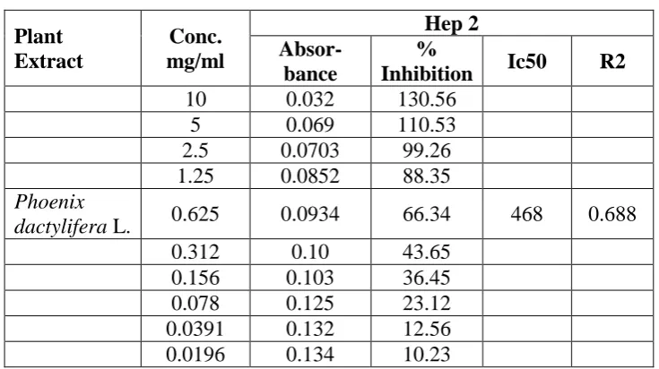
Table 3: Determination of Cytotoxicity by SRB assay.
Plant Extract
Conc. mg/ml
Hep 2
Absor-bance
%
Inhibition Ic50 R2
10 0.032 130.56
5 0.069 110.53
2.5 0.0703 99.26
1.25 0.0852 88.35
Phoenix
dactylifera L. 0.625 0.0934 66.34 468 0.688
0.312 0.10 43.65
0.156 0.103 36.45
0.078 0.125 23.12
0.0391 0.132 12.56
0.0196 0.134 10.23
Cancer therapy in the form of surgery or radiotherapy is effective when the disease is early
detected but many cancers are still diagnosed when cells from a primary tumor have already
metastasized to other parts of the body and the main form of treatment at this point is
chemotherapy.[16] Chemotherapy entails delivering drugs systemically so that they can reach
and kill the tumor cells, but most of these drugs cause severe side effects in patients and,
therefore, need to be used at suboptimal levels.[17] Plants have served as a rich source of
therapeutic agents for many centuries, being used themselves or as the basis for synthetic
drugs[18] and despite the great developments in organic synthesis, 55% of recent
chemotherapeutic drugs are derived from or based upon natural products.[19] The use of plants
as food and in folk and traditional medicine has made these natural resources one of the main
[image:7.595.114.482.319.525.2]www.wjpr.net Vol 7, Issue 01, 2018. 1569
alternative therapies using natural products is increasing, especially those derived from
plants, due to the increasingly high number of cancer cases worldwide. The effect of RPMI
1640 media (control), cyclophosphamide (standard), ethanolic seeds extract of Phoenix
dactylifera L on the growth of HL-60, HT-29, A 431, A 549 and MCF-7 cell lines were
examined by MTT assay. Dose response curves constructed between the range of 10-80
μg/ml, Results indicate that the antiproliferative effect strengthens with increase in the
concentration of ethanolic seeds extract of Phoenix dactylifera L. Highest cytotoxicity was
found against HL-60 and HT-29. However ethanolic extract have moderate activity on A 549,
A 431 and MCF-7. The percentage growth inhibition was found to be increasing with
increasing concentration of test compounds, and that show Phoenix dactylifera L. effect on
Hep 2 cell line upto 0.0196 mg/ml and that IC50 value on Hep 2 cell line was 468, Phoenix
dactylifera L has no effect on normal healthy body cell So Phoenix dactylifera Lsecond hand
as antitumour action to diminish the side effect.
REFERENCES
1. M. R. P. Rao, U. R. Adagale, A. Shetty, P. Namjoshi, P. Gaitonde and P. Jain, Cancer
Immunotherapy, 2007.
2. M. Mubeen and S. G. Kini, “A review on the design and development of EGFR tyrosine
kinase inhibitors in cancer therapy,” International Journal of Therapeutic Applications,
2012; 5: 29–37.
3. S. U. Park, “Anticancer compounds from plants,” EXCLI Journal, 2012; 11: 386–389.
4. Croce CM. Oncogenes and cancer. The New England Journal of Medicine, 2008; 358:
502-511.
5. Croce CM. Oncogenes and cancer. The New England Journal of Medicine, 2008; 358:
502-511.
6. G. M. Cragg and D. J. Newman, “Medicinals for the millennia: the historical
record,” Annals of the New York Academy of Sciences, 2001; 953: 3–25.
7. D. J. Newman, G. M. Cragg, and K. M. Snader, “The influence of natural products upon
drug discovery,” Natural Product Reports, 2000; 17(3): 219–220.
8. G. Schwartsmann, M. J. Ratain, G. M. Cragg et al., “Anticancer drug discovery and
development throughout the world,” Journal of Clinical Oncology, 2002; 20(18):
www.wjpr.net Vol 7, Issue 01, 2018. 1570
9. Zohary, D., Hopf, M. Domestication of plants in the old world: The origin and spread of
cultivated plants in West Asia, Europe, and the Nile Valley (Oxford University
Press, Oxon, UK), 2000.
10.Wrigley, G. in Evolution of crop plants, Date palm, eds Smartt J., Simmonds N.W.
(Longman Group, Essex, UK), 2nd ed, 1995; 399–403.
11.http://islamqa.info/en/6831.
12.Patel S, Gheewala N, Suthar A, Shah A. In-vitro Cytotoxicity Activity of Solanum
nigrum Extract Against HELA Cell Line and VERO Cell Line. International Journal of
Pharmacy and Pharmaceutical science, 2009; 1(1): 38-46.
13.Patel SR, Suthar AP, Patel RM. In Vitro Cytotoxicity Activity of Semecarpus anacardium
Extract Against HEP 2 Cell Line and VERO Cell Line. International Journal of Pharm
Tech Research, 2009; 1(4): 1429-1433.
14.Raval BP, Shah TG, Patel JD, Patel BA, Patel RK. Potent Anticancer Activity of Nigella
Sativa Seeds. Scholars Research Library, 2010; 2(1): 52-56.
15.Masters R.W., Animal cell culture, Cytotoxicity and viability assays, Third edition,
202-203.
16.C. Martin-Cordero, A. J. Leon-Gonzalez, J. M. Calderon-Montano, E. Burgos-Moron,
and M. Lopez-Lazaro, “Pro-oxidant natural products as anticancer agents,” Current Drug
Targets, 2012; 13(8): 1006–1028.
17.A. Jemal, R. Siegel, J. Xu, and E. Ward, “Cancer statistics, 2010,” CA Cancer Journal for
Clinicians, 2010; 60(5): 277–300.
18.G. M. Marchetti, K. A. Silva, A. N. Santos et al., “The anticancer activity of
dichloromethane crude extract obtained from Calea pinnatifida,” Journal of Experimental
Pharmacology, 2012; 4: 157–162.
19.D. J. Newman and G. M. Cragg, “Natural products as sources of new drugs over the 30
years from 1981 to 2010,” Journal of Natural Products, 2012; 75(3): 311–335.
20.G. M. Cragg and D. J. Newman, “Nature: a vital source of leads for anticancer drug
development,” Phytochemistry Reviews, 2009; 8(2): 313–331.
21.F. R. F. Nascimento, G. V. B. Cruz, P. V. S. Pereira et al., “Ascitic and solid Ehrlich
tumor inhibition by Chenopodium ambrosioides L. treatment,” Life Sciences, 2006;
78(22): 2650–2653.
22.A. Jemal, R. Siegel, E. Ward, Y. Hao, J. Xu and M. J. Thun, “Cancer statistics,