www.wjpr.net Vol 4, Issue 07, 2015
398
A CLINICAL COMPARATIVE STUDY TO EVALUATE THE
EFFICACY AND SAFETY OF LIV.52 DS TABLETS IN
NON-ALCOHOLIC STEATOHEPATITIS (NASH)
Dr. S.G. Maity1 and Dr. Asim Kumar Mandal*2
1
Consultant Gastroenterologist, Kothari Medical College, Alipore Road, Alipore, Kolkata, – 700 027, India.
*2
Medical Superintendant, Institute of Post Graduate Research & Education in Ayurveda and SVSP Hospital, Kolkota-700009, India.
ABSTRACT
The present study was planned to evaluate the clinical efficacy and
safety of Liv.52 DS tablets with UDCA in patients with Non-Alcoholic Steatohepatitis in a randomized, comparative clinical study. A total of
35 patients aged between 18-65 years with Non-Alcoholic Steatohepatitis characterized by elevated liver enzymes and hepatomegaly with pain and discomfort in the right upper abdomen and those who were willing to give informed consent were included in the study. At the initial randomization visit, a detailed medical history and symptomatic evaluation was done. In addition, examination specific to the steatohepatitis with Hepatomegaly was also done. All patients received liver screen (set of blood tests) including complete haemogram, liver function tests and ultrasound abdomen. All the patients were routinely screened and subjected to biochemical investigations. Biochemical investigations included SGPT, SGOT, ALT, Serum Bilirubin, Total Protein, Albumin and Globulin. Clinical examinations were repeated every four weeks for 12 weeks and biochemical investigations were repeated only after 12 weeks. All the patients were randomized arbitrarily using random table into either Liv.52 DS group (n=19) or UDCA (n=16). The two groups were similar with regards to the demographic data, baseline parameters and biochemical investigations. The Liv.52 DS group received 2 Tablets twice daily and UDCA group received 600 mg thrice daily for 12 weeks. Statistical analysis was carried out using GraphPad Prism Software, Version 4.01. All the patients completed the
Volume 4, Issue 7, 398-414. Research Article ISSN 2277– 7105
Article Received on 25 April 2015,
Revised on 15 May 2015, Accepted on 03 June 2015
*Correspondence for
Author
Dr. Asim Kumar Mandal
Medical Superintendant, Institute of Post Graduate Research & Education in Ayurveda and SVSP Hospital, Kolkota-700009, India.
www.wjpr.net Vol 4, Issue 07, 2015
399 study and their data was available for analysis. Significant evidence of hepatoprotective effects of Liv.52 DS in patients with Non-Alcoholic Steatohepatitis in terms of clinical response and reduction in biochemical parameters were observed after 12 weeks of treatment. The clinical and biochemical recovery in Liv.52 DS group was faster as compared to UDCA group indicating the beneficial effect of Liv.52 DS tablets in patients with Non-Alcoholic Steatohepatitis. There were no clinically significant adverse reactions either reported or observed during the entire study period. The overall compliance to the treatment was good and no treatment discontinuations were reported. The results of the present study showing
clinical benefit of Liv.52 DS appear promising in the management of Non-Alcoholic Steatohepatitis.
KEYWORDS: Non-Alcoholic Steatohepatitis (NASH), Liv.52 DS, Ursdeoxycholic acid (UDCA), Non-Alcoholic Fatty Liver Disease (NAFLD).
INTRODUCTION
Hepatic steatosis with or without hepatitis, in the absence of alcohol use, was first described by Ludwig et al. and is referred to as non-alcoholic steatosis or Non-Alcoholic Steatohepatitis.[1] Non-Alcoholic Steatohepatitis (NASH) is a metabolic liver disorder that is seen in 2-6% of the general population. Non-Alcoholic Fatty Liver Disease (NAFLD) and its various manifestations are seen in all ethnic groups, across the globe and in both genders.[2, 3] The prevalence of NASH is 2.1–6.3% in the general population, rising to 9–40% in obese individuals with a body mass index (BMI) of 30 kg/m2 or more.[4,5] The disease
predominantly occurs between the ages of 40 and 60 although there have been reports in children over the age of 10.[6-10] NASH progresses to Cirrhosis in 20% of these patients. Once developed, 30-40% of patients with NASH cirrhosis will experience liver-related death over a 10-year period.[11] In its initial phases, during which fat accumulates in the liver, no clinical symptoms are evident. In advanced stages, fibrosis (eventually progressing to established cirrhosis in some patients) is detectable histologically.[12]
www.wjpr.net Vol 4, Issue 07, 2015
400 hepatic steatosis.[16] Most patients are asymptomatic, have mild-to-moderate elevations of serum aminotransferase levels, clinical hepatomegaly and features of fatty liver on imaging.[17] Once patients with simple steatosis develop NASH, up to 50% of them could develop advanced fibrosis. Patients with NASH and cirrhosis are at risk of complications of portal hypertension and hepatocellular carcinoma. These complications contribute to lower life expectancy for cirrhotic NASH patients.[18] Increased fructose consumption has also been shown as a risk factor for development for NASH and that increased fructose consumption correlates with the severity of fibrosis in patients with nonalcoholic fatty liver disease.[19, 20]
Current pathogenic hypothesis of NASH include insulin resistance, oxidative stress and abnormal production of pro-inflammatory cytokines as key factors in the development and progression of NASH.[21-24] NASH patients will have increased levels of oxidative stress as compared with patients with steatosis alone.[25,26] The most common risk factor for NASH is obesity.[27] Obesity is a pro-inflammatory state and leads to insulin resistance via adipocytokines. Insulin resistance leads to increased lipolysis and exponentially high delivery
of free fatty acids (FFA) to liver. Accumulation of free fatty acids leads to hepatic steatosis and FFA-mediated lipotoxicity that eventually progresses to fibrosis/cirrhosis.[28]
Nonspecific constitutional symptoms of weakness fatigue and malaise precede in a third of NASH patients.[6] Despite similarities the patients of NASH are mostly asymptomatic whereas patients of alcoholic hepatitis are always symptomatic. NASH due to drugs viz. nucleoside analogs antimitotic agents or tetracyclines,[29] can present dramatically with rapid
onset of fulminant hepatic failure. Hepatomegaly (75%) and splenomegaly (25%) [30] are the most common signs in NASH patients. Presence of ascites and spider angiomata, indicates development of cirrhosis.[31] Apart from age, diabetes is a strong independent predictor of fibrosis in NASH.[29]
Liver biopsy is the preferred method for establishing the diagnosis of nonalcoholic fatty liver disease and determining the histological stage, chiefly the existence of NASH. The most
www.wjpr.net Vol 4, Issue 07, 2015
401 Treatment aims to control the conditions that are associated with NASH, such as obesity, diabetes, and hyperlipidemia. The goals of management include: Correction of the underlying risk factors, avoidance of factors that promote progression of liver disease and specific treatment of NASH.[34] General measures of risk reduction such as weight loss in obese patients, effective treatment of hyperlipidemia and diabetes mellitus are usually recommended. Other methods currently in use are: weight loss drugs (Orlistat), physical activity, oral antidiabetic medications (metformin, troglitazone, pioglitazone, and rosiglitazone), cytoprotective agents (taurine, ursodeoxycholic acid [UDCA]),
hypolipidemics (clofibrate, gemfibrozil, bezafibrate, atorvastatin, and other HMG-CoA reductase inhibitors), several antioxidants and a combination of different therapies (diet and UDCA, vitamin E and pioglitazone).[11, 35-38] Although a wide range of drugs are available for fatty liver, but are not devoid of adverse effects.
Ursodeoxycholic acid (UDCA) has several mechanisms of action that justify its use in NASH: hydrophilic effect (resulting in the displacement of toxic hydrophobic biliary salts),
and immunomodulatory and cytoprotective properties. An oral dose of 13–15 mg/day for 12 months was efficacious in improving liver biochemistry alterations and steatosis, although no favorable changes occurred in the rest of the histological lesions of NASH. However, recently reported results of a randomized multicenter study in which NASH patients received between 13 and 15 mg/ kg/day of ursodeoxycholic acid for 2 years showed no improvement of liver disease with respect to placebo treated patients.39 Ayurveda, an indigenous system of medicine in India, has a long tradition of treating liver disorders with plant drugs. In the present study Liv.52 DS Capsule was evaluated for the efficacy and safety in management of nonalcoholic steatohepatitis. Liv.52 DS Tablet - is a herbal formulation consisting of extracts of Capparis spinosa, Cichorium intybus, Mandhura bhasma, Solanum nigrum, Terminalia arjuna, Cassia occidentalis, Achillea millefolium and Tamarix gallica.
AIM OF THE STUDY
To compare the clinical efficacy and safety of Liv.52 DS tablets with Ursdeoxycholic acid (UDCA) in patients with Non-Alcoholic Steatohepatitis.
STUDY DESIGN
www.wjpr.net Vol 4, Issue 07, 2015
402 documents, product related information and informed consent form were submitted to the “Institutional Ethics Committee” and were approved by the same.
MATERIAL AND METHODS Inclusion Criteria
A total of 35 male and female patients aged between 18-65 years with Non-Alcoholic Steatohepatitis characterized by elevated liver enzymes and hepatomegaly with pain and discomfort in the right upper abdomen, patients who has not participated in a similar investigation in past four weeks and those who were willing to give informed consent were included in the study.
Exclusion Criteria
Patients with severe metabolic disorders, carcinoma of liver or pancreas, patients with a known history of present condition of allergic response to similar pharmaceutical products, its components or ingredients in the test products, patients with pre-existing systemic disease necessitating long-term medications, genetic and endocrinal disorders, patients who has participated in a similar clinical investigation in the past four weeks and those who refused to sign the informed consent form were excluded. Women of child bearing age and lactating women were also excluded from the trial.
Study Procedure
At the initial randomization visit, a detailed medical history and symptomatic evaluation was done. In addition, examination specific to the steatohepatitis with Hepatomegaly was also done. All patients underwent complete haemogram, ultrasound abdomen, liver function tests and biochemical investigations including SGPT, SGOT, ALT, Serum Bilirubin, Total Protein, Albumin and Globulin. All the patients were routinely screened and subjected to biochemical investigations. Biochemical investigations included SGPT, SGOT, ALT, Serum
Bilirubin, Total Protein, Albumin and Globulin. Clinical examinations were repeated every four weeks for 12 weeks and Liver function tests were repeated only after 12 weeks. All the
www.wjpr.net Vol 4, Issue 07, 2015
403 Primary and Secondary End-points
The primary end-points were relief from abdominal discomfort, nausea/vomiting, reduction in elevated liver function parameters, improvement in hepatomegaly and infiltration sonographically. The secondary end point measures were incidence of adverse events and overall compliance to the treatment.
Statistical Analysis
Statistical analysis was carried out using Fishers exact test for between the group comparison for evaluation of nausea/vomiting parameter. Biochemical parameters were analysed using paired „t‟ test for within the group comparison and unpaired t-test for between the group
comparisons. The values are expressed as Mean ± SD. Hepatomegaly and fatty infiltration were analysed using Wilcoxan signed rank test for within the group comparison and Mann Whitney test for between the group comparisons. Statistical analysis was carried out using GraphPad Prism Software, Version 4.01.
Adverse Events
All adverse events, either reported or observed by patients, were recorded with information about severity, date of onset, duration, and action taken regarding the study drug. Relation of adverse events to the study medication was predefined as “Unrelated” (follows a reasonable
temporal sequence from the administration of the drug), “Possible” (follows a known response pattern to the suspected drug, but could have been produced by the patient‟s clinical state or other modes of therapy administered to the patient), and “Probable” (follows a known
response pattern to the suspected drug that could not be reasonably explained by the known characteristics of the patient‟s clinical state).
Patients were allowed to voluntarily withdraw from the study, if they so desired without assigning reasons. For patients withdrawing from the study, efforts were made to ascertain
the reason for dropout. Non compliance (defined as failure to take less than 80% of the medication) was not regarded as treatment failure, and reasons for non compliance were
noted.
RESULTS
www.wjpr.net Vol 4, Issue 07, 2015
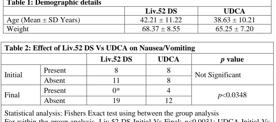
404 randomized comparative clinical study. There was no statistical difference in the demographic characters between the Liv.52 DS group and UDCA groups (Table 1).
All the patients completed the study and their data was available for analysis. The effect of Liv.52 DS Tablet and UDCA on nausea/vomiting was evaluated in table 2. In Liv.52 DS group, 8 out of 19 patients had nausea/vomiting at entry and at the end of the study all the patients in the group were free from nausea/vomiting symptoms with a significance of
p<0.0031. In the UDCA group, 8 out of 16 patients had nausea/vomiting symptom at entry and at the end of the study, 4 patients still persisted to have symptoms of nausea/vomiting. For between the groups, the level of significance was found to be p<0.0348 in Liv.52 DS group as compared to the UDCA group.
The effect of Liv.52 DS and UDCA on liver function test is shown in table 3. Statistical analysis was performed to compare the effect of Liv.52 DS and UDCA on the liver function test parameters. Results for the mean SGPT in Liv.52 DS shows significant changes from 68.89 ± 21.25 IU/L to 45.50 ± 19.87 IU/L at the end of the study as compared to the initial values. In the UDCA group also, SGPT reduced from 76.13 ± 34.95 IU/L to 48.13 ± 16.00 IU/L at the end of the study as compared to initial values. Statistical analysis performed within the group has shown that the level of significance was p<0.0004 in Liv.52 DS group and p<0.0005 in UDCA group as compared to the initial values in both the groups, which implies that there was no significant variation among the groups for SGPT values. Mean SGOT in Liv.52 DS group reduced from 56.74 ± 31.77 IU/L to 39.00 ± 11.47 IU/L at the end
of the study with a significance of p<0.033. In the UDCA group, mean SGOT values were 44.63 ± 19.38 IU/L and 42.13 ± 16.34 IU/L before and after the treatment respectively with no significant changes. Mean ALP value in Liv.52 DS group also reduced from 211.2 ± 49.85 µg/L to 198.2 ± 48.67 µg/L with a significance of p<0.008. Whereas, mean ALP value in the UDCA group was 186.1 ± 61.10 µg/L and 178.3 ± 50.98 µg/L before and after treatment with no significant changes. In the Liv.52 DS group and UDCA group, changes were not significant for other four variables like serum bilirubin, total protein, albumin and globulin with respect to mean values before and after the trial.
www.wjpr.net Vol 4, Issue 07, 2015
405 as compared to initial values. Statistical analysis performed within the group has shown that the level of significance was p<0.0003 in Liv.52 DS group and p<0.0010 in UDCA group as compared to the initial values in both the groups, which implies that there was no significant variation among the groups for hepatomegaly and infiltration values.
Overall response to treatment was assessed by interviewing the patients at the end of the study period. In the Liv.52 DS group, 2 patients (10.53%) showed marked improvement, 4 patients (21.05%) showed moderate improvement and 13 patients (68.42%) showed slight improvement. Whereas, in the UDCA group, 7 patients (43.75%) showed moderate improvement, 7 patients (43.75%) showed slight improvement and 2 patients (12.5%) had no change after treatment (Table 5).
DISCUSSION
NASH is a progressive disease in more than one in four and has spontaneous regression in less than one in six. Therapy options include weight reduction in obese, good control in diabetics and exercise.[17] Although there is no consensus for the treatment for NASH, effort needs to be made to prevent development of fibrosis, which results in cirrhosis and portal hypertension. As the pathogenesis of this condition is not clear, treatment has been largely empirical.[40] Treatment should be focused on correction of the underlying metabolic syndrome. The role of specific pharmacologic treatment continues to evolve. Several large clinical trials using a variety of agents are currently under way and should provide additional treatment option for those with nonalcoholic steatohepatitis.[41]
The beneficial results observed in this study in Liv.52 DS group compared to UDCA could be due to synergistic actions of the herbs, P-Methoxy benzoic acid from Capparis spinosa has potent hepatoprotective activity against chemically-induced hepatotoxicity, prevents elevation of malondialdehyde levels (plasma and hepatic) and enzyme levels (AST and
ALT).[42-44] It improves the functional efficiency of the liver and spleen, with protective action on the histological architecture of the liver, and a salutary effect on liver glycogen and
www.wjpr.net Vol 4, Issue 07, 2015
406 DNA in tissue debris,[49] It also has potent antioxidant action, as evident by its free radical scavenging effects, inhibition of hydrogen peroxide and iron chelation,[50, 51]Solanum nigrum
protects DNA against oxidative damage,[52] and also acts as a potent scavenger of hydroxyl and diphenyl picryl hydrazyl radicals,[53] The cytoprotective effect of Solanum nigrum
against gentamicin-induced toxicity showed a significant inhibition of cytotoxicity, and hydroxyl radical scavenging potential,[54] Terminalia arjuna reduces cholesterol levels and is also useful in liver disorders,[55, 56] It has potent antioxidant activity, which is due to its effects on lipid peroxidation,[57] Arjunaphthanoloside from Terminalia arjuna inhibits nitric
oxide production, and terminoside A decreases inducible nitric oxide synthase levels in lipopolysaccharide-stimulated peritoneal macrophages,[58] It has strong antiviral activity, inhibiting viral attachment and penetration,[59] It also has supportive antibacterial activity,[60]
[image:9.595.79.519.504.699.2]Cassia occidentalis has significant hepatoprotective effects in chemically-induced liver damage.[61] It modulates hepatic enzymes, which provides hepatoprotection.[62] Achillea millefolium is beneficial in chronic hepatitis,[63] and has anti-hepatoma activity[64] Tamarix gallica is a hepatic stimulant, digestive and hepatoprotective, and has a salutary effect on liver glycogen and serum proteins.[65] Mandura bhasma has hepatoprotective property, and is beneficial in chemically-induced hepatotoxicity as it prevents changes in liver enzyme activities.[66] Mandura bhasma is a powerful hematinic and tonic.[67] The results of the present study showing clinical benefit of Liv.52 DS appear promising in the management of non-alcoholic steatohepatitis when compared to UDCA.
Table 1: Demographic details
Liv.52 DS UDCA
Age (Mean ± SD Years) 42.21 ± 11.22 38.63 ± 10.21
Weight 68.37 ± 8.55 65.25 ± 7.20
Table 2: Effect of Liv.52 DS Vs UDCA on Nausea/Vomiting
Liv.52 DS UDCA p value
Initial Present 8 8 Not Significant
Absent 11 8
Final Present 0* 4 p<0.0348
Absent 19 12
Statistical analysis: Fishers Exact test using between the group analysis
www.wjpr.net Vol 4, Issue 07, 2015
[image:10.595.77.526.76.621.2]407 Table 3: Effect of Liv.52 DS Vs UDCA on various biochemical parameters
S.
No. Parameter
Liv.52 DS UDCA
Initial Final Significance Initial Final Significance
1. SGPT IU/L 68.89 ± 21.25
45.56 ± 19.87
p<0.0004 76.13 ± 34.95
48.13 ±
16.00 p<0.0005
2. SGOT IU/L 56.74 ± 31.77
39.00 ± 11.47
p<0.033 44.63 ± 19.38
42.13 ±
16.34 NS
3. ALP µg/L 211.2 ± 49.85
198.2 ± 48.67
p<0.008 186.1 ± 61.10
178.3 ±
50.98 NS
4. Serum Bilirubin mg/dl 0.82 ± 0.15 0.80 ±
0.12 NS
1.02 ± 0.67
1.00 ±
0.72 NS
5. Total Protein mg/dl
7.12 ± 0.56
7.15 ±
0.53 NS
7.29 ± 0.52
7.28 ±
0.63 NS
6. Albumin mg/dl
4.26 ± 0.37
4.35 ±
0.42 NS
4.35 ± 0.36
4.43 ±
0.40 NS
7. Globulin mg/dl
2.81 ± 0.45
2.80 ±
0.26 NS
2.94 ± 0.43
2.85 ±
0.49 NS
Statistical Analysis: Paired t-test for within the group analysis
Between the group analysis using the change in the biochemical parameter from initial to final was not significant for Liv.52 DS Vs UDCA
Table 4: Effect of Liv.52 DS Vs UDCA on Hepatomegaly and Infiltration
Drug Initial Final Significance
Liv.52 DS 3.53 ± 0.84 2.21 ± 0.80 p<0.0003
UDCA 3.43 ± 0.96 2.62 ± 0.89 p<0.0010
Statistical Analysis: Wilcoxan signed rank test for within the group analysis Analysis for between the groups using Mann Whitney test was not significant
Table 5: Overall Impression
Description Score Liv.52 DS UDCA
Symptoms became worse 1 -
-No change 2 - 2 (12.5%)
Slight Improvement 3 13 (68.42%) 7 (43.75%)
Moderate Improvement 4 4 (21.05 %) 7 (43.75%)
Marked Improvement 5 2 (10.53%) -
www.wjpr.net Vol 4, Issue 07, 2015
www.wjpr.net Vol 4, Issue 07, 2015
409
CONCLUSION
www.wjpr.net Vol 4, Issue 07, 2015
410 group. This indicates the activity of the Liv.52 DS in patients infected with steatohepatitis. There were no clinically significant adverse reactions either reported or observed during the entire study period. The overall compliance to the treatment was good and no treatment discontinuations were reported. The results of the present study showing clinical benefit of Liv.52 DS appear promising in the management of steatohepatitis.
REFERENCES
1. Ludwig J, Viggiano TR, McGill DB, Ott BJ. Nonalcoholic steatohepatitis. Mayo Clinic experiences with a hitherto unnamed disease. Mayo Clinic Proceedings, 1980; 55(7): 434-438.
2. Whalley S, Puvanachandra P, Desia A, Kennedy H. Hepatology outpatient service provision in secondary care: a study of liver disease incidence and resource costs. Clin Med,2007; 7: 119-124.
3. Chalasani N, Younossi Z, Lavine JE. The diagnosis and management of non-alcoholic fatty liver disease: Practice guideline by the American Association for the study of liver
diseases, American College of Gastroenterology, and the American Gastroenterological Association. Hepatology2012.
4. Reid AE. Nonalcoholic steatohepatitis. Gastroenterology, 2001; 121(3): 710-723.
5. Youssef WI, McCullough AJ. Steatohepatitis in obese individuals. Best practice and research. Clin Gastroenterol, 2002; 16(5): 733-747.
6. Bacon B, Faravash MJ and Janney CG. Nonalcoholic steatohepatitis: An expanded clinical entity. Gastroenterology,1994; 107: 1103-1106.
7. Lee RG: Nonalcoholic steatohepatitis: A study of 49 patients. Hum Pathol, 1989; 11: 594-598.
8. Nonomura A, Mizukami Y and Unoura M. Clinicopathologic study of alcohol-like liver disease in non-alcoholics; Nonalcoholic steatohepatitis and fibrosis. Gastroenterol Jpn, 1992; 27: 521.
9. Baldrige AD, Perez-Atayde AR and Graeme-Cook F. Idiopathic steatohepatitis in childhood: A multicenter retrospective study. J Pediatr,1995; 127: 700.
10.Moran JR, Ghishan FK and Halter SA. Steatohepatitis in obese children: A cause of chronic liver dysfunction. Am J Gastroenterol,1983; 78: 374.
www.wjpr.net Vol 4, Issue 07, 2015
411 12.Neuschwander-Tetri BA, Caldwell S. Nonalcoholic steatohepatitis: summary of an
AASLD. Single Topic Conference Hepatology,2003; 37: 1202-1219.
13.Adams LA, Feldstein AE. Nonalcoholic steatohepatitis: risk factors and diagnosis: metabolic risk factors for NASH. Expert Rev Gastroenterol Hepatol, 2010; 4: 623-635. 14.Day CCP, James OFW. Steatohepatitis: a tale of two hits? Gastroenterology, 1998; 114:
842-845.
15.Powell EE, Cooksley WG, Hanson R, et al. The natural history of nonalcoholic steatohepatitis: a follow-up study of forty-two patients for up to 21 years. Hepatology ,
1990; 11: 74-80.
16.Adams LA, Angulo P, Lindor KD. Nonalcoholic fatty liver disease. CMAJ, 2005; 172: 899-905.
17.Das K, Kar P, Non-Alcoholic Steatohepatitis JAPI, 2005; 53: 195-199.
18.Propst A, Propst T, Judmaier G, Vogel W. Prognosis in nonalcoholic steatohepatitis. Gastroenterology, 1995; 108: 1607.
19.Abdelmalek MF, Suzuki A, Guy C. Increased fructose consumption is associated with fibrosis severity in patients with nonalcoholic fatty liver disease. Hepatology, 2010; 51(6): 1961-1971.
20.Ouyang X, Cirillo P, Sautin Y. Fructose consumption as a risk factor for non-alcoholic fatty liver disease. Journal of Hepatology, 2008; 48(6): 993-999.
21.Propts, Propts T, Zangerl G, Ofner D, Judmaier G, Vogel W et al. Prognosis and Life Expectancy in Chronic Liver Diseases. Dig Dis Sci, 1995; 40: 1607.
22.Farrell GC. Non-alcoholic Steatohepatitis: What is it, and Why is it Important in the Asia-Pacific Region? J Gastroenterol Hepatol, 2003; 18: 124-138.
23.Oneta CM, Dufour JF. Non-alcoholic Fatty Liver Disease: Treatment Options Based on Pathogenic Considerations. Swiss Med Wkly, 2002; 132: 403-505.
24.Day CP, James OFW. Steatohepatitis: A Tale of Two „Hits‟? Gastroenterology, 1998;
114: 842-845.
25.AJ Sanyal, Campbell-Sargent C, Mirshahi F. Nonalcoholic steatohepatitis: association of
insulin resistance and mitochondrial abnormalities. Gastroenterology, 2001; 120: 1183-1192.
26.Videla LA, Rodrigo R, Orelland M. Oxidation stress-related parameters in the liver of nonalcoholic fatty liver disease patients. Clin Sci, 2004; 106: 261-268.
www.wjpr.net Vol 4, Issue 07, 2015
412 28.Praveen G, Andrea G. Etiopathogenesis of nonalcoholic steatohepatitis: Role of obesity, insulin resistance and mechanisms of hepatotoxicity. International Journal of Hepatology, 2012: 1-8.
29.Angulo P, Keach JC, Butts KP. Independent predictors of liver fibrosis in patients with nonalcoholic steatohepatitis Hepatology,1999; 30: 1356-1362.
30.Leevy CM. Fatty liver: A study of 270 patients with biopsy proven fatty liver and a review of the literature. Medicine, 1962; 41: 249.
31.Itoh S, Yougel T, Kawagoe K. Comparison between alcoholic and nonalcoholic hepatitis.
Am J Gastroenterol,1987; 82: 650.
32.Berasain C, Bete´s M, Panizo A, Ruiz J, Herrero JI, Civeira MP, Prieto J et al. Pathological and virological findings in patients with persistent hypertransaminesemia of unknown aetiology. Gut, 2000; 47: 429-435.
33.Skelly MM, James PD, Ryder SD. Findings on liver biopsy to investigate abnormal liver function tests in the absence of diagnostic serology. J Hepatol, 2001; 35: 195-199.
34.Ramesh S, Sanyal AJ. Evaluation and management of non-alcoholic steatohepatitis Journal of Hepatology, 2005; 42: 2-12.
35.Angelico F, Burattin M, Alessandri C, Del Ben M, Lirussi F. Drugs improving insulin resistance for non-alcoholic fatty liver disease and/or non-alcoholic steatohepatitis. Co-chrane Database Syst Rev,2007; 1: CD005166.
36.Angulo P, Lindor KD. Treatment of non-alcoholic steatohepatitis. Best Pract Res Clin Gastroenterol,2002; 16(5): 797-810.
37.Younossi ZM, Diehl AM, Ong JP. Nonalcoholic fatty liver disease: an agenda for clinical research. Hepatology,2002; 35(4): 746-752.
38.Wang RT, Koretz RL, Yee HF Jr. Is weight reduction an effective therapy for nonalcoholic fatty liver? A systematic review. Am J Med, 2003; 115(7): 554-559.
39.Laurin J, Lindor K, Crippin J, Gossard A, Gores G, Ludwig J, Rakela J, McGill D et al.
Ursodeoxycholic acid or clofibrate in the treatment of non-alcohol-induced steatohepatitis: a pilot study. Hepatology, 1996; 23: 1464-1467.
40.Kerkar N. Non-alcoholic steatohepatitis in children. Pediatr Transplantation, 2004; 8: 613-618.
41.Seela R, Sanyal AJ. Evaluation and management of non-alcoholic steatohepatitis. Journal of Hepatology, 2005; 42: 2-12.
42.Gadgoli C, Mishra SH. Antihepatotoxic activity of p-methoxy benzoic acid from
www.wjpr.net Vol 4, Issue 07, 2015
413 43.Mantawy MM, Hamed MA, Sammour EM, Sanad M. Influence of Capparis spinosa and Acacia arabica on certain biochemical haemolymph parameters of Biomphalaria alexandrina. J Egyptian Society Parasitol, 2004; 34(2): 659-677.
44.Asolkar LV, Kakkar KK, Chakre OJ. Capparis spinosa: Glossary of Indian Medicinal Plants with Active Principles. Second supplement, Part I (A–K), (1965–1981), Publications & Information Directorate (CSIR), New Delhi., 1992: 167.
45.Huseini HF, Alavian SM, Heshmat R, Heydari MR, Abolmaali K. The efficacy of Liv.52 on liver cirrhotic patients: A randomized, double-blind, placebo-controlled first approach.
Phytomedicine, 2005; 12(9): 619-624.
46.Germano MP, Pasquale DR, D'Angelo V, Catania S, Silvari V, Costa C. Evaluation of extracts and isolated fraction from Capparis spinosa L. buds as an antioxidant source. J Agri Food Chem, 2002; 50(5): 1168-1171.
47.Amirghofran Z, Azadbakht M, Karimi MH. Evaluation of the immunomodulatory effects of five herbal plants. J Ethnopharmacol, 2000; 72(1–2): 167–172.
48.Kim JH, Mun YJ, Woo WH, Jeon KS, An NH, Park JS. Effects of the ethanol extract of
Cichorium intybus on the immunotoxicity by ethanol in mice. Intl Immunol, 2002; 2(6): 733-744.
49.Gazzani G, Daglia M, Papetti A, Gregotti C. In vitro and ex vivo anti- and prooxidant components of Cichorium intybus. J Pharmaceut Biomed Analysis, 2000; 23(1): 127-133. 50.Llorach R, Barberan TFA, Ferreres F. Lettuce and chicory by products as a source of
antioxidant phenolic extracts. J Agri Food Chem, 2004; 52(16): 5109-5116.
51.El SN, Karakaya S. Radical scavenging and iron-chelating activities of some greens used as traditional dishes in Mediterranean diet. Intl J Food Sci Nutrition, 2004; 55(1): 67-74. 52.Son YO, Kim J, Lim JC, Chung Y, Chung GH, Lee JC et al. Ripe fruit of Solanum
nigrum L. inhibits cell growth and induces apoptosis in MCF-7 cells. Food Chem Toxicol, 2003; 41(10): 1421-1428.
53.Heo KS, Lim KT. Antioxidative effects of glycoprotein isolated from Solanum nigrum L. J Med Food, 2004; 7(3): 349-357.
54.Prashanth Kumar V, Shashidhara S, Kumar MM, Sridhara BY. Cytoprotective role of
Solanum nigrum against gentamicin-induced kidney cell (Vero cells) damage in vitro. Fitoterapia, 2001; 72(5): 481-486.
55.Ram A, Lauria P, Gupta R, Kumar P, Sharma VN. Hypocholesterolaemic effects of
www.wjpr.net Vol 4, Issue 07, 2015
414 56.Manna P, Sinha M, Sil PC. Aqueous extract of Terminalia arjuna prevents carbon tetrachloride induced hepatic and renal disorders. BMC Complement Alternative Med, 2006; 6: 33.
57.Munasinghe TC, Seneviratne CK, Thabrew MI, Abeysekera AM. Antiradical and antilipoperoxidative effects of some plant extracts used by Sri Lankan traditional medical practitioners for cardioprotection. Phytotherapy Res, 2001; 15(6): 519-523.
58.Ali A, Kaur G, Hayat K, Ali M, Ather M. A novel naphthanol glycoside from Terminalia arjuna with antioxidant and nitric oxide inhibitory activities. Pharmazie, 2003; 58(12):
932-934.
59.Cheng HY, Lin CC, Lin TC. Antiherpes simplex virus type 2 activity of casuarinin from the bark of Terminalia arjuna Linn. Antiviral Res, 2002; 55(3): 447-455.
60.Samy PR, Ignacimuthu S, Sen A. Screening of 34 Indian medicinal plants for antibacterial properties. J Ethnopharmacol, 1998; 62(2): 173-182.
61.Bin-Hafeez B, Ahmad I, Haque R, Raisuddin S. Protective effect of Cassia occidentalis
L. on cyclophosphamide-induced suppression of humoral immunity in mice. J Ethnopharmacol, 2001; 75(1): 13-18.
62.Jafri MA, Jalis Subhani M, Javed K, Singh S. Hepatoprotective activity of leaves of
Cassia occidentalis against paracetamol and ethyl alcohol intoxication in rats. J Ethnopharmacol, 1999; 66(3): 355-361.
63.Yaeesh S, Jamal Q, Khan AU, Gilani AH. Studies on hepatoprotective, antispasmodic, and calcium antagonist activities of the aqueous-methanol extract of Achillea millefolium. Phytotherapy Res, 2006; 20(7): 546-551.
64.Lin LT, Liu LT, Chiang LC, Lin CC. In vitro anti-hepatoma activity of fifteen natural medicines from Canada. Phytotherapy Res. 2002; 16(5): 440-444.
65.Sehrawat A, Sultana S. Tamarix gallica ameliorates thioacetamide-induced hepatic oxidative stress and hyperproliferative response in Wistar rats. J Enzyme Inhibition Med
Chem, 2006; 21(2): 215-223.
66.Kanase A, Patil S, Thorat B. Curative effects of Mandura Bhasma on liver and kidney of
albino rats after induction of acute hepatitis by CCl4. Indian J Exp Biol, 1997; 35(7): 754-764.