COMPARATIVE STUDY ON THE EFFECT OF NANO-CURCUMIN
COMPLEXES AS ANTITUMOR IN MICE
Radwa Wahid Mohamed1, Tahani Elsayed Mohamed1, Gehan Salah Eldin Moram1*,
Abdelfattah Mohsen Badawy2 and Mohamad Fahmy Zaky2
1
Biochemistry and Nutrition Department, Women College, Ain Shams University, Cairo,
Egypt. 2
Department of Petrochemicals, EgyptainPetroleum Research Institute, Cairo, Egypt.
ABSTRACT
This study was undertaken tocompare the antitumor activity of copper
oxide nanoparticles (CuO-NPs), hydrazine sulphate-copper
nanoparticle (HS-Cu-NPs), nano-curcumin (Nano-Cur), curcumin
capped copper nanoparticles (Cur-Cu-NPs) and native curcumin
(Native-Cur) with ascorbate in vivo and invitro using breast carcinoma
cell line (MCF-7). Methods: CuO-NPs and HS-Cu-NPs were injected
intratumorally at dose 10 mg/Kg b.wt., Cur-Cu-NPs was injected
intratumorally at dose 50 mg/Kg b.wt., Nano-Cur and Native-Cur were
injected intratumorally (G6, G7) or administered orally (G8, G9) at
dose 50 mg/Kg b.wt. In vivo, Female mice were divided into 9 groups:
G1: Healthy control. G2: tumor bearing mice (TBM). G3:
TBM+CuO-NPs + ascorbate. G4: TBM+ HS-Cu-NPs + ascorbate. G5: TBM+Cur-Cu-NPs +
ascorbate. G6, G8: TBM+Nano-Cur + ascorbate G7, G9: TBM+Native-Cur +ascorbate. In
vitro, Cell lines were treated with the different nano complexes for 48h. Results: In vivo,
Different nano-complexes significantly reduced tumor markers and inflammation
immunologic markers and significantly elevated the caspase-3 activity and P53 gene
expression. These results were confirmed by histopathological examination and
immunohistochemistry. In vitro, all nano-complexes showed a clear cytotoxic effect on
(MCF-7). In conclusion: All nano-complexes administration significantly inhibited tumor
growth and reduced tumor markers but Cur-Cu nano-complex was the most effective
treatment.
Volume 7, Issue 19, 1404-1436. Research Article ISSN 2277– 7105
Article Received on 21 October 2018,
Revised on 09 Nov. 2018, Accepted on 30 Nov. 2018
DOI: 10.20959/wjpr201819-13845
*Corresponding Author
Prof. Dr. Gehan Salah
Eldin Moram
Department of Biochemistry
and Nutrition, Women's
College, Ain Shams
KEYWORDS: Curcumin, Nanocurcumin, Curcumin capped copper nanoparticles, EAC,
tumor bearing mice.
INTRODUCTION
Cancer is the second most common cause of disease-related death in the United States.[1]
Recent advances in nanoparticle-based cancer drug delivery present a promising strategy to
achieve high therapeutic efficiency of anticancer agents by providing protection during
circulation and enhancing their bioavailability.[2] Nanoparticles (NPs) are typically defined as
particles with diameter from 1 to 100 nm and have been exploited for both diagnostic and
therapeutic purposes. Therefore, NPs must be sufficiently small to perfuse out of the blood
stream, penetrate the vessels, and reach the tumor site.[3]
Turmeric (Curcuma longa) belongs to the family Zingiberaceae. The most important part of
the turmeric tuber is a group of bioflavonoids, i.e. curcumins. Curcumin (Cur) has significant
anti-inflammatory, antioxidant, chemoprotective, anticancer, and gastroprotective
properties.[4] Reducing the particle size of Cur in the nanometer range not only improves its
aqueous phase solubility and cellular uptake but also enhances its activity as anticancer
agent.[5]
Cur forms strong complexes with most of the known metal ions. It has been observed that
complexation with Cur reduces the toxicity of metals and some Cur complexes with metals
like Cu2+, act as new metal-based antioxidants.[6] Previous studies have shown that the
different metal oxide nanoparticles induce cytotoxicity in cancer cells, but not in normal
cells.[7] Recent study demonstrated that copper oxide nanoparticles (CuO-NPs) selectively
induce apoptosis of tumor cells in vitro.[8]
Hydrazine sulphate (HS) is a chemical compound that has been studied as a treatment for
cancer and certain side effects caused by cancer. HS may block the tumor from taking
glucose that tumor cells need to grow.[9]
Vitamin C (Vit C) is a naturally occurring potent antioxidant and cofactor for many enzymes.
Vit C has been demonstrated to induce apoptosis in cancer cells by creating oxidative stress
via upregulation of reactive oxygen species (ROS) release.[10] High dosage of vit C as an
MATERIALS AND METHODS
1. Materials
1.1. Curcumin: Curcumin was purchased from HiMedia Laboratories, Mumbai, India.
1.2. Tumor cell line: MCF-7 (breast carcinoma cell line) was obtained frozen in liquid
nitrogen (-180ᵒC) from the American Type Culture Collection. The murine Ehrlich ascites
carcinoma (EAC) cells were provided by the oncology unit, from the National cancer institute
(NCI), Cairo University, Egypt.
1.3. Animals: One hundred and eighty healthy adult female Swiss albino mice weighing
between 20-25g were supplied from oncology unit – (NCI), Cairo University, Egypt. Mice
were maintained on standard commercial pellets diet [12] and tap water ad libitum, and kept
individually in stainless cages in constant conditions.
1.4. Chemicals: Copper oxide nanoparticles, Hydrazine sulphate, Vitamin C, Poly vinyl
alcohol and Sodium Borohydride were purchased from (Sigma, USA). All other chemicals
were purchased from El- Gomhouria Company, Cairo, Egypt.
2. Methods
2.1. Synthesis of nanocomplexes: The present study includes 4 nanocomplex (NC)
compounds:
2.1.1. Copper oxide Nanocomplex (CuO-NCs): Copper oxide Nanocomplex was
synthesized using copper oxide nanoparticles (CuO-NPs) with ascorbate as described by
Kimoto et al.[13]
2.1.2. Hydrazine Sulphate-Copper Nanocomplex (HS-Cu-NCs): The (HS-Cu-NPs) with
ascorbate was prepared according to the method described by Morsy et al.[14]
2.1.3. Curcumin capped copper Nanocomplex (Cur-Cu-NCs): The synthesis of
(Cur-Cu-NPs) with ascorbate was carried out according to the method described by Kamble et al.[15]
2.1.4. Curcumin Nanocomplex (Cur-NCs): the highly basic nanocurcumin salt with
2.2.Nanoparticles characterization: Transmission electron microscopy (TEM):
Observations in the Nano scale to the corresponding nanoparticles for determination of the
size and shape were shown from TEM as described by Basniwal et al.[17]
2.3. In vitro study: (Cytotoxicity assay): Evaluation of the cytotoxicity of different
nano-complexes against breast carcinoma cell line (MCF-7) was carried out by
Sulphorhodamine-B assay of cytotoxic activity as described by Muthuraman et al.[18]
2.4. In vivo study
2.4.1. Assessment of LD50 for different nanocomplexes: LD50 was examined for all
different nanocomplexes using healthy adult female Swiss albino mice according to the
method described by Narang and Desavi.[19]
2.4.2. Ehrlich Ascites Carcinoma (EAC) cells preparation: EAC cells were maintained in
vivo according to the method recommended by the Egyptian National Cancer Institute, Cairo
University. Cells were harvested and the desired concentration of tumor cells was obtained by
dilution with saline (0.9% NaCl).
2.5. Experimental design: Throughout this study, a total number of 180 adult female Swiss
albino mice were subjected to experimentation. The animals were divided into 9 groups (20
mice /group) as follows:
Group I: Healthy Control group (HC): healthy mice were fed on standard pellet diet and
water ad libitum until the end of the experiment.
Group II: Tumor bearing mice group (TBM): mice in this group were inoculated with a
single intramuscular (IM) injection of 2.5 x10⁶ EAC/ml in the right thigh to form a solid
tumor as described by Abd ElDayem et al.[20]
Group III: Copper oxide nanocomplexes group (CuO-NCs): mice in this group were
inoculated with a single intramuscular injection of 2.5 x10⁶ EAC/ml in the right thigh, then
treated with CuO-NCs intratumorally (10 mg/Kg b.wt.) suspensed in 0.3 ml distilled water
three times per week for two weeks.
Group IV: Hydrazine sulphate-copper nanocomplexes group (HS-Cu-NCs): mice in this
intratumorally (10 mg/Kg b.wt.) suspensed in 0.3 ml distilled water solution three times per
week for two weeks.
Group V: Curcumin Capped Copper nanocomplexes group (Cur-Cu-NCs): mice in this
group were inoculated with EAC as mentioned before, then treated with Cur-Cu-NCs
intratumorally (50 mg/Kg b.wt.) dissolved in 0.3ml distilled water solution three times per
week for two weeks.
Group VI: Curcumin nanocomplexes group (Cur-NCs): mice in this group were
inoculated with EAC as mentioned before, then treated with Cur-NCs intratumorally (50
mg/Kg b.wt.) suspensed in 0.3ml distilled water solution three times per week for two weeks.
Group VII: Native Curcumin complexes group (Native-Cur-Cs): mice in this group were
inoculated with EAC as mentioned before, then treated with Native-Cur-Cs intratumorally
(50 mg/Kg b.wt.) suspensed in 0.3ml distilled water three times per week for two weeks.
Group VIII: Oral curcumin nanocomplexes group (O-Cur-NCs): mice in this group were
inoculated with EAC as mentioned before, then treated orally with Cur-NCs (50 mg/Kg
b.wt.) suspensed in 0.3ml distilled water by gavage tube three times per week until the end of
the experiment.
Group IX: Oral native Curcumin complexes group (O-native-Cur-Cs): mice in this
group were inoculated with EAC as mentioned before, then treated orally with (50 mg/Kg
b.wt.) of native curcumin complexes suspensed in 0.3ml distilled water by gavage tube three
times per week until the end of the experiment.
2.6. Blood sample collection: At the end of experimental period, 10 mice from each group
were scarified after 12 hours fasting with water ad libitum. Blood was allowed to stand for
the separation of serum. Serum was kept in plastic vials at -20ᵒC until used for biochemical
analyses.
2.7. Tissues sampling: Liver and tumor were separated and cleaned, rinsed and washed by
saline solution. Part of the liver and tumor were stored frozen at -20ᵒC until used for tissue
biochemical analyses. Another portion of tumor tissues were stored frozen at -80ᵒC for gene
2.8. Tumor assessment: The effect of different NCs on tumor growth inhibition and host’s
survival time was examined by studying the following parameters:
2.8.1. Assessment of tumor weight: After solid tumors were separated and cleaned, tumor
weight (g) was recorded immediately using sensitive digital scale.
2.8.2. Assessment of tumor volume: After dissection, the size of solid tumor was measured
using Vernier caliper to measure the two axes; the tumor volume was calculated using the
following formula as described by Jensen et al.[21]
Tumor volume (mm3) = 0.52 X (length x width2)
Where length is the greatest longitudinal diameter and width is the greatest transverse
diameter.
2.8.3. Measurement of tumor growth inhibition
Tumor growth inhibition ratio (T/G %) was recorded as described by Abd El Dayemet al.[20]
using the following formula:
T/G (%) = (Mean tumor weight of TBM group – Mean tumor weight of treated group/ Mean
tumor weight of TBM group) X 100
2.8.4. Measurement of life span
Ten mice of each group were kept alive to measure the mean survival time (MST). Mice were
monitored by daily recording the mortality. The percentage of increased life span (%IL) was
calculated using the following equations as described by Ayyad et al.[22]
Mean survival time (MST) = [(day of first death + day of last death)/2]
ILS (%) = [(MST of treated group/MST of EAC group) -1] X 100
2. 9. Biochemical measurements
2.9.1. Assessment of tumor markers
2.9.1.1. Assessment of serum CA15-3 (carbohydrate Antigen 15-3)
CA15-3 was determined quantitatively in serum samples using sandwich enzyme-linked
immune-sorbent assay according to the method of Luftner et al.[23] by CA15-3 ELISA kit.
2.9.1.2. Assessment of serum Alkaline Phosphatase (Alp) activitiy
2.9.2. Assessment of cell apoptosis
a. Apoptotic assay In vitro
Apoptosis Detection with Hoschet 33342 staining assay: Apoptosis in the breast cancer cell
lines (MCF-7) was screened through fluorescence microscopy visualization. Hoschet 33342
is a fluorochrome for nuclear staining which permits uniqueness between viable, apoptotic
and necrotic cells. In this work, the breast cancer cell lines (MCF-7) were subjected to
Hoschet staining after treatment with different nanocomplexes, Images of the cells were
taken by a UV-fluorescence microscope within 30 min to study the apoptosis-induction
potential according to the method described by Lovine et al.[25]
b. Apoptotic assay In vivo
1- Assessment of Caspase-3 activity in tumor tissues
The activity of caspase-3 was determined quantitatively in tumor tissues using Caspase-3
ELISA kit according to the method described by Kaushal et al.[26]
2- P53 gene expression analysis RNA extraction
Total cellular RNA was extracted from frozen tissue samples of solid Ehrlich tumor. First
strand cDNA was generated from 1µ of total RNA according to the method described by
Bassiony et al.[27]
Quantitative real time (qRT-PCR): Synthesized cDNA was quantified using SYBR
green-based real-time PCR according to the method described by Bassionyet al.[27]
3- P53 Immunohistochemical analysis
Immunohistochemical examination of p53 was performed using Streptavidin-Biotin method
by Histostain-plus kit (Zymed, USA). For counterstaining, sections were stained with
hematoxylin, then dehydrated and mounted. Sections were examined using light microscope
(Olympus, CX41, Japan) to evaluate p53 immunostaining. Positive nuclei for p53
accumulation were stained brown.[28]
2.9.3. Assessment of inflammation/immunologic markers
2.9.3.1. Assessment of serum interleukin-6 (IL-6)
The determination of serum IL-6 was performed following the quantitative immunoassay
2.9.3.2. Assessment of C-reactive protein (CRP)
CRP in serum was measured by Enzyme-Linked Immunosorbent Assay (ELIA) according to
the method described by Ridker et al.[30]
2.9.4. Assessment of oxidative stress markers
Oxidative stress markers measured in liver tissues included: (GSH) concentration,[31] (CAT)
enzyme activity,[32] and (MDA) level.[33]
2.10. Statistical analysis
Data were analyzed using the Statistical Package for Social Science (SPSS) program, version
17.0. The data were expressed as mean ± standard deviation (S.D) of the mean. Statistical
differences between groups were performed using one way analysis of variance (ANOVA).
The mean difference was significant at P < 0.05 level according to Levesque.[34]
RESULTS
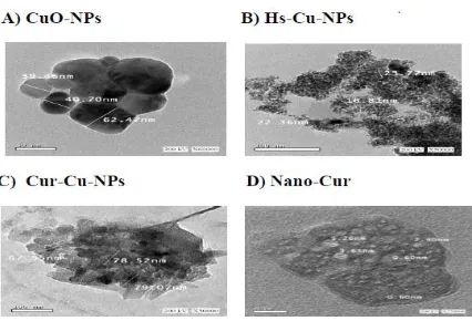
1. Nanoparticles characterization
High Resolution Transmission Electron Microscopy (HR-TEM): The morphology and
diameter of Nanoparticles were analyzed using HR-TEM. TEM average diameter was
calculated from measuring over 100 particles in random field of TEM view. TEM image
revealed that average particle size of CuO-NPs, HS-Cu-NPs, Cur-Cu-NPs and the formed
Nano-cur sodium salt were approximately around 39.46 - 62.47 nm, 16.81 - 23.77 nm, 67.55
-79.07 nm and 0.6 - 5.26 nm, respectively (fig.1).
2. In vitro study
Cytotoxic assay: Cell lines were treated with the different concentrations (25, 50, 100, 200
ug/ml) of different nano-complexes for 48h.The different nano complexes showed a clear
cytotoxic effect on breast carcinoma cell line (MCF-7) and a clear concentration-response
relationship. As shown in (fig.2), the calculated IC50 indicated that CuO-NCs and
HS-Cu-NCs were more cytotoxic to MCF-7, then Cur-HS-Cu-NCs followed by native-Cur-Cs. The least
effect exerted by Cur-Cu-NCs.
3. In vivo study
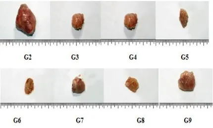
3.1. Effect of different nano-complexes on tumor assessment: The incidence of solid
and fast growing in G2, While G3, G4, G5, G6, G7, G8 and G9 showed a markedly smaller
and slower tumor growth compared to G2 (Fig.3).
Remarkably, as shown in table (1), the significant reduction in tumor weight were -78.7% for
Cur-Cu-NCs treatment followed by -69.1% for Cur-NCs treatment then G3 and G4 (chemical
treatments) recorded -64.2% and -61.8% respectively and finally G8 recorded -60% when
compared with G2. Whereas, groups administrated native-Cur-Cs either injection or oral
showed less impact on tumor weight reduction reached -58.2% and -43.6% respectively as
compared with G2 (P<0.05).
Moreover, significant and progressive tumor suppression in G3, G4, G5, G6, G7, G8 and G9
was also recorded with a percentage of significant reduction in tumor volume, 98.6%,
-98.6%, -99.2%, -98.9%, -98.5%, -98.5% and -97.5% respectively, when compared with G2
(P<0.05) (table.1).
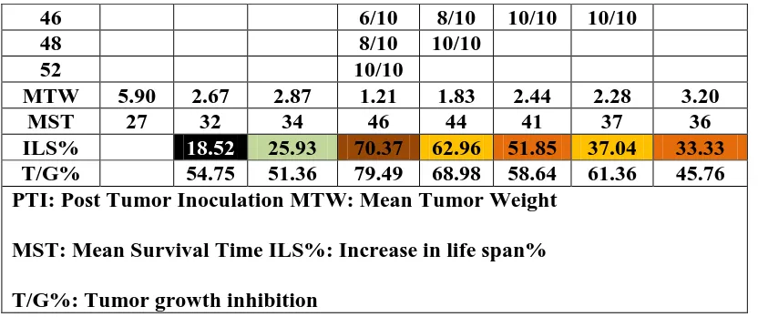
Similarly, results illustrated in table (2), show the inhibition of tumor growth (T/G %) in
different nano-complexes treatments in G3, G4, G5, G6, G7, G8 and G9. It was 54.8%,
51.4%, 70.37%, 62.9%, 51.9%, 37.04% and 33.3% when compared to G2 in the same order.
The previous results indicated that there was a significant increase in mean survival time
(MST) and increased life span (%ILS) in all TBM treated groups especially in G5. Cur-Cu
NCs treatment showed the highest prolongation of MST; (46 days) after that G6, G7 caused a
remarkable prolongation of MST (44 days) and (41days) respectively, then G8, G9 recorded
(37 days) and (36 days) respectively and finally G3 and G4 (chemical treatments) caused a
slight prolongation of MST; (32 days) and (34 days) respectively as compared with G2 (27
days), P<0.05.
3.2. Effect of different nano-complexes on serum tumor marker (CA 15-3) level and
(ALP) activity
Table (3) shows the Serum (CA 15-3) levels in different nano-complexes groups. Cur-Cu-
NCs treatment in G5 was the most effective treatment that caused a significant reduction in
serum CA 15-3 level by -87.1% as compared to G2. whereas, There was a significant
decrease in serum (CA 15-3) levels in G3 and G4 by -79.3% and -78.8% respectively while
Cur-NCs and native-Cur-Cs treatments caused a significant decrease in serum (CA 15-3)
levels in G6, G7, G8 and G9 by -79.6%, -41.7%, -63.7% and -23.1% respectively as
As indicated in table (3), regarding to ALP activity, ALP activity was significantly and
noticeably decrease in G5 (Cur-Cu- NCs) and G6 (Cur-NCs) to the control level by -61.2%
and -60.3% when compared to G2 (P<0.05). Chemical treatments in G3 and G4 caused a
significant decrease in ALP activity by -45.9% and -42.6% respectively while Cur-NCs and
native-Cur-Cs treatments in G7, G8 and G9 caused a significant decrease by -37.2%, -47.5%
and -30.6% respectively as compared to G2 (P<0.05).
3.3. Effect of different nano-complexes on cell apoptosis
a. In vitro study
As shown in figure (4), the alterations in nuclear morphology in response to different
nano-complexes treatments were assessed by nuclear counterstain (Hoschet 33342). Different
nano-complexes significantly reduced the number of colonies of breast carcinoma cell line
(MCF-7). Cur-Cu-NCs was the most effective cytotoxic treatment to MCF-7 as there were
less colonies and more nuclear condensation as compared to the control. Noteworthy,
CuO-NCs and HS-Cu-CuO-NCs treatments were more toxic to (MCF-7), then Cur-CuO-NCs and
native-Cur-Cs compared to the control.
b. In vivo study
1- Effect of different nano-complexes on Caspase-3 activity (Casp-3) in tumor tissues
Table (4) shows the results of caspase-3 activity in tumor tissues. There was a highly
significant increase of 481.4%, 431.4%, 550%, 262.9%, 190%, 171.4% and 111.4% of
caspase activity in tumor tissues of treated groups in G3, G4, G5, G6, G7, G8 and G9
respectively compared to G2 (P<0.05). Noticeably, Cur-Cu-NCs treatment was highly
apoptotic that caused a significant increase in caspase-3 activity in G5 compared to untreated
TBM group.
2-Effect of different nano-complexes on P53 gene expression in tumor tissues
Changes of the gene expression by different nano-complexes were shown in table (4).
Expression of P53 was examined by real time PCR. Results showed that the expression levels
of P53 were down regulated in TBM (G2) as compared to the control muscle (G1).
Furthermore, the levels of P53 was significantly increased more than the control level in
TBM treated with Cur-Cu-NCs in G5 and Cur-NCs in G6 by 2686.7% and 2240.7%
respectively as compared to G2 (P<0.05). In addition, increase in expression of P53 was
respectively as compared to G2, however, this increase still significantly less than the control
level.
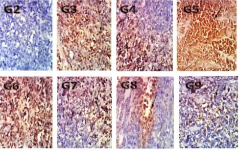
3- P53 Immunohistochemistry of Ehrlich solid tumor
Results of P53 gene expression were significantly confirmed by immunohistochemical
analysis where TBM received Cur-Cu-NCs and Cur-NCs highly expressed P53 (score 3,
+++) in G5 and G6, respectively (Fig. 5) compared to moderate expression (score 2, ++) of
P53 in TBM received CuO-NCs, HS-Cur-NCs, native-Cur-Cs and Cur-NCs in G3, G4, G7
and G8 respectively. Weak positive expression of P53 (score 1, +) was observed in G9
treated with oral Native-Cur-Cs as compared to negative staining of P53 in untreated TBM.
3.4. The effect of different nano-complexes on serum inflammation/immunologic
markers C-reactive protein (CRP) and interleukin-6 (IL-6) in experimental groups
Regarding to the inflammation immunologic markers, there was a significant increase in CRP
and IL-6 levels in untreated TBM. In contrast, Mice in G5 treated with Cur-Cu-NCs exhibited
a significant decrease in CRP and IL-6 levels by -82.8% and -39.5%, respectively, while
CuO NCs and HSCurNCs treatments caused a significant decrease in CRP levels by
-72.2% and -73.1% respectively and a marked significant decrease in IL-6 levels by -30.4%
and -31.02% respectively as compared to untreated group. A statistically significant reduction
in CRP levels by -69.2%, -43.6%, -59.03% and -26.4% in G6, G7, G8 and G9 as compared to
G2. Also there was a significant reduction in IL6 levels by 31.7%, 22.5%, 26.7% and
-10.6% in G6, G7, G8 and G9 as compared to G2 (table. 5).
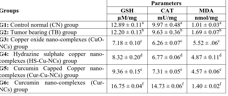
3.5. The effect of different nano-complexes on oxidative stress markers in liver tissues
Table (6) illustrates oxidative stress markers measured in liver tissue homogenates. GSH
content was considerably decreased in G2 by 5.4% relative to G1. Remarkably, CuO-NCs,
HS-Cu-NCs and Cur-Cu-NCs treatments caused a significant decrease in GSH content in G3,
G4 and G5 by -41.1%, -31.8% and -23.3%, respectively compared to G2. Cur-NCs and
native-Cur-Cs treatments caused a substantial elevation of GSH content in groups 6, 7, 8 and
9 by 37.3%, 33.6%, 29.9% and 28.2%, respectively as compared to G2, (P<0.05).
Regarding to CAT enzyme activity, it was found to be significantly lowered in G3, G4 and
G5 by- 34.9%, -29.7% and -24.1% correspondingly, when compared with G2. In G6, G7, G8
and G9, CAT activity in liver tissue homogenates was expressively increased by 52.9%,
As exhibited in table 6, a significant increase in MDA levels in G3, G4 and G5 by 226.6%,
188.2% and 170.4%, respectively which associated with a significant decrease in GSH
content and catalase activity. Predictably, Hepatic MDA levels in Cur-NCs and
native-Cur-Cs treatments were significantly lowered by -17.2%, -11.2%, -11.2% and -13.6% for G6, G7,
G8 and G9, respectively as compared to G2 (P<0.05).
Figure 1: HR-TEM image of the prepared NPs shows that: A) CuO-NPs with average
size 39.46 - 62.47 nm. B) HS-Cu-NPs with average size 16.81 - 23.77nm. C) Cur-Cu-NPs
[image:12.595.91.517.199.489.2]
Figure 2: Cytotoxic assay indicates the effect of different nano-complexes on surviving
fraction of MCH-7 cell line after 48 h at different concentrations.
Figure 3: Representative images illustrating the effect of different nano-complexes
treatments on solid tumor volume in different experimental groups (G2: TB, G3:
CuO-NCs, G4: HS-Cu-CuO-NCs, G5: Cur-Cu-CuO-NCs, G6: Cur-CuO-NCs, G7: Native-Cur-Cs, G8: O-
[image:13.595.85.515.409.668.2]
Figure 4: Effect of different nano-complexes on cell nuclei. Cells were stained with
Hoechst 33342 to image the nuclei. Condensed nuclei of (MCF-7) cell line indicate
apoptotic cells.
Figure 5: Photomicrographs represent immunohistochemistry staining of p53
expression of Ehrlich solid tumor from mice. G2: Shows negative immunohistochemical
reaction (no expression of P53) (score 0) in TBM. G3, G4, G7, G8: shows moderate positive
expression of P53 (score 2, ++) in TBM treated IT with CuO-NCs, HS-Cu-NCs, Native
[image:14.595.77.544.73.283.2] [image:14.595.78.546.359.656.2]
(score 3, +++) in TBM treated (IT) with Cur-Cu-NCs and Cur-NCs. G9 shows weak positive
expression of P53 (score 1, +) in TBM treated with Native O-Cur-Cs. Magnification is
(X400).
Table 1: Effect of different nanocomplexes on tumor weight (g) and tumor volume
(mm3) in experimental groups.
Groups
Parameters
Tumor weight Tumor volume
(g) (mm3)
G2: Tumor bearing (TB) group 3.85± 0.35a 4785.00 ± 539.86a
G3: Copper oxide nano-complexes (CuO-NCs)
group 1.38±0.29
b
66.77 ± 36.17b
G4: Hydrazine sulphate copper nano-complexes
(HS-Cu-NCs) group 1.47±0.26
bc
66.98 ± 35.02b
G5: Curcumin Capped Copper nano-complexes
(Cur-Cu-NCs) group 0.82±0.30
d
38.38 ± 12.32b
G6: Curcumin nano-complexes (Cur-NCs) group 1.19±0.32b 54.96 ± 45.83 b
G7: Native curcumin complexes (Native-Cur-Cs)
group 1.61±0.23
bc
73.79 ± 45.29b
G8:Oral curcumin nano-complexes (O-Cur-NCs)
group 1.54±0.19
bc
70.10 ± 42.37b
G9: native Oral curcumin complexes
(Native-O-Cur-Cs) group 2.17±0.30
c
117.42 ± 83.64b
• Values are mean ±SD.
•There is no significant difference between means having the same letter in the same column (p ≤0.05).
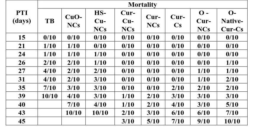
Table 2: Effect of different nanocomplexes on life span and tumor growth inhibition in
tumor bearing mice groups.
PTI (days)
Mortality
TB
CuO-NCs HS- Cu-NCs Cur- Cu-NCs Cur-NCs Cur-Cs O - Cur-NCs O- Native-Cur-Cs
15 0/10 0/10 0/10 0/10 0/10 0/10 0/10 0/10
21 1/10 1/10 0/10 0/10 0/10 0/10 0/10 0/10
24 1/10 1/10 1/10 0/10 0/10 0/10 0/10 0/10
26 2/10 2/10 1/10 0/10 0/10 0/10 0/10 1/10
27 4/10 2/10 2/10 0/10 0/10 0/10 1/10 1/10
31 4/10 2/10 3/10 0/10 0/10 0/10 1/10 2/10
35 7/10 3/10 3/10 0/10 0/10 2/10 2/10 2/10
39 10/10 4/10 3/10 1/10 2/10 3/10 3/10 3/10
40 7/10 4/10 1/10 2/10 4/10 3/10 5/10
43 10/10 10/10 2/10 3/10 6/10 6/10 7/10
[image:15.595.83.515.547.779.2]
46 6/10 8/10 10/10 10/10
48 8/10 10/10
52 10/10
MTW 5.90 2.67 2.87 1.21 1.83 2.44 2.28 3.20
MST 27 32 34 46 44 41 37 36
ILS% 18.52 25.93 70.37 62.96 51.85 37.04 33.33
T/G% 54.75 51.36 79.49 68.98 58.64 61.36 45.76
PTI: Post Tumor Inoculation MTW: Mean Tumor Weight
MST: Mean Survival Time ILS%: Increase in life span%
T/G%: Tumor growth inhibition
Table 3: The effect of different nano-complexes on serum tumor markers (CA 15-3) and
Alkaline phosphatase (ALP) activity in different experimental groups.
Groups
Parameter
CA 15-3 ALP
ng/ml U/L
G1: Control normal (CN) group 0.05 ± 0.05a 46.70 ± 2.22a
G2: Tumor bearing (TB) group 3.72 ± 0.11b 116.40 ± 9.53b
G3: Copper oxide nano-complexes (CuO-NCs) group 0.77 ± 0.09c 62.90 ± 5.63c
G4: Hydrazine sulphate copper nano-complexes
(HS-Cu-NCs) group 0.79 ± 0.07
cd
66.85 ± 5.64c
G5: Curcumin Capped Copper nano-complexes
(Cur-Cu-NCs) group 0.48 ± 0.03
e
45.20 ± 7.50a
G6: Curcumin nano-complexes (Cur-NCs) group 0.76 ± 0.08cd 46.25 ± 7.01a
G7: Native curcumin complexes (Native-Cur-Cs) group 2.17 ± 0.09f 73.10 ± 1.72d
G8:Oral curcumin nano-complexes (O-Cur-NCs) group 1.35 ± 0.07g 61.10 ± 5.09c
G9: native Oral curcumin complexes (Native-O-Cur-Cs)
group 2.86 ± 0.05
h
80.80 ± 9.27e
• Values are mean ±SD.
•There is no significant difference between means having the same letter in the same column (p ≤0.05).
Table 4: Effect of different nanocomplexes on P53 gene expression in experimental groups. Groups Parameter P53 gene expression Caspase-3 ng/100 mg
G1: Control normal (CN) group 1.00 ± 0.00a ---
G2: Tumor bearing (TB) group 0.15 ± 0.15a 0.70 ± 0.01a
G3: Copper oxide nano-complexes (CuO-NCs) group 0.56 ± 0.18a 4.07 ± 0.10b
G4: Hydrazine sulphate copper nano-complexes
(HS-Cu-NCs) group 0.58 ± 0.34
a
3.72 ± .09c
G5: Curcumin Capped Copper nano-complexes
(Cur-Cu-NCs) group 4.18 ± 1.21
b
[image:16.595.92.508.70.244.2]
G6: Curcumin nano-complexes (Cur-NCs) group 3.51 ± 1.99b 2.54 ± 0.06e
G7: Native curcumin complexes (Native-Cur-Cs) group 0.94 ± 0.40a 2.03 ± 0.06f
G8:Oral curcumin nano-complexes (O-Cur-NCs) group 0.98 ± 0.01a 1.90 ± 0.01g
G9: native Oral curcumin complexes (Native-O-Cur-Cs)
group 0.37 ± 0.25
a
1.48 ± 0.02h
• Values are mean ±SD.
•There is no significant difference between means having the same letter in the same column (p ≤0.05).
Table 5: The effect of different nano-complexes on serum inflammation/immunologic
markers C-reactive protein (CRP) and interleukin-6 (IL-6) in experimental groups.
Groups
Parameters
CRP IL-6
ng/ml Pg/ml
G1: Control normal (CN) group 0.11 ± 0.01a 115.10 ± 9.09a
G2: Tumor bearing (TB) group 2.27 ± 0.13b 254.07 ± 11.60b
G3: Copper oxide nano-complexes (CuO-NCs) group 0.63 ± 0.08c 176.82 ± 5.30c
G4: Hydrazine sulphate copper nano-complexes
(HS-Cu-NCs) group 0.61 ± 0.05
c
175.25 ± 1.30cd
G5: Curcumin Capped Copper nano-complexes
(Cur-Cu-NCs) group 0.39 ± 0.01
d
153.81 ± 6.60e
G6: Curcumin nano-complexes (Cur-NCs) group 0 .70 ± 0.04e 173.51 ± 4.38cd
G7: Native curcumin complexes (Native Cur-Cs) group 1.28 ± 0.05f 196.95 ± 2.92f
G8:Oral curcumin nano-complexes (O-Cur-NCs) group 0.93 ± 0.07g 186.30 ± 5.68g
G9: native Oral curcumin complexes (Native O-Cur-Cs)
group 1.67 ± 0.10
h
227.08 ± 2.62h
• Values are mean ±SD.
•There is no significant difference between means having the same letter in the same column (p ≤0.05).
Table 6: The effect of different nano-complexes on oxidative stress markers in liver tissues of experimental groups.
Groups
Parameters
GSH CAT MDA
µM/mg mU/mg nmol/mg
G1: Control normal (CN) group 12.89 ± 0.11a 9.97 ± 0.48a 1.01 ± 0.03a
G2: Tumor bearing (TB) group 12.20 ± 0.13b 9.63 ± 0.36b 1.69 ± 0.07b
G3: Copper oxide nano-complexes
(CuO-NCs) group 7.18 ± 0.10
c
6.26 ± 0.07c 5.52 ± .06c
G4: Hydrazine sulphate copper
nano-complexes (HS-Cu-NCs) group 8.32 ± 0.20
d
6.77 ± 0.06d 4.87 ± 0.11d
G5: Curcumin Capped Copper
nano-complexes (Cur-Cu-NCs) group 9.36 ± 0.15
e
7.31 ± 0.05e 4.57 ± 0.06e
G6: Curcumin nano-complexes
(Cur-NCs) group 16.75 ± 0.04
f
[image:17.595.82.525.579.767.2]
G7: Native curcumin complexes
(Native-Cur-Cs) group 16.30 ± 0.01
g
13.67 ± 0.14g 1.49 ± 0.02g
G8:Oral curcumin nano-complexes
(O-Cur-NCs) group 15.85 ± 0.15
h
12.80 ± 0.57h 1.50 ± 0.03gh
G9: native Oral curcumin complexes
(Native-O-Cur-Cs) group 15.64 ± 0.22
i
13.80 ± 0.31g 1.46 ± 0.03gh
• Values are mean ±SD.
•There is no significant difference between means having the same letter in the same column (p ≤0.05).
DISCUSSION
1. Nanoparticles Characterization
Bioavailability of a drug to the cells, whether in vitro or in vivo, is critical for its optimal
efficacy. To enhance the solubility of drugs in aqueous solvents, increase their
bioavailability, enhance serum half-life, for tumor cell targeting and bioimaging,
nanotechnology has recently emerged as a new technology of choice.[35]
Characterization of different NCs was done using HR-TEM technique in order to provide
clear insight into morphology and particle size. The present study revealed that average
particle size of CuO-NPs were approximately around 39.46-62.47 nm and spherical in shape
with a smooth surface. This is very similar to those described in the previous studies.[36]
Whereas, TEM image showed that average particle size of HS-Cu-NPs were 16.81-23.77 nm
and decahedran in shape with irregular surface. It was reported that Cu activates hydrazines
to free radical species.[37]
Our study revealed that TEM image for Cur-Cu-NPs and the formed Nano-cur sodium salt
were 67.55-79.07 nm and 0.6-5.26 nm respectively. The formed nanocurcumin sodium salts
were nanorods with smooth surface. Basicity showed high pH = 9.5 due to the formed
Nano-Cur sodium salt and excess of sodium bicarbonate. This high basicity has been characterized
by quick solubility in water and its high penetration through cell wall. Confirming this result,
Basniwal et al. (2011) found that Nano-Cur prepared by wet-milling technique, in size range
of 2–40 nm was shown to express stronger antimicrobial potential and anti-cancer activity as
2. Cytotoxic effect of different nanocomplexes
Data presented in the current study demonstrated that different nano complexes showed a
clear cytotoxic effect on breast carcinoma cell line (MCF-7) and a clear
concentration-response relationship.
In a similar study, Khosropanah et al. (2016) formulated Cur nanoparticles to increase its
bioavailability and to study the effect on breast cancer cells. More than 50% of the tumor
cells died within 48 hours after the administration of Cur. The dosage of Nano-Cur in this
study was effective in half the dosage of the regular preparation of native-Cur.[39]
In a previous study, Breast cancer cells treated with different concentrations of native-Cur
resulted in the inhibition of cell proliferation in a dose- and time-dependent manner. The
literature reveled that Cur is a potent anticancer agent because of its ability to obstruct
various biochemical pathways which are associated with the proliferation of cancer cells by
binding with the various targets.[40]
It is evident from the current study that chemical treatment by CuO-NPs generated
cytotoxicity. CuO-NPs Oxidative stress has been suggested to play an important role in the
toxicity mechanisms of nanoparticles. This has been attributed due to their small size and
large surface area which is generally thought to generate ROS. ROS such as superoxide anion
(O2-), hydroxyl radical (HO•) and hydrogen peroxide (H2O2) elicit a variety of physiological
and cellular events including inflammation, DNA damage and apoptosis.[7] CuO-NPs were
found to induce cytotoxicity in a human liver carcinoma cell line (HepG2) in a
dose-dependent manner, which was probably mediated through ROS generation and oxidative
stress.[41]
Regarding to HS-Cu-NCs treatment, an important distinguishing feature of cancer cells is
their propensity to obtain energy through the anaerobic metabolism of glucose. The enzyme
phosphoenol pyruvate carboxykinase played very important role in gluconeogenesis and
proposed that inhibition of this enzyme would impede gluconeogenesis and reduce the
severity of cachexia. Hydrazine sulfate is thought to interfere with gluconeogenesis.
Hydrazine, a metabolite of HS, has been reported to have cytotoxic effects on hepatocyte cell
Our result demonstrates the efficacy of Cur with promising antiangiogenic and
antiproliferative potential as compared to Cur-Cu-NPs. While describing the possible
mechanism, it has been illustrated that Cur analogues are found to be less active than Cur in
suppressing nuclear factor kappa-light-chain-enhancer of activated B cells (NF-kB) activation
and tetrahydro-curcumin was found to be less active than Cur in preventing chemical-induced
skin tumor promotion in mice due to the reduction of the carbonyl group.[43]
3. Antitumor effect of different nanocomplexes
Our data also showed that there was significant reduction in tumor weight and volume in all
treated TBM groups. Moreover, the previous results indicated that there was a significant
increase in mean survival time (MST) and increased life span (%ILS) in all TBM treated
groups especially in G5. Cur-Cu-NCs were the most effective treatment followed by Cur-
NCs then CuO-NCs then HS-Cu-NCs and finally Native-Cur-Cs. These results summarized
that intratumoral (IT) treatment with Cur-Cu-NCs and Cur-NCs have a stronger antitumor
effect than chemical IT treatments (CuO-NCs and HS-Cu-NCs) and native curcumin
treatment either IT or oral.
The transcription factor (NF-κB) has the main role in the creation of tumors and
inflammation, and the goal of most pharmaceutical and entomological preparations is to
reduce its hyper productivity. Cur is a natural product which reacts with a large variety of
compounds in the downstream of the NF-κB pathway. Cur blocks IKB kinase (IKK)
activation, phosphorylation, and the degradation of nuclear factor kappa light polypeptide
gene enhancer in B cells inhibitor, alpha (IĸBα). A large number of Cur analogues were
investigated in order to improve its efficiency in blocking NF-ĸB.[44]
The current study goes with the study of Shahani et al. who found that A number of previous
studies have shown that Cur exhibit poor bioavailability in the human body when
administered orally. It is due to their rapid degradation and poor absorption in the
gastrointestinal tract, which results in low plasma concentrations and a very low distribution
in tissues. In addition to oral intake, injection entry was investigated and has proven effective
in keeping curcumin in tissues for a longer period. Intravenous intake nanoparticles has been
proven to be effective for the treatment of tumors in animals.[45]
In a similar study, Wanninger et al. also reported that Cur and the curcuminoids should be
complexes. Cu (II) curcumin complexes turned out to exhibit the highest selective
cytotoxicity in vitro.[46] The Cu (II) complexes have increased solubility and crystallinity due
to blocking of the phenolic –OH groups through alkylation. These complexes showed
significantly enhanced antitumor activity against human cancer cell lines in comparison with
the free ligands.[47] These findings indicate why Cur-Cu NCs was the effective treatment as
antitumor in our study.
Cu is an essential trace element that is widely distributed throughout the body and forms the
essential redox–active reaction center in a variety of metalloenzymes. Cu concentration is
obviously altered in tumors, and that serum concentrations are correlated with tumor
incidence, progression and recurrence in a large number of human tumors.[49] Previous study
reported that CuO-NPs can induce cancer cells apoptosis through a mitochondrion-mediated
apoptosis pathway, which raises the possibility that CuO-NPs could be used to cure
melanoma and other cancers.[48]
Gratefully, tumor hypoxia can be exploited to develop prodrugs that become activated in the
reducing environment of cancer cells. In this concern, Cu is very appealing because it can
exist under two different oxidation states in cells. The anoxic character of cancer cells
promotes the reduction of Cu (II) to Cu (I), which is not possible in normal healthy cells and
thus provides a therapeutic opportunity to target tumors.[49] Cu (I) can catalyze the formation
of reactive oxygen and nitrogen species (ROS and RNS), to induce a pro-apoptotic oxidative
stress.[50]
HS is active metabolite and that it may normalize the carbohydrate metabolism of cancer
patients with cachexia. Our results are consistent with previous result suggesting that HS
administered to rats with transplanted tumors inhibited tumor growth and increased
survival.[51]
Regarding to tumor marker, in the current study, Cur-Cu-NCs treatment in G5 was the most
effective treatment that caused a significant reduction in serum CA 15-3 level. Chemical
treatmens (G3 and G4) recorded a significant decrease in serum (CA 15-3) levels less than
Cur-Cu-NCs treatment in G5. Also, Cur NCs treatment in G6 caused a significant decrease in
serum (CA 15-3) levels very close to chemical treatments levels compared to TBM group.
compared to Cur NCs and other NCs treatments. These results were supported by our
results.[52]
CA15-3 (also known as mucin 1) is overexpressed in human breast cancers and in their
subsequent metastases.[53] CA15-3 promotes tumor invasion and metastasis through
activation of the mitogen-activated protein kinase signaling pathway[53] and down regulation
of E-cadherin.[54] Previous studies suggested that carbohydrate antigen 15-3 (CA15-3) is
predictive marker of radiological response in metastatic breast cancer.[55]
Regarding to ALP activity, ALP activity was significantly and noticeably decreased due to
Cur-Cu-NCs and Cur-NCs treatments to the control level. Chemical treatments (CuO-NCs
and HS-Cu NCs) caused a significant decrease in ALP activity but not to the control level
while Cur NCs (oral) and native Cur Cs (IT or oral) treatments caused less decrease in ALP
activity as compared to TBM.
Our results are in agreement with an in vivo study reported that Nano-Cur supplementation
prevented the increase in such hepatic enzymes, especially in groups received Nano-Cur after
tumor induction, suggesting that Nano-Cur may have a potential protective effect against
liver damage.[56]
It is clear from the current study that Cur-Cu-NCs was the most effective treatment and
decreased ALP activity to the control level, that may be due to the protective and antioxidant
effect of nanocurcumin which abolished almost the harmful effects of CuO-NPs treatments.
Our results are in agreement with previous results investigating oxidative stress role of
N-acetyl-cystein (NAC) in the cytotoxicity of CuO NPs, results showed that NAC abolished
almost fully the harmful effect of CuO NPs at all concentrations studied when HepG2 cells
were exposed to CuO NPs in the presence of the NAC.[41]
Previous studies demonstrated that vit C induce apoptosis in cancer cells by creating
oxidative stress via upregulation of reactive oxygen species (ROS) release.[57] Furthermore,
using high dosage of vit C as an anti-cancer therapy has shown to reduce cancer cell growth
and lessen chemo-therapy side effects such as nausea, fatigue, pain and depression.[11]
4. Apoptotic effect of different nanocomplexes
suppressor gene is a frequent event in tumorigenesis. Interestingly, mutations in the p53 gene
were shown to occur at different phases of malignant transformation, thus contributing
differentially to tumor initiation, promotion, aggressiveness, and metastasis.[58]
The role of Cur in triggering apoptosis has been investigated in numerous studies, and there is
a range of evidence demonstrating its potential to activate different pathways related to
apoptosis. Interestingly, it has been revealed that Cur-mediated apoptosis induction in cancer
cells occurs in a p53-dependent mode.[59]
Moreover, Balasubramanyam et al. previously demonstrated that Cur could inhibit
p300-specific acetylation of p53, which may be helpful in the acetylation-dependent regulation of
p53 function; this causes Cur, which targets p300 to serve as a lead compound in cancer
suppression [60]. Therefore, it is concluded that one of the Cur pathways which play a role in
cancer suppression is modulations of the transcriptional co-activating proteins mediating the
p53 gene level, which promotes invasion, metastasis and a metabolic shift to an anaerobic
process known as the 'Warburg effect'. Gratefully, tumor hypoxia can be exploited to develop
pro-drugs that become activated in the reducing environment of cancer cells.[61]
Current study illustrated that the expression levels of P53 were down regulated in TBM (G2)
as compared to the control muscle (G1). Furthermore, the levels of P53 were significantly
increased more than the control level in TBM treated intratumorally with Cur-Cu NCs and
Cur NCs in G5 and G6, respectively as compared to G2. Moreover, increase in P53
expression was noticed in TBM treated with chemicals (G3, G4), TBM treated with Cur NCs
orally and TBM treated with Cur Cs (IT or oral) as compared to G2, but, the increase in P53
expression was less than the control level.
Results of P53 gene expression were significantly confirmed by immunohistochemical
analysis where TBM received Cur-Cu NCs and Cur NCs highly expressed P53 in G5 and G6
respectively compared to moderate expression of P53 in TBM received chemical treatments
(CuO-NCs and HS-Cur-NCs), native Cur-Cs (IT) and Cur-NCs (oral) in G3, G4, G7 and G8
respectively. Whereas, G9 treated with oral native Cur-Cs observed minimized alteration in
expression of p53 as compred to negative staining of P53 in untreated TBM.
Moreover, our results are consistent with the previous results suggesting that the expressions
cleaved caspase-3) were up-regulated while the expression of anti-apoptotic gene bcl-2 was
downregulated in HepG2 cells treated with CuO-NPs. It was suggested that bax is
up-regulated by p53. Since an increase in bax expression was noticed, the role of p53 in the
upregulation of bax upon CuO-NPs exposure can be postulated. The insertion of bax into the
mitochondrial membrane possibly leads to p53-mediated apoptosis.[62] Caspases are activated
during apoptosis in many cells and are known to play a vital role in both initiation and
execution of apoptosis.[41]
Furthermore, other authors observed that CuO-NPs targeted the mitochondria of HeLa cells
in vitro, which resulted in the release of cytochrome C from the mitochondria and the
activation of caspase-3 and caspase-9 after the CuO-NPs entered the cells.
Our result revealed that there was a highly significant increase of caspase-3 activity in tumor
tissues of treated groups in G3, G4, G5, G6, G7, G8 and G9 respectively compared to G2.
Noticeably, Cur-Cu-NCs treatment was highly apoptotic that caused a significant increase in
caspase-3 activity in G5 more than other treatments as compared to untreated TBM group.
Increase in caspase-3 activity in tumor tissues of TBM treated with chemicals was
significantly more than its activity in TBM treated with Cur-NCs (IT or oral) and
native-Cur-Cs (IT or oral). Furthermore, Cur-Nnative-Cur-Cs (IT) treatment caused a significant increase in
caspase-3 activity very close to its activity exerted by chemical treatments.
In the present study, it was observed that the changes in nucleus following NCs treatment on
MCF-7 cell line. Hoschet 33342 is cell-permeant nuclear counterstain that emits blue
fluorescence when bound to dsDNA. For this reason, this dye was used to distinguish
condensed pycnotic nuclei in apoptotic cells. It is clear that Cur-Cu-NCs was the most
effective cytotoxic treatment to MCF-7 as there were less colonies and more nuclear
condensation as compared to the control. Noteworthy, CuO-NCs and HS-Cu-NCs treatments
were more toxic to (MCF-7), then Nano-Cur-NCs and Native-Cur-Cs compared to the
control. This observation is consistent with the data obtained by Wang et al.[48]
5. Anti-inflammatory activity of different nanocomplexes
Regarding to the inflammation immunologic markers, there was a significant increase in CRP
and IL-6 levels in untreated TBM. In contrast, Mice in G5 treated with Cur-Cu-NCs exhibited
a significant decrease in CRP and IL-6 levels. Then, CuO-NCs and HS-Cur-NCs treatments
but the reduction was less than Cur-Cu-NCs treated group. A statistically significant
reduction in CRP and IL-6 levels was observed in all groups treated with Cur-NCs or native
Cur-Cs as compared to G2.
Cur is described as a potent inhibitor of angiogenesis. It is reported that Cur can
down-regulate all positive regulators of angiogenesis including cytokines, such as IL-6 can act
either as stimulators or inhibitors, depending on their amounts, the tumor site, and the tumor
microenvironment. These cytokines play a pivotal role in promotion of tumor growth,
particularly by recruiting massive vasculature.[63]
Moreover, our results are consistent with previous results suggesting that Nano-Cur,
polymeric nanoparticle encapsulated Cur, readily dispersed in aqueous media and with
confirmed anti-cancer potentials in preclinical in vivo models. Nano-Cur retained the
mechanistic specificity of free Cur, inhibiting the activation of the seminal transcription
factor NF-κB and reducing steady state levels of pro-inflammatory cytokines like ILs and
TNF-α.[64]
Previous study demonstrated that one of the main metabolic pathways for hydrazine
derivatives leads to formation of various free radical species in vitro and in vivo. Cu, is
known to activate hydrazines to free radical species and have been shown to induce DNA
damage. Thus, the oxidative metabolism of hydrazines by copper and consequent formation
of reactive species may contribute significantly to the pathophysiology of hydrazines in
humans.[37]
6. Antioxidant effect of different nanocomplexes
Regarding to GSH content and CAT enzymes activity, it was found to be significantly
lowered in chemically treated groups (G3, G4) and G5 which is treated with Cur-Cu NPs.
when compared with G2. Also, there was a significant increase in MDA levels in G3, G4 and
G5 which associated with a significant decrease in GSH content and catalase activity.
Predictably, Hepatic MDA levels in Cur-NCs and Native Cur-Cs treated groups were
significantly lowered.
Cur has been found to be an excellent scavenger of most ROS.[65] The reaction of peroxyl
radicals with Cur produces Cur phenoxyl radicals, which are less reactive than the peroxyl
reaction of phenoxyl radicals back to Cur by water soluble antioxidants like ascorbic acid,
impart the molecule with a chain breaking antioxidant ability.[66] It was also documented that
Cur has a property of donating electrons in order to neutralize free radicals by creating stable
products, and thus breaking a chain reaction of creating free radicals in a living organism.
Cur’s ability of capturing hydrogen peroxide is higher than that of the commercial
antioxidants at the same concentration.[67]
Recently, Assadian et al., also reported that in vitro cytotoxicity of CuO-NPs was associated
with significant increase at intracellular ROS level with effective induction of oxidative
stress.[68] The capability of NP to produce free radicals is one of the primary mechanisms of
NPs toxicity. It may result in oxidative stress, inflammation, and consequent damage to
proteins, membranes, and DNA.[69]
In a recent study, Thit et al. aimed to determine the role of ROS release and establish the
sequence of events during CuO-NP toxicity. Results showed that CuO-NPs were more toxic
than Cu2+, due to the increase in the generation of ROS, DNA damage and decrease levels of
GSH compared to control.[70]
It is well documented that free radical species are very reactive and bind irreversibly to
cellular macromolecules, causing inhibition of cellular functions and inducing profound
cellular damage. Primary free radicals, e.g., alkyl radicals, react with molecular O2, leading to
the formation of reactive oxygen-derived species, superoxide anion radical (O2.-), hydrogen
peroxide (H2O2) and, eventually, to the highly reactive hydroxyl radical (.OH). Alternatively,
primary radicals can eliminate hydrogen atoms from membrane lipids, inducing peroxidation
and decomposition of lipid membranes and compromising cellular functions. Reactive
oxygen species formation has been shown to induce “oxidative stress” where the production
of oxidant overwhelms antioxidant defense mechanisms. Oxidative stress is known to exhaust
reduced glutathione in cells, compromising cellular integrity.[71]
Hydrazine derivatives have been shown to deplete glutathione and cause oxidative stress.
Therefore, formation of free radical species during the biotransformation of hydrazines may
be very important in the toxicity and pathophysiology of hydrazines. The formation of
oxygen radicals and metal/peroxo species from hydralazine has been involved in DNA strand
scission, and it has been suggested that these reactive species may also form oxidation
CONCLUSION
Drug conveyance frameworks utilizing nanoparticles are appreciated as a promising
methodology for enhancing the safety and bioavailability of curcumin. The basic aim of the
present study was to compare the efficacy of different nanocomplexes as a potential
anticancer and antiangiogenic agent in concert with native curcumin. Cur-Cu-NCs treatment
was the most effective one as antitumor. Our results confirmed that Cu (II) curcumin
complexes turned out to exhibit the highest selective cytotoxicity and also showed significant
reduction in solid tumor volume in ascites tumor-bearing mice without harmful effects on
liver and kidney function. Also, Nano-Cur appeared to be an effective free radical quencher
with antioxidant activities, and capable of inhibiting oxidative stress, as it could protect mice
liver from chemical treatments induced altered hepatic functioning. Our study provided
valuable insights into the possible mechanism of CuO-NCs and HS-Cu-NCs cytotoxicity in
tumor cells. Furthermore, the antitumor effect of chemical treatments was very close to
Nano-Cur effect, but chemical treatment adversely affected liver and kidney function. We
suggest that Cur-Cu-NCs, which are a potentially safe and inexpensive for clinical use, may
be considered as an effective chemopreventive agent against tumors with more protective
rather than therapeutic action.
ACKNOWLEDGEMENT
I am deeply grateful to Allah. Coming from the intense gratification of my supervisors whom
their contribution has raised the quality of this research. They have always supported me and
have given me enthusiasm for science. They have patiently guided me. I owe them the
greatest degree of appreciation. I am thankful to Prof.Dr/ Tahani Elsayed, professor of
biochemistry, for her assistance throughout the research work. I am sincerely thankful to
Pro.Dr/ Gehan Salah, professor of nutrition, for her assistance and her kind support. My
thankfulness is also to Pro.Dr/Abdelfattah Badawi, professor of applied surfactant laboratory,
for his great support.
REFERENCES
1. Siegel RL, Miller KD, Jemal A. Cancer statistics, 2017. CA Cancer J Clin, 2017; 67(1):
7-30.
2. Amreddy N, Babu A, Muralidharan R, et al. Recent Advances in Nanoparticle-Based
Cancer Drug and Gene Delivery. In: Advances in Cancer Research. Elsevier, 2018; 137:
3. Ruan S, He Q, Gao H. Matrix metalloproteinase triggered size-shrinkable gelatin-gold
fabricated nanoparticles for tumor microenvironment sensitive penetration and diagnosis
of glioma. Nanoscale, 2015; 7(21): 9487-9496.
4. Jovičić D, Jozinović A, Grčević M, Spaseska Aleksovska E, Šubarić D. Nutritional and
health benefits of curcumin. Hrana u Zdr i Boles Znan časopis za Nutr i dijetetiku, 2017;
6(1): 22-27.
5. Basniwal RK, Khosla R, Jain N. Improving the anticancer activity of curcumin using
nanocurcumin dispersion in water. Nutr Cancer, 2014; 66(6): 1015-1022.
6. Asti M, Ferrari E, Croci S, et al. Synthesis and characterization of 68Ga-labeled
curcumin and curcuminoid complexes as potential radiotracers for imaging of cancer
and Alzheimer disease. Inorg Chem, 2014; 53(10): 4922-4933.
7. Vinardell M, Mitjans M. Antitumor activities of metal oxide nanoparticles.
Nanomaterials, 2015; 5(2): 1004-1021.
8. Nagajyothi PC, Muthuraman P, Sreekanth TVM, Kim DH, Shim J. Green synthesis:
in-vitro anticancer activity of copper oxide nanoparticles against human cervical carcinoma
cells. Arab J Chem, 2017; 10(2): 215-225.
9. Sinha BK, Mason RP. Biotransformation of hydrazine dervatives in the mechanism of
toxicity. J Drug Metab Toxicol, 2014; 5(168171): 8.
10. Park S. The effects of high concentrations of vitamin C on cancer cells. Nutrients, 2013;
5(9): 3496-3505.
11. Fritz H, Flower G, Weeks L, et al. Intravenous vitamin C and cancer: a systematic
review. Integr Cancer Ther, 2014; 13(4): 280-300.
12. Council NR, others. Nutrient Requirements of Laboratory Animals. National Research
Council, 1962.
13. Kimoto E, Tanaka H, Gyotoku J, Morishige F, Pauling L. Enhancement of antitumor
activity of ascorbate against Ehrlich ascites tumor cells by the copper:
glycylglycylhistidine complex. Cancer Res, 1983; 43(2): 824-828.
14. Negm NA, Morsy SMI, Said MM. Corrosion inhibition of some novel hydrazone
derivatives. J Surfactants Deterg, 2005; 8(1): 95-98.
15. Kamble S, Utage B, Mogle P, et al. Evaluation of curcumin capped copper nanoparticles
as possible inhibitors of human breast cancer cells and angiogenesis: a comparative
study with native curcumin. AAPS PharmSciTech, 2016; 17(5): 1030-1041.
hepatocellular carcinoma in rats. Int J Pharm Pharm Sci, 2014; 6(3): 54.
17. Basniwal RK uma., Khosla R, Jain N. Improving the anticancer activity of curcumin
using nanocurcumin dispersion in water. Nutr Cancer, 2014; 66(6): 1015-1022.
doi:10.1080/01635581.2014.936948.
18. Muthuraman P, Enkhtaivan G, Bhupendra M, Chandrasekaran M, Rafi N, Kim DH.
Investigation of the role of aspartame on apoptosis process in HeLa cells, Saudi J. In:
Biol. Sci, 2015.
19. Narang AS, Desai DS. Anticancer drug development. In: Pharmaceutical Perspectives
of Cancer Therapeutics. Springer, 2009; 49-92.
20. Dayem SAE, Foda F, Helal M, Zaazaa A. The role of catechin against
doxorubicin--induced cardiotoxicity in Ehrlich Ascites Carcinoma Cells (EAC) bearing mice. J Am
Sci, 2010; 6(4).
21. Jensen MM, Jørgensen JT, Binderup T, Kjær A. Tumor volume in subcutaneous mouse
xenografts measured by microCT is more accurate and reproducible than determined by
18 F-FDG-microPET or external caliper. BMC Med Imaging, 2008; 8(1): 16.
22. Ayyad S-EN, Abdel-Lateff A, Alarif WM, Patacchioli FR, Badria FA, Ezmirly ST. In
vitro and in vivo study of cucurbitacins-type triterpene glucoside from Citrullus
colocynthis growing in Saudi Arabia against hepatocellular carcinoma. Environ Toxicol
Pharmacol, 2012; 33(2): 245-251.
23. Lüftner D, Cheli C, Mickelson K, Sampson E, Possinger K. ADVIA
Centaur®HER-2/neu shows value in monitoring patients with metastatic breast cancer. Int J Biol
Markers, 2004; 19(3): 175-182.
24. Zawta B, Klein G, Bablok W. Temperature conversion in clinical enzymology. Klin lab,
1994; 40: 33-42.
25. Sandhu LC, Warters RL, Dethlefsen LA. Fluorescence studies of Hoechst 33342 with
supercoiled and relaxed plasmid pBR322 DNA. Cytom J Int Soc Anal Cytol, 1985; 6(3):
191-194.
26. Kaushal V, Herzog C, Haun RS, Kaushal GP. Caspase protocols in mice. In: Caspases,
Paracaspases, and Metacaspases. Springer, 2014; 141-154.
27. Bassiony H, Sabet S, El-Din TAS, Mohamed MM, El-Ghor AA. Magnetite
nanoparticles inhibit tumor growth and upregulate the expression of P53/P16 in Ehrlich
solid carcinoma bearing mice. PLoS One, 2014; 9(11): e111960.
28. Kabel AM. Effect of combination between methotrexate and histone deacetylase
29. Das REG, Poole S. The international standard for interleukin-6: evaluation in an
international collaborative study. J Immunol Methods, 1993; 160(2): 147-153.
30. Ridker PM, Buring JE, Shih J, Matias M, Hennekens CH. Prospective study of
C-reactive protein and the risk of future cardiovascular events among apparently healthy
women. Circulation, 1998; 98(8): 731-733.
31. Beutler E. Improved method for the determination of blood glutathione. J lab clin Med,
1963; 61: 882-888.
32. Aebi H. Catalase in vitro Methods Enzymol 105: 121--126. Find this Artic online, 1984.
33. Draper HH, Hadley M. A review of recent studies on the metabolism of exogenous and
endogenous malondialdehyde. Xenobiotica, 1990; 20(9): 901-907.
34. Levesque R. Programming and Data Management for SPSS Statistics 17.0: A Guide for
SPSS Statistics and SAS Users. SPSS, Chicago, 2007.
35. Ravindran J, Nair HB, Sung B, Prasad S, Tekmal RR, Aggarwal BB. RETRACTED:
Thymoquinone poly (lactide-co-glycolide) nanoparticles exhibit enhanced
anti-proliferative, anti-inflammatory, and chemosensitization potential, 2010.
36. Nasrollahzadeh M, Sajadi SM, Rostami-Vartooni A, Khalaj M. Green synthesis of
Pd/Fe3O4 nanoparticles using Euphorbia condylocarpa M. bieb root extract and their
catalytic applications as magnetically recoverable and stable recyclable catalysts for the
phosphine-free Sonogashira and Suzuki coupling reactions. J Mol Catal A Chem, 2015;
396: 31-39.
37. Sinha BK, Mason RP. Biotransformation of Hydrazine Dervatives in the Mechanism of
Toxicity. J Drug Metab Toxicol, 2014; 52(3): 1-6. doi:10.4172/2157-7609.1000168.
38. Basniwal RK, Buttar HS, Jain VK, Jain N. Curcumin nanoparticles: preparation,
characterization, and antimicrobial study. J Agric Food Chem, 2011; 59(5): 2056-2061.
39. Khosropanah MH, Dinarvand A, Nezhadhosseini A, et al. Analysis of the
antiproliferative effects of curcumin and nanocurcumin in MDA-MB231 as a breast
cancer cell line. Iran J Pharm Res, 2016; 15(1): 231-239.
40. Kumaravel M, Sankar P, Rukkumani R, others. Antiproliferative effect of an analog of
curcumin bis-1, 7-(2-hydroxyphenyl)-hepta-1, 6-diene-3, 5-dione in human breast
cancer cells. Eur Rev Med Pharmacol Sci, 2012; 16(14): 1900-1907.
41. Siddiqui MA, Alhadlaq HA, Ahmad J, Al-Khedhairy AA, Musarrat J, Ahamed M.
Copper Oxide Nanoparticles Induced Mitochondria Mediated Apoptosis in Human