STUDY OF STRESS ON CYTOKINE MEDIATED AUTOIMMUNE
THYROIDITIS
Vinod Singh*1, Mayuri Khare1, M. K. Sahu2, Poonam Singh3
1
Department of Microbiology, Barkatullah University, Bhopal, (M.P), India. 2Jawaharlal
Nehru Cancer Hospital, Bhopal, (M.P), India. 3
ClinicalPsychologist, Bhopal, (M.P), India.
ABSTRACT
Autoimmune thyroid diseases are the most common organ-specific
autoimmune disorders. Approximately 5% of the overall population
suffers from this disorder. Hashimoto's thyroiditis (HT) or
hypothyroidism is the most common clinical expressions of thyroid
dysfunction but differ clinically as well as in pathophysiology. It is the
major autoimmune endocrine disorder which is linked to human
immune system under psychological stress. The study focuses on the
level of thyroid and stress hormone and its impact on the immune
system. Two hundred human subjects along with normal healthy
population were randomly selected on the basis of clinical
examinations. Hormonal and cytokine analysis was conducted of both
thyroid and stress physiology, both pro and anti-inflammatory status was performed using
serum samples. Statistical analysis was performed of all the parameters.The study depicts
significant increase in TSH and decrease in T3 and T4 hormones also rise in stress hormones
was observed. Rise in the hormones resulted in significant increase in the level of
pro-inflammatory cytokines TNF-α and IFN-γ and decrease in anti-pro-inflammatory cytokines IL-4,
IL-6 and IL-10. Individuals‟ long term effects of relationship between psychosocial and
physiological stress conditions (i.e., genetics, constitutional factors) and disease is affected by
the biological nature, number, and persistence which may influence the course of chronic
disease. Therapies in development of biopsychosocial model provide a basis for
understanding and treatment of disease, the impact of illness from a societal perspective.
There exists a considerable amount of data on the psychosocial factors related to thyroid
disorders.
Volume 4, Issue 11, 1800-1815. Research Article ISSN 2277– 7105
*Correspondence for Author
Vinod Singh
Department of
Microbiology, Barkatullah University, Bhopal, (M.P), India.
Article Received on 16 Sept. 2015,
KEYWORDS: autoimmunity, anti-inflammatory, cytokines, hashimoto‟s thyroiditis, pro-inflammatory, psychological-physiological stress.
INTRODUCTION
All living organisms maintain a complex dynamic equilibrium, or homeostasis, which is
constantly challenged by internal or external adverse effects. Stress is defined as a state in
which homeostasis is actually threatened or perceived to be so.[1] Homeostasis is
reestablished by a complex repertoire of behavioral and physiological adaptive responses of
the organism.[2]
Stress may cause immunodepression but may also exert an immunoenhancing effect on cell
numbers same stressor may have a positive effect rather than a negative one, depending on its
duration or intensity.[3] According to investigations, stress has been classified as acute
laboratory stress and natural stress. Stressor induced neurosensory signals are processed in
the paraventricular nucleus (PVN) of the hypothalamus. In response to stressors,
hypothalamus secrets corticotrophin-releasing hormone (CRH) and arginine vasopressin
(AVP). CRH-containing neurons have different pathways and projections to noradrenergic
centers in the brain stem and spinal cord.[4] CRH activates hypothalamic pituitary axis (HPA),
leading to release of peptides from the pituitary and adrenocorticotropic hormone,
enkephalins, and endrophins. Adrenocorticotropic hormone induces release of
glucocorticoids from the adrenal cortex and CRH and central nervous system (CNS) together
stimulate noradrenergic neurons resulting in secretion of norepinephrine (NE) by peripheral
sympathetical nervous system (SNS) and release of epinephrine (EPI) from the adrenal
medulla. The activation of these two neurochemical pathways and release of hormones and
transmitters have profound effects of immune function.[5]
Stress hormones influence numerous physiologic processes; they regulate inflammatory
diseases, their effects maintaining balance between cell-mediate and humoral immunity and
on neurogenic inflammation in peripheral tissues. The hypothalamic-pituitary-adrenal (HPA)
axis and SNS represent the peripheral stress system, its activation occurs in CNS in response
to distinct blood-borne, neurosensory signals.[6, 7] Homeostasis within the immune system is
largely dependent on cytokines, the chemical messengers between immune cells, which play
crucial roles in mediating inflammatory and immune responses, for instance, immune
and IL-6 and activates HPA axis and stimulates the hypothalamic stress response.[8] The HPA
axis regulates a wide variety of immune functions affecting cell trafficking, migration,
maturation and differentiation; this regulation is the result of several neuroendocrine
pathways including hormones.
Thyroid dysfunction is an important cause of depression.[9] Hypothyroidism is considered a
potentially reversible cause of depression, and both disorders have symptoms that may
complicate studies attempting to clarify the relationship between them.[10,11] The changes
occur due to hormone involving both stress and thyroid hormones, among these cortisol,
prolactin, thyroid stimulating hormone (TSH), triiodothyronine (T3), and thyroxine (T4) are
the major.[12]
Stress induces changes in the secretion of several hormones, which affect immune function
by either increasing or decreasing immune activity. The thyroid hormones are essential for
the maintenance of neurotransmitters associated with stress, and have also a significant
impact on the immune response.[13] The changes occurring both in stress and thyroid
hormones are the major cause for such changes in the body immune system. Study has been
done to analyze these changes in the hormone level and the difference with the euthyroid
subjects.[14]
Cytokines encompasses all the immunomodulating agents that trigger inflammation and
respond to infections. It includes two classes pro-inflammatory and anti-inflammatory. Stress
hormones changes stress system activity through modulating pro or anti-inflammatory
cytokines, TNF-α, IFNγ, IL-2, IL-6, IL-4 and IL-10, balance by stimulating or suppressing
the progression of this autoimmune disease.[15]
Present study emphasizes on the effect caused due to stress on an autoimmune
hypothyroidism and changes in the immune system by the stressors, also study will be a
stepping stone in understanding the thyroid–depression interaction, people suffering from
hypothyroidism and the one with psychological disorder.
MATERIALS AND METHODS Study Participants
200 samples, 100 as patients and 100 as normal healthy control, age ranging from 20-60
included height and body weight measurements, and body mass index (BMI). Blood pressure,
medical histories, bleeding and smoking habits, were recorded, heart disease, diabetes, stroke
or other neurological disorders or depression; significant medication use beta-blockers,
inhaled beta agonists, hormonal contraceptives, corticosteroid use within prior three months,
psychotropic medication use within prior eight weeks; psychiatric hospitalization within past
year; was confirmed at the beginning of the study.
Blood collection and sample preparation
After the Institutional Ethical committee (IEC) cleareance, 10ml blood was withdrawn in
serum separation vials from selected subjects after overnight fasting with dry disposable
syringe and needle by venous puncture under aseptic conditions. Serum was separated after
30 minutes by centrifuging at 3000 rpm for 10 minutes; this sample was then used for all the
assays.
Hormonal Analysis
All the tests were performed using commercially available enzyme immunoassay kits (from
Krishgen Biosystems, Mumbai, India). The level of the hormones in serum sample of the
subjects was determined by ELISA.
Thyroid hormone analysis
The specific thyroid hormone (TSH, T3, and T4) enzyme linked immunosorbent assay
(ELISA) applies quantitative sandwich immunoassay. The microtiter plate was pre-coated
with a monoclonal antibody specific for the hormone. Standards, samples and control (25µL)
were added to the microtiter plate wells and the hormone if present binds to the antibody
pre-coated wells. In order to quantitatively determine the amount of hormone present in the
sample, a standardized preparation of horseradish peroxidase (HRP)-conjugated polyclonal
antibody, specific for the hormone was added (100 µL) to each well to sandwich the hormone
immobilized on the plate. The microtiter plate was incubated (60 minutes), and then the wells
were thoroughly washed by working washing solution 5 times (300 µL) to remove all
unbound components. TMB (3, 3‟, 5, 5‟ tetramethyl-benzidine) substrate solution was then
added (100 µL) to each well. The enzyme (HRP) and substrate were allowed to react for a
short incubation period in dark (20 minutes). Only those wells that contain the specific
hormone and enzyme-conjugated antibody for it exhibit a change in color. The enzyme
sulphuric acid solution) and the color change was measured by the ELISA reader at a
wavelength of 450 nm.[16, 17]
Stress hormone analysis
The stress hormones (cortisol, prolactin) immunoassay was performed using competitive
microplate enzyme immunoassay. Plate coated with anti-cortisol antibodies was used. Serum
reference, patient specimens and control (25 µL) was first added to the microplate well.
Enzyme- conjugate (100µL) was added. The conjugate binds with antibody coated microplate
to form an antigen-antibody complex. Unbound conjugate was removed by working washing
solution 5 times (300 µl each time). The enzyme activity in the antibody-bound fraction is
inversely proportional to the native stress hormone concentration. The enzyme activity was
revealed by a color change in TMB-Substrate solution (100µL). The plate was incubated for
20-30 minutes at room temperature in the dark. Stop Reagent (150μl) was added into each
well at the same timed intervals and absorbance was taken by the ELISA reader at
450nm.[18,19]
Cytokine Analysis
Proinflammatory Cytokines
The procedure is an enzyme-linked immunosorbent assay for quantitative detection of human
proinflammatory cytokine (TNF-α, IFN-γ) in cell culture supernatants, human plasma
(EDTA, heparin and citrate), serum, cerebrospinal fluid, urine, synovial fluid or other body
fluids. Two-fold serial dilution of standards (2000pg/ml, 1000pg/ml, 500pg/ml, 250pg/ml
125pg/ml, 62.5pg/ml, and 31.3pg/ml) and samples (100 µl) was pipette into the wells. The
plate was incubated for 2 hours at room temperature then washed with working washing
solution 4 times (300µl each time) to remove unbound labeled antibodies. Detection antibody
(100 µl) was pipette to the wells. The plate was incubated for 2 hours at room temperature
and were again washed using same working washing solution following same procedure.
Streptavidin-HRP (100 µl) was pipette to the wells. The plate was incubated for 30 minutes at
room temperature and were again washed using same working washing solution. TMB
substrate solution was added (100 µl) to the wells, resulting in color development
proportional to the amount of specific cytokine bound. The plate was incubated for 15
minutes at room temperature in dark. The stop reagent changes the color from blue to
Anti-inflammatory cytokines
Human anti-inflammatory cytokines (IL-4, IL-6, IL-10) ELISA assay employs an antibody
specific for human anti-inflammatory cytokine coated on microtiter plate. Two-fold serial
dilution of standards (2000 pg/ml, 1000pg/ml, 500pg/ml, 250pg/ml, 125pg/ml, 62.5pg/ml and
31.5pg/m), samples (50 µl) and biotinylated anti-human specific cytokine (50µl) was pipette
into the wells. The plate was incubated for 1 hour 30 minutes at room temperature. Cytokine
present in a sample is captured by the antibody immobilized to the wells and by the
biotinylated specific detection antibody wells were washed with working washing solution 5
times (300µl each time) to remove unbound labeled antibodies. HRP-conjugated streptavidin
(100 µl) was pipetted to the wells. The plate was again incubated for 30 minutes at room
temperature and were again washed using same working washing solution following same
procedure. Following the second wash step, TMB substrate solution (50 µl) was added to the
wells, resulting in color development propoportional to the amount of cytokine bound. The
plate was incubated for 20 minutes at room temperature in dark. The stop solution (25µl)
changes the color from blue to yellow, and the intensity of the color is measured at 450 nm.
STATISTICAL ANALYSIS
Statistical analysis were carried out by using the statistical packages for GraphPad Prism 6.0
for Windows (GraphPad Software Inc. California, CA, USA). Mean and standard deviation
(SD) were calculated for continuous variables. The group size was small t-test was used to
assess the differences of the variables. One tailed p values were considered statistically
significant at p<0.0001.
RESULTS
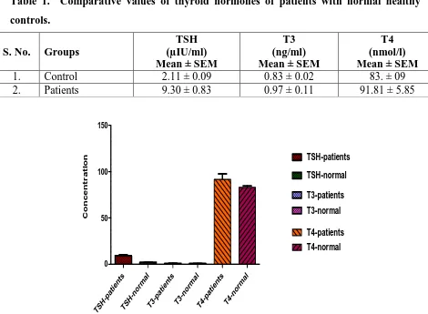
Thyroid Hormones
The changes in thyroid hormones of patients are compared with the control. Data is
represented in Mean ± SEM (n = 100). Values among thyroid patients are significantly higher
in case of TSH (P< 0.0001) and significantly not different in case of T3 (P< 0.1116) and T4
(P< 0.0773) from control Table. 1. The values are illustrated in Fig. 1.
Table 1. Comparative values of thyroid hormones of patients with normal healthy controls.
S. No. Groups
TSH (µIU/ml) Mean ± SEM
T3 (ng/ml) Mean ± SEM
T4 (nmol/l) Mean ± SEM
1. Control 2.11 ± 0.09 0.83 ± 0.02 83. ± 09
2. Patients 9.30 ± 0.83 0.97 ± 0.11 91.81 ± 5.85
TS H-p atie nts TS H-n orma l T 3-pat ien ts T 3-norma
l T 4-pat ien ts T 4-norma
[image:7.595.61.531.78.423.2]l 0 50 100 150 TSH-patients TSH-normal T3-patients T3-normal T4-patients T4-normal Co n c e n t r a t io n
Fig. 1. Mean concentration of thyroid hormones Stress Hormones
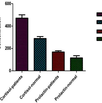
The changes in stress hormones cortisol among patients as compared with normal healthy
control in Table. 2. Data is represented in Mean ± SEM (n = 100) are highly significantly
[image:7.595.148.446.206.418.2]higher (P< 0.0001) from normal healthy control. (Fig. 2. represents the mean ± sem of the hormone).
Table 2. Data representing values of stress hormones S. No. Groups
Cortisol (nmol/l) Mean ± SEM
Prolactin (mIU/l) Mean ± SEM
1. Control 289.30 ± 17.12 115.50 ± 19.69
Cort isol -pat ient s Co rtis ol-n orma l Prol actin -pat ient s Prol actin -nor mal 0 200 400 600 Cortisol-patients Cortisol-normal Prolactin-patients Prolactin-normal C o n c e n tr a ti o n
Fig. 2. Mean ± sem values of stress hormones. Cytokines
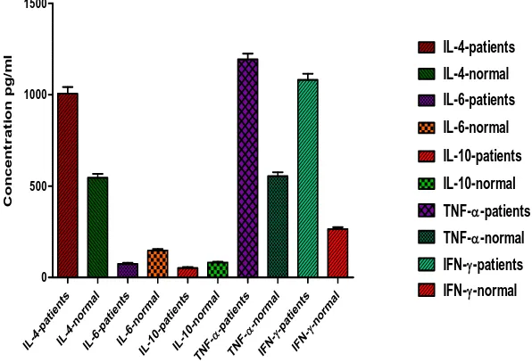
Anti-Inflammatory Cytokines and Pro-inflammatory Cytokines
The level of changes in cytokines of thyroid patients as compared with that of the normal
healthy control. (Data represented in Mean ± SEM (n = 100)) are highly significantly
different (P< 0.0001) from normal healthy control Table. 3.
It has been also observed that the level of pro-inflammatory cytokines value (TNF-α, IFN-γ)
was significantly raised as comparison to anti-inflammatory cytokines (IL-4, IL-6 and IL-10)
Fig. 3.
Table 3. Values of anti-inflammatory and pro-inflammatory cytokines S. No. Groups
†
IL – 4 (pg/ml) Mean ± SEM
†
IL – 6 (pg/ml) Mean ± SEM
†
IL – 10 (pg/ml) Mean ± SEM
*
TNF – α (pg/ml) Mean ± SEM
*
IFN – γ (pg/ml) Mean ± SEM
1. Control 545.6 ± 21.64 147.70 ± 8.11 82.03 ± 3.59 553.9 ± 21.75 265.20 ± 10.4
2. Patients 1006 ± 36.64 73.78 ± 6.56 51.63 ± 5.62 1193.0 ± 32.51 1081.00 ± 33.8
†
Anti-inflammatory cytokines, *Pro-inflammatory cytokines
[image:8.595.157.367.81.292.2]IL- 4-patient s IL-4-no rmal IL- 6-patient s IL-6-no rmal IL-10-patient s IL-10-no rmal -pat ient s TNF --norma l TNF - -p atie nts IFN--norma l IFN-0 500 1000 1500 IL-4-patients IL-4-normal IL-6-patients IL-6-normal IL-10-normal IL-10-patients
TNF--patients TNF--normal IFN--patients IFN--normal
[image:9.595.149.447.84.287.2]C o n c e n tr a ti o n p g /m l
Fig. 3. Concentration of anti and pro-inflammatory cytokines DISCUSSION
The elevated basal levels of stress hormones like cortisol and prolactin have shown
association with chronic stress suppress immunity by directly affecting cytokine profiles.
Cytokines are communicatory molecules produced primarily by immune cells.[20]
Proinflammatory cytokines mediate acute inflammatory reactions. Th1 cytokines mediate
cellular immunity by stimulating natural killer cells and cytotoxic T cells, immune cells that
target intracellular pathogens (e.g., viruses). Finally, Th2 cytokines mediate humoral
immunity by stimulating B cells to produce antibody, which “tags” extracellular pathogens
(e.g., bacteria) for removal. In a meta-analysis of over 30 years of research, intermediate that
stressors in life style could promote a Th2 shift (i.e., an increase in Th2 cytokines relative to
Th1 cytokines).[21] A Th2 shift has the effect of suppressing cellular immunity in favor of
humoral immunity. In response to more chronic stressors (e.g., long-term caregiving for a
dementia patient), proinflammatory, Th1, and Th2 cytokines become dysregulated and lead
both to suppressed humoral and cellular immunity.[22] Intermediate and chronic stressors are
associated with slower wound healing and recovery from surgery, poorer antibody responses
to vaccination, and antiviral deficits that are believed to contribute to increased vulnerability
to viral infections (e.g., reductions in natural killer cell cytotoxicity.[23]
Immune challenges such as infections with bacteria release bacterial lipopolysaccharides
(LPS), which induce the nuclear factor (NF) kB mediated secretion of IL-1 and IL-6, which
presenting cells (APC), such as monocytes/macrophages, dendritic cells, and other
phagocytic cells that are components of innate immunity, and by the helper T-lymphocytes
subclasses Th1, Th2, and Treg that are components of adaptive immunity. Homeostatis
within the immune system is largely dependent on cytokines, the chemical messengers
between immune cells, which play crucial roles in mediating inflammatory and immune
responses. It has also been found that increase in cytokine like (IFN)-gamma, TNF-alpha lead
to cell-mediated immunity; whereas increase in IL-4, TL-10 leads to the cell stimulation
enhance humoral immunity.[25, 26] Naïve T cells (Th0) are precursors of Th1 and Th2 cells,
and IL-12 (produced by APCs) is the major inducer of Th1 differentiation and hence, cellular
immunity. Thus, IFN-gamma inhibit Th2, whereas IL-4 and IL-10 inhibit Th1 cell activities.
IL-4 and IL-10 promote humoral immunity by stimulating the growth and activation of mast
cells and eosinophils, the differentiation of B cells into antibody secreting B cells, and
immunoglobulin switching to IgE. Importantly, these cytokines also inhibit macrophage
activation, T-cell proliferation, and the production of proinflammatory cytokines.[27, 28]
We have found the significant increase in level of TSH and decrease status in T3 and T4
hormone. As thyroid hormones makes and stores hormones that help regulate the heart rate,
blood pressure, body temperature, and the rate at which food is converted into energy.[29] It
achieves this by manufacturing the hormones, thyroxine (T4) and triiodothyronine (T3) and
secreting them into the blood stream. Thyroid stimulating hormone (TSH) secreted by the
pituitary gland stimulates the release of other thyroid hormones T4 and T3 which further
enhances all the cells of the body to metabolize at a faster rate.[30] On a broad extent two
types of hypothyroidism has been studied, primary hypothyroidism caused by decreased
production of T4 and T3 due to thyroid dysfunction increase production of TSH; by pituitary
(TSH) or hypothalamic (TRH) disease.[31, 1]
Cellular and metabolic processes of the body modulate inflammatory processes of the
immune system and can induce major changes in the downstream cytokine. They are effector
molecules that can instantly alter the quality of the immune response in the autoimmune
diseases. Intervention increase stress hormone levels which interfere beneficial effects of
stress-related mediators, such as protection from cytokine-mediated shock syndrome or
prevention of autoimmunity.[32]
involving both stress and thyroid hormones, among these prolactin, cortisol, thyroid
stimulating hormone (TSH), triiodothyronine (T3), and thyroxine (T4) are the major.[34]
Hypothyroidism is routinely considered in the differential diagnosis of depressive and anergic
states, and is screened for with determinations of serum thyroxine (T4), trio dothyronine (T3),
and basal thyroid-stimulating hormone (TSH).[35] Thyroid failure with its predilection for
behavioral presentation is much more likely to manifest as depression or lack of energy to a
psychiatrist.
In HT, cell-mediated immunity promotes the induction of auto-antibodies and self-reactive T
cells against Tg, and other auto-antigens, including thyroid peroxidase. HT is characterized
by infiltration of lymphocytes and other immune cells, thyroid enlargement and fibrosis, and
progressive destruction of thyrocytes that eventually results in hypothyroidism.[36] Upon
initiation of the immune response to Tg, thyroid-specific T lymphocytes migrate to the
thyroid and through interferon (IFN)-g production induce thyrocyte expression of major
histocompatibility complex (MHC) class-II molecules. This results in further expansion of
autoreactive T cells and the inflammatory response leading to the accumulation of activated
CD4 + and CD8 + T cells, B cells, plasma cells, and macrophages in the thyroid.
Cytokines are involved in common endocrine diseases, such as diabetes mellitus and
autoimmune thyroid disease (ATD). The hormones mediate the differentiation of Th0 (naïve
T Helper cells) towards the Th2 humoral immune response to the detriment of the Th1
cell-mediated response. APC‟s secrete cytokines that mediate Th1 differentiation, however the
presence of bacterial products such as LPS that bind to Toll-like Receptors induce the
production of IL-1 and IL-6, which cross the blood-brain barrier and trigger the hypothalamic
CRH-stress response.[37] In this manner, a blood borne stressor of infectious nature can
activate the HPA axis. Th1 effects are mediated by the cytokines IL-12,18,2 and γInterferon
and T cells and Macrophages. Th2 responses are mediated by IL-4,6,13 and B Cells,
Eosinophils and Mast Cells. CRH: Corticotropin releasing Hormone; NE: Norepinephrin;
Th0: Naïve Helper cells; APC: Antigen Presenting Cell; LPS: Lipopolysaccharide; HPA:
Hypothalamic-Pituitary-Adrenal Axis.[38]
The defense system is an example of subtle autoregulation which is intervened with
medications. The therapeutical use of this intervention is called „immunomodulation‟.[39]
prolactin and cortisol play a prominent role in stimulation and modulation of immune
function. An immediate immune response occurs through small cell signaling by numerous
cells of the immune system. Cytokines encompasses all these cells and a large and diverse
family of regulators produced throughout the body by cells. Cytokines refer to
immunomodulating agents, trigger inflammation and respond to infections.[40]
Cytokines include two classes of it pro and anti inflammatory. We have found stress hormone
changes stress system activity through modulating pro or anti inflammatory cytokines.
Increase in significant level of pro-inflammatrative cytokines of TNF-α and IFN-γ and
significant decrease in IL-4 and IL-10. Stress hormones changes stress system activity
through modulating pro or anti-inflammatory cytokines, TNF-α, IL-6, IFN-γ, IL-4 and IL-10,
balance by stimulating or suppressing the progression of autoimmune diseases.
Studies from humans and animal models have revealed significant new insights into the
complex role of cytokines in the pathogenesis of AITD. Modulating cytokine responses have
yielded highly encouraging results and they hold considerable promise in the treatment of
autoimmune diseases. Pro-inflammatory cytokines such as GM-CSF and IL1b can contribute
to Foxp3 + Treg expansion, whereas a regulatory suppressor cytokine such as TGF-b can
initiate a pathogenic Th17 T cell response. These observations highlight the paradoxical
effects of cytokines and their critical roles in maintaining a delicate balance between health
and disease. Therefore, additional studies to understand the complex interplay between
different cytokines and their effects on the different components of the immune system in the
context of a particular disease are essential.
CONCLUSION
Present era is full of stress that to of environmental, psychological, physiological and
physical stress and all of it compound to produce severe impact. Any of above if dominates
leads to affect our body physiology directly. Surprisingly out of the above psychological
stress, life style pattern and physiological interaction leads to progressive disturbance in
producing disease syndrome.
Study interprets a significant rise in stress hormones like cortisol and prolactin, leading to
immune regulation of body homeostasis. We have found that stress hormone after
recognizing the immune cells which promote the mother T and B cells and Th cells type Th1
autoimmune process. Increase in cytokines alpha-TNF and gamma interferon attacked on
thyroid cells destruction and predominates for Th2 mediated immune response promoting
antigen specific B cells. These B cells produce anti-TSH receptor antibodies leading to
hypothyroidism.
In addition to above we also found that compound effect of stress, immunity and endocrine
system, they are directly linked and in adverse conditions these leads to autoimmune diseases
like HT. Due to above combination a lower response of T4 hormone leads to low BMR and
glucose absorption rate in digestive tract, due to low BMR and inappropriate glucose
absorption has lead to hyperlipidemia posing high response blood pressure and other
cardiovascular diseases.
The study concludes that to regulate the hypothyroidism like autoimmune disorder one has to
take proper care of compound stress increasing day by day particularly in immune
suppression autoimmune conditions. Supportive and suggestive combination therapy in
addition to regular treatment will benefit the patient of hypothyroidism because stress,
immunity and infection play a major role not only hypothyroidism but may lead to other
autoimmune diseases.
ACKNOWLEDGEMENTS
Authors extend immense gratitude towards M.P. Council of Science and Technology
(M.P.C.S.T) Bhopal for funding the project and Department of Microbiology, Barkatullah
University, Bhopal for laboratory facility. Also would like to acknowledge the Physicians and
other members of the different hospitals of Bhopal for allowing the collection and providing
the serum samples from the patients.
CONFLICT OF INTEREST
There are nonfinancial competing interests (political, personal, religious, ideological,
academic, intellectual, commercial, or any other) to declare in relation to this manuscript.
REFERENCES
1. Chrousos GP. Stress and disorders of the stress system. Nature Rev- Endocrinology,
2009; 5: 374- 381.
2. Kua EH, Tsoi WF, Cheah JS, Thai AC, Yeo PPB. Stress, Personality and
3. Korneva ET, Shkmek EK. Hormones and the immune system. Moscow. Nauka., 1988.
4. Moynihan JA, Stevens SY. Mechanisms of stress-induced modulation of immunity in
animals. In: Ader R, Felten DL, Cohen N, eds. Psychoneuroimmunology.; New York:
Academic Press, 2001; 227-249.
5. Khansari DN, Murgo AJ, Faith RE. Effects of stress on the immune system. Immunol
Today, 1990; 11: 170-175.
6. Chrousos GP. The hypothyalamic-pituitary-adrenal axis and immune-mediated
inflammation. N Engl J Med., 1995; 332: 1351-1362.
7. Elenkov IJ, Wilder RL, Chrousos GP, Vizi ES. The sympathetic nerve-an integrative
interface between two super systems: the brain and the immune system. Pharmacol Rev.,
2000; 52: 595-638.
8. Tausk F, Elenkov I, Moynihan J. Psychoneuroimmunology. Dermatology Therapy, 2008;
21: 22-31.
9. Marques-Deak A, Cizza G, Sternberg E. Brain-immune interactions and disease
susceptibility. Molecular Psychiatry, 2005; 10: 239-250.
10. Cleare AJ, McGeorge A, O‟Keane V. Neuroendocrine evidence for an association
between hypothyroidism, reduced central 5-HT activity and depression. Clin. Endocrinol
(Oxford), 1995; 43: 713-719.
11. Boswell EB, Anfinson TH, Nemeroff CB. Depression associated with endocrine disorder.
In: Depression and Physical Illness;Robertson MM, Katona CLE eds. Willey: Chichester,
England, 1997; 256-292.
12. Hangalapura BN, Nieuwland MGB, Buyse J, Kemp B, Parmentier HK. Effect of Duration
of Cold Stress on Plasma Adrenal and Thyroid Hormone Levels and Immune Responses
in Chicken Lines Divergently Selected for Antibody Responses. Poul Sci., 2004; 83:
1644–1649.
13. Wang HC, Klein JR. Immune function of thyroid stimulating hormone and receptor. Crit.
Rev. Immunol., 2001; 21: 323-337.
14. Witherspoon RL, Shuler ES, Gilbert S. Estimation of Thyroxine, Triiodothyronine,
Thyrotropin, Free Thyroxine and Triiodothyronine Uptake by Use of Magnetic-Particle
Solid Phase. Clinical Chemistry, 1985; 31(3): 415-419.
15. Maier SF, Watkins LR. Cytokines for psychologists: implications of bidirectional
immune-to-brain communication for understanding behavior, mood, and cognition.
16. Fisher DA. Physiological variation in thyroid hormones. Physiological and
pathophysiological considerations. Clin Chem., 1996; 42: 135-139.
17. Spencer CA. Interlaborate/Intermethod differences in functional sensitivity of
immunometric assay of thyrotropin (TSH) and impact on reliability of measurement of
subnormal concentration of TSH. Clinical chemistry, 1995; 41: 367.
18. Peters JR. Clin Edocrinol., 1982; 17: 583.
19. Check JH. Falsely elevated steroidal assay levels related to heterophile antibodies against
various animal species. Gynecol Obstet Invest., 1995; 40: 139-140.
20. Roitt I, Brostoff J, Male D. Immunology, 5th ed. Mosby Int: London, 1998.
21. Segerstrom SC, Miller GE. Psychological stress and the human immune system: a
meta-analysis of 30 years of inquiry. Psychol. Bull., 2004; 130: 601–630.
22. Kiecolt-Glaser JK, McGuire L, Robles TF, Glaser R. Psychoneuroimmunology:
psychological influences on immune function and health. J. Consult. Clin. Psychol., 2002;
70: 537–547.
23. Besedovsky HO, del Rey AE, Sorkin E. What do the immune system and the brain know
about each other. Immunol Today, 1986; 4: 342-346.
24. Fearon DT, Locksley RM. The instructive role of innate immunity in the acquired
immune response. Science, 1997; 272: 50-53.
25. Trinchieri G. Interleukin-12 and the regulation of innate resistance and adaptive
immunity. Nature Rev., 2003; 3: 133-146.
26. Asvold BO, Vatten LJ, Nilsen Tom IL, Bjoro T. The association between TSH within the
reference range and serum lipid concentration in a population-based study. The HUNT
Study. European J Endocrinology, 2007; 156: 181-186.
27. Mizokami T, Wu Li A, El-Kaissi S, Wall Jack R. Stress and Thyroid Autoimmunity.
Thyroid, 2004; 14 (12): 1047-1055.
28. Braverman LE, Werner, Ingbar‟s. The Thyroid. A Fundamental and Clinical Text. 8th ed.
2000.
29. Chuang E, Molitch ME. (Prolactin and autoimmune diseases in humans). Acta Biomed.,
2007; 78 (1): 255-261.
30. Sanders VM, Baker RA, Ramer-Quinn DS, Kasprowicz DJ, Fuchs BA, Street NE.
(Differential expression of the beta2- adrenergic receptor by Th1 and Th2 clones:
31. Celik I, Akalin S, Erbas T. (Serum levels of interleukin 6 and tumor necrosis factor-alpha
in hyperthyroid patients before and after propylthiouracil treatment). Eur J Endocrinol.,
1995; 132: 668–672.
32. Lee YM, Fujiwara J, Munakata Y, Ishii T, Sugawara A, Kak M. (A mutation of the
glucocorticoid receptor gene in patients with systemic lupus erythematosus). Tohoku J
Exp Med., 2004; 203: 69–76.
33. Weetman AP. (Autoimmune thyroid disease: propagation and progression). Eur J
Endocrinol., 2003; 148(1): 1–9.
34. Mikos H, Mikos M, Pietrzak RB, Niedziela M. (The clinical role of serum concentration
of selected cytokines: IL-1β, IL-6 in diagnosis of autoimmune thyroid disease (AITD) in
children). Autoimmunity, 2014: May 7.
35. Francisco Tausk, Ilia Elenkov, Jan Moynihan. (Psychoneuroimmunology). Dermatologic
Therapy, 2008; 21: 22-31.
36. Kiecolt-Glaser JK, Loving TJ, Stowell JR. (Hostile marital interactions, proinflammatory
cytokine production, and wound healing). Arch Gen Psychiatry, 2005; 62: 1377–84.
37. Dickens C, Jackson J, Tomenson B, Hay E, Creed F. (Association of depression and
rheumatoid arthritis). Psychosomatics, 2003; 44: 209–15.
38. Mosmann TR, Sad S. (The expanding universe of T-cell subsets: Th1, Th2 and more).
Immunol Today, 1996; 17: 138–46.
39. Herbert TB, Cohen S. (Stress and immunity in humans: A Meta-Analytic Review). Psy
Med., 1993; 55: 364-79.
40. Tsatsoulis A. (The role of stress in the clinical expression of thyroid autoimmunity).