INTERNAL HYDROSTATIC PRESSURE OF THE
FUNDULUS EGG
I. THE ACTIVATED EGG*
BY CHIEN-YUAN KAO AND ROBERT CHAMBERS Lilly Research Laboratories, Marine Biological Laboratory,
Woods Hole, Mass.
{Received 13 June 1953)
I. INTRODUCTION
The egg of the killer minnow, Fundulus, has presented the physiologist with an exceptional problem because of the claim still extant that it is impermeable to water. J. Loeb found that the fertilized Fundulus egg could develop equally well in sea water of varying tonicities and even in tap water, as well as in distilled water. He noticed (1912) that when placed in hypertonic solutions of balanced mixtures of salts, the whole eggs remained floating and continued developing for 10 days, whereas newly hatched larvae died in a few minutes. These results indicated to him that the egg did not lose or gain either salt or water to affect the density or development of the ovum. When, however, the eggs were transferred to hypertonic solutions of single or unbalanced combinations of salts, they sank within a short while. This was to him a sign of permeability to water as an indication of damage. The apparent impermeability to water and electrolytes was attributed by Loeb exclusively to the tough chorion surrounding the egg, which became permeable only when injured by unbalanced salt solutions. Death of newly hatched larvae in hypertonic media was considered as confirmatory of the protection given by the chorion.
Later works of Bodine (1928) and Armstrong (1928), working with whole eggs as well as eggs with the chorion surgically removed, indicated that the presence or absence of the chorion did not materially alter the lethal action of unbalanced salt solutions on the egg. Yagle (1930), using whole and dechorionated eggs, confirmed Loeb's flotation experiments and came to the conclusion that not only the chorion was impermeable to water and electrolytes but also the vitelline membrane on the surface of the egg. In any event, the Fundulus egg has been considered as unique because of the apparent impermeability to water and electrolytes during its healthy state (Krogh, 1939; Brooks & Brooks, 1941). Indeed, this condition has been spoken of as a regulatory mechanism by which the Fundulus egg maintains its osmotic concentration in environments of various tonicities from that of sea water to that of brackish water (Prosser, 1951).
A counterpart of the marine Fundulus egg has been suggested to be the egg of the fresh-water trout. Gray (1932) claimed that the healthy trout egg was completely
impermeable to water and electrolytes. Impermeability to water was indicated by the absence of any appreciable change in the weight of the egg transferred to Ringer's solutions of greatly hypertonic strengths. On the other hand, impermeability to electrolytes, i.e. lack of escape of electrolytes, was indicated by no increase in con-ductivity of the distilled water in which healthy trout eggs had been immersed. Svetlov (1929) by freezing-point determinations of the various components of the trout egg had claimed that the chorion of the trout egg was freely permeable to both water and electrolytes. He found that by changing the composition of the external medium, the freezing-point of the whole egg (egg proper and perivitelline fluid) altered by an amount which could be accounted for by the replacement of the perivitelline fluid by the new external medium. Gray accepted Svetlov's con-clusions, but claimed the impermeability to be due to ' the static properties of the vitelline membrane which excluded the passage of water and electrolytes' from the egg proper. This impermeability he claimed to be responsible for the maintenance of the relatively high osmotic concentration within the egg (f.p. = — 0-48° C.) as compared to its native fresh-water environment (f.p. = —0-02° C ) . Similar to the Fundulus egg, the trout egg was supposed to develop permeability to water and electrolytes only when it was injured, either mechanically or through ageing.
Except for the work of Krogh & Ussing who found no evidence for the penetra-tion of heavy water (1937), the investigapenetra-tions indicating lack of water permeability of the Fundulus egg and of the trout egg have depended chiefly on changes in weight or density of the eggs placed in different environments.* It was assumed that if the eggs were permeable to water, then their weights in hypertonic media would decrease and their densities increase because of loss of water to the external environment. This was the basis of Loeb's flotation experiments and Gray's observation of the trout egg proper floating within its own chorion in hypertonic media. These observations indicated no net changes in weight and density, but cannot indicate transitory changes at any specific time, which is needed to appreciate progressive changes with time. In view of the importance of the conclusion that these two species of eggs are completely impermeable to water, the problem was reinvestigated on the Fundulus egg with different techniques designed to show transitory changes as well as net alterations in water exchange. The method employed consists of simultaneous determinations of pressure and volume of the egg during early development and under different experimental conditions. Whenever movement of water occurs, there is either a volume change or a pressure change or more likely a combination of the two. In sea-urchin eggs permeability to various substances has been studied by means of volume changes (Lucke & McCutcheon 1932). Information would be more complete if it were possible to combine a study of both pressure and volume changes. The results of this and succeeding papers on the Fundulus egg present evidence indicating free permeability of the egg to water.
This has been accomplished by means of simultaneous pressure and volume studies. In the present paper is described the method of study, and the internal hydrostatic pressure of the unfertilized Fundulus egg upon artificial activation and during its subsequent development in sea water.
II. METHOD
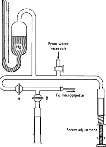
[image:3.595.198.403.280.563.2]The apparatus used for recording pressure was a mercury manometer. As shown in Fig. 1, there were two outlets to the manometer: one of these was connected to a micropipette, and the other to a syringe with a screw adjustment. The lower part
Fig. 1. Schematic diagram of the manometer used in the experiments. Except for the portion occupied by mercury, the entire system is filled with water. The side syringe is connected through valve B. When the micropipette has been inserted into an egg, valve B is closed and valve A opened. Pressure changes are effected by the screw adjustment.
142 C H I E N - Y U A N KAO AND ROBERT CHAMBERS
The technique of operation in detail is as follows: Convenient lengths of capillary tubing, o-8 ram. outside diameter, were washed, dried and one end drawn over a micro-flame, into a suitable micropipette having a gradually sloping, conical shaftlet terminating in a closed tapering tip. The coarser, open end of the micropipette at the opposite end of the microtip was then sealed in a flame and several such pipettes set aside for use. When ready for operation, the sealed, coarser end of the micropipette was broken open and dipped into a drop of thin paraffin oil coloured orange with Sudan III. A very short length of the oil entered the open end of the lumen by capillarity, while the greater part of the lumen of the micropipette, extending to its tapering tip, remained filled with air. The main shaft of the micropipette, with the column of oil at the open end, was then coupled to a standard, water-filled, metal pipette holder of the micromanipulator, the holder being connected to the hydraulic system of the apparatus. No air was allowed to interpose between the water in the metal holder and the oil in the coarse end of the micropipette. After a tight connexion had been achieved, a slight pressure was exerted on the side syringe, driving the water, with the oil ahead of it, along the shaft of the micropipette and towards the microtip. Meanwhile the tip of the micropipette was care-fully broken to the desired diameter of 5-10/x while under observation through the microscope. Further pressure displaced water in the shaft with the oil column in advance of it towards the microtip. When the oil reached the tip and the entire micropipette was filled with fluid, the hydraulic system was ready for use. Valve B (Fig. 1) of the side arm was then closed and valve A opened, thereby connecting the micropipette with the mercury manometer.
At the beginning of each experiment, the mercury column was adjusted to zero reading through the screw adjustment, with the tip of the micropipette just beneath the surface of the sea water in the dish containing the eggs to be operated upon. T h e pipette was then inserted into an egg which would displace a small amount of its material into the micropipette provided there was an appropriately lower pressure in the pipette. When this occurred, a meniscus was created between the oil in the pipette and the fluid exuding from the egg. By moving the syringe to manipulate pressure this meniscus was maintained close to the inner surface of the chorion of the egg. Under these conditions the egg was thus at the nearest physiologic state of pressure found practicable. If there were any increase or decrease of pressure in the egg, and if the meniscus were carefully maintained at this level by turning the screw adjustment, all pressure changes so caused were transmitted to the mercury column. T h e height of the column in the mercury manometer represented the pressure in the egg at any given time.
Eggs were stripped from a female into a dish of sea water. The bottom of the dish had been covered with a layer of paraffin wax into which were cut several depressions, slightly larger than a Fundulus egg. These depressions serve to hold the eggs while being punctured under microscopic observation. Punctures were made with the tapering tips of diagonally directed micropipettes having a bore of 15-20/x fora distance of \ mm. from the tip which averaged 5-10/x across. These sizes were found to be most convenient for recording pressures, since with narrower pipettes, the movement of the meniscus became sluggish, making it difficult to observe rapid changes in pressure.
intervals beginning from the moment of puncture, (2) internal hydrostatic pressure of the egg, (3) the outside diameter of the chorion, and (4) the diameter of the egg proper.
III. OBSERVATIONS AND RESULTS
A. Morphology of the Fundulus egg and effect of puncture
The egg of the Fundulus found in Woods Hole waters is spherical, fairly constant in size from the same batch, and averages i-8 mm. in diameter. It possesses an outer relatively thick coat, the chorion (c. io^i), which is flaccid during the first few minutes after it has been stripped into sea water. From then on, the chorion is inelastic and maintains a constant diameter. The eggs rapidly become turgid. Closely applied to the under-surface of the chorion lies the egg proper which consists chiefly of yolk material and several relatively large oil globules. The proto-plasmic portion of the egg is spread out in a thin layer over its periphery. Just below the surface of the egg proper is a layer of closely packed, small, colourless spherules, the so-called platelets. When the unfertilized egg is left standing in sea water, it becomes spontaneously activated, undergoing a sequence of events similar to that occurring after insemination (Kagan, 1935). These consist of disappearance of the platelets, development of a perivitelline space, and formation of a localized elevation on the egg surface, the blastodisc. The activation process begins within 15-20 min. after standing in sea water. The platelets disappear suddenly and completely, all within 30-45 sec. Following this, the perivitelline space forms and gradually enlarges, separating the under-surface of the chorion from the outer surface of the egg proper. Throughout all the experiments recorded, the diameter of the chorion, from the first few minutes after the eggs had been released into sea water, has been found to remain constant. The formation of the perivitelline space is accompanied by a corresponding shrinkage of the egg proper. Meanwhile, the peripheral proto-plasmic layer slowly moves towards one pole of the egg, so that, by the end of an hour, there is a distinctly formed ovoid blastodisc on the surface of the egg in pre-paration for cleavage and subsequent development.
144 C H I E N - Y U A N KAO AND ROBERT CHAMBERS
the fact that the punctured eggs remained yellow and translucent, were indications that puncturing had no deleterious effect. In one control experiment, an egg was punctured shortly after it was inseminated at the time of stripping. The egg was kept under observation till after five normal cleavages had occurred, while all through this time the puncturing pipette had been kept in place.
In a successfully performed puncture, a small amount of coagulum always formed on the surface of the egg around the shaft of the pipette. This coagulum served as an airtight seal and also to hold the pipette in place. It was this tight seal and the inelasticity of the chorion which made the recording of pressure possible. Of incidental interest was the existence of a relation between the site of puncture and that of the subsequently forming blastodisc. In either NaCl or KC1 alone, the blastodisc always formed at the site of puncture. In sea water and in a mixture of NaCl and KC1 (19:1), isotonic with sea water, there was no relationship (Kao, 1951).
B. Internal hydrostatic pressure
In Fig. 2 are shown the results of ten experiments on the eggs from different females on different days. Immediately upon puncture there was recorded a negative pressure in the egg averaging about — 10 mm. Hg. This negative pressure
150
100-50
0
-20 10 20 30 40 50 60 70Time after puncture (min.)
Fig. 2. Internal hydrostatic pressure of puncture-activated eggs in sea water. Each dot represents a measurement on one egg consistently kept in sea water. Beyond 40-50 min., only a few as recorded were kept in sea water. Other eggs used at this time were subjected to other media. The results obtained are recorded in a second paper of this series. (Chambers, Chambers & Kao, 1951).
speaking then, the formation of the internal hydrostatic pressure can be divided into two stages, a rising stage during the first 50 min. after activation, and a long-lasting plateau stage which follows (Kao, Chambers & Chambers, 1951).
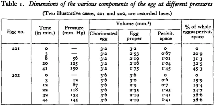
[image:7.595.125.473.236.409.2]Associated with the rise in pressure was the progressive enlargement of the perivitelline space. Dimensions of the various parts of the egg are presented in Table 1. Measurements, by means of an ocular micrometer, were made of the
Table 1. Dimensions of the various components of the egg at different pressures
(Two illustrative cases, aoi and aoa, are recorded here.)
Egg no.
3 0 1
aoa Time (in min.) 0 3 8 2 0 41 0 3 1 3 3 3 3a 44 Pressure (mm. Hg) — 56 «as 150 — 1 3 87
I l 8 133 145 Volume (mm.* Chorionated egg 3-a 3-3 3-a 3 3 3 3 3 6 3 6 3-6 3 6 3 6 3 6 Egg proper 3 a a-53 3-19
2 1 6 1 7 5
3 6
3 0 3 9
a-35 3-19
3 1 9
Perivit space 0 0-67 I'OI 1-04 1'45 0 0 6 0-7 1 35 1-41 1-41
% of whole egg as peri vit.
space 0 30-9 31-3 32-5 4S"3 0 1 5 9
19-4 34-7 38-6
3 8 6
diameter of the whole egg (including the chorion) and that of the egg proper. Volumes of these two parts were then calculated, and the volume of the perivitel-line space was obtained by subtraction. Since the volumes of the different parts in different eggs varied appreciably, the volume of the perivitelline space was finally expressed as a percentage of the volume of the entire egg including the chorion. This method of measurement, i.e. calculating volumes from diameters, was used because the various components measured were practically spheres. During the early phase in which the experiments were performed, the blastodisc had not accumulated enough to distort the spherical appearance of the egg proper. From Table 1 it can be seen that the chorion does not alter its dimensions, and that the enlargement of the perivitelline space was due to a decrease in volume of the egg proper.
Correlating the pressure curve (Fig. 2) with Table 1, a question arose as to whether the rise of the internal hydrostatic pressure was due to compression of the egg proper by the expanding perivitelline space. To ascertain the possibility of some perivitelline compression, a method was employed to remove the pressure. Eggs stripped into sea water were allowed to activate spontaneously for 1 hr. After the perivitelline space was fully developed the chorion was snipped* so as to permit a free communication between the perivitelline space and the external medium. When micropipettes were inserted into such eggs, the recorded pressures were never
• We are much indebted to Dr J. P. Trinkaus (Department of Zoology, Yale University) for his generous help in this operation.
146 C H I E N - Y U A N KAO AND ROBERT CHAMBERS
above 2-4 mm. Hg. Normally, without snipping the chorion, the pressure at this time would have been in the vicinity of 150 mm. Hg.
Another series of experiments was performed for the purpose of continuous recording of pressure changes. These are represented in Fig. 3, in which one egg was punctured in sea water in the usual manner. All the visible changes, including
mm. mm. Hg 0-15- -150
0-10- -100
005
-10 20 30 40
Time after puncture (mln.)
50 60
Fig. 3. The progressive changes in pressure and perivitelline space of one egg in sea water and effect of subsequent immersion in sucrose hypertonic (i -9 M) to sea water. Sea water was removed and sucrose added when the pressure and perivitelline space have reached a nearly full state of development. The scale on the left side of the ordinate indicates the average width of the perivitelline space as measured by an ocular micrometer. The scale on the right side of the ordinate indicates pressure in mm. of mercury.
the enlargement of the perivitelline space, were the same as those described above. When the pressure and the perivitelline space had reached nearly the full state of development, the external medium was changed from sea water to 1-9 M-sucrose (twice the tonicity of Woods Hole sea water). Immediately the perivitelline space disappeared as the egg proper swelled. Accompanying these morphologic changes was the precipitous fall in pressure which persisted at zero.
IV. DISCUSSION AND CONCLUSION A. Terminology
B. Discussion of results
The interpretation of the experimental results herein reported are based on the progressive morphologic changes, the progressive pressure change, and the experi-ments in which the pressure was reduced. The Fundulus egg is isotonic with the body fluids of the fish, and when first laid is hypotonic to sea water. Owing to the relative hypertonicity of sea water, some water is lost by the egg during the first few minutes. This would account for the initial negative pressure recorded (Fig. 2). The amount lost, however, is not large enough to induce any appreciable volume change of the egg proper. As the activation process commences with the sudden disappearance of the platelets, the pressure begins to rise. The correlation between the disappearance of the platelets and the development of pressure is further supported by the observation that in those eggs in which the platelets did not disappear, no pressure ever developed. Furthermore, when the platelets dis-appeared more slowly than normal, the pressure also climbed at a correspondingly slower rate. This condition of slower disappearance of platelets can be produced by immersing the puncture-activated unfertilized eggs from the beginning in isotonic NaCl.
148 C H I E N - Y U A N KAO AND ROBERT CHAMBERS
substances of the marine Fundulus egg and of the fresh-water trout egg may be closely related or even be identical substances.
The vitelline membrane, whatever it is, of the Fundulus egg proper has been claimed to be impermeable to water. It is the purpose of this paper and following papers to show that the methods employed by previous workers showed only an absence of a net exchange of water, and did not indicate non-penetration. Gray (1932), in discussing the impermeability of the trout egg, had already stated that if the vitelline membrane were permeable to water, then water exclusion by the egg must be due either to the development of a hydraulic pressure in the opposite direction or to some active metabolic process. He concluded, however, that neither mechanism was at play, emphasizing especially that no pressure could develop in the egg during life. The reasons for his conclusion were that the chorion was a non-viable component of the egg, and that being freely permeable to water and electrolytes of the external medium, the chorion could not serve as a semipermeable membrane for any osmotic function. The results reported here show clearly that a pressure does develop in the Fundulus egg during life, although the pressure is dependent only on the parts external to the living egg proper, viz. the chorion and the colloid in the perivitelline space. Conceivably a similar situation may exist in the trout egg. In any case, results herein reported have shown the development of an opposing hydrostatic force which, owing to its circumferential location around the egg proper, could prevent further ingress or egress of water into or from the egg proper.
Table 1 shows that the shrinkage of the egg proper from the rigid chorion amounts to approximately 40 % of the original volume. This can be accounted for only by the loss of something from the egg proper, presumably water, which by moving into the perivitelline space, and kept there by the jelly-like material, con-tributes to the development of the pressure. Further evidence that the egg proper is permeable to water is provided by the sucrose experiment. The fact that in the sucrose experiment the perivitelline space disappears indicates that not only is the chorion freely permeable to water, but because the diameter of the chorionated egg remains constant, and because the change in volume of the perivitelline space corresponds strictly to the change in volume of the egg proper, it is to be concluded that water transfer to and from the perivitelline space must be through the egg surface out of and into the egg proper.
SUMMARY
perivitelline space. It was concluded that the perivitelline substance is the chief factor contributing to the pressure. The changes in volume of the egg proper are inversely proportional to the size of the perivitelline space, indicating that the surface of the egg proper is permeable to water.
REFERENCES
ARMSTRONG, P. B. (1928). The antagonism between acetic acid and the chlorides of sodium, potassium, and calcium as manifested in developing Fundulus embryos. J. Gen. Phytioi. 11,
5I5-23-BODINE, J. H. (1928). Action of salts on Fundulus eggs. Biol. Bull., Woods Hole, 34, 396-404. BROOKS, S. C. & BROOKS, M. M. (1941). Permeability of Living Cells. Protoplasma Monograph. CHAMBERS, R., CHAMBERS, E. & KAO, C. Y. (1951). The internal hydrostatic pressure of the
un-fertilized activated fundulus egg exposed to various experimental conditions. Bid. Bull., Woods Hole, 101, 206.
GRAY, J. (1932). The osmotic properties of the eggs of the trout (Salmo fario). J. Exp. Biol. 9,
277-209.
KAGAN, B. M. (1935). The fertilizable period of the eggs of Fundulus heteroclitus and associated phenomenon. Biol. Bull., Woods Hole, 69, 185-201.
KAO, C. Y. (1951). Micropuncture and the localization of the subsequently-forming blastodisc in the unfertilized Fundulus egg. Biol. Bull., Woods Hole, 101, 222.
KAO, C. Y., CHAMBERS, R. & CHAMBERS, E. (1951). The internal hydrostatic pressure of the un-fertilized Fundulus egg activated by puncture. Biol. Bull., Woods Hole, 101, 210.
KROGH, A. (1939). Osmotic Regulation of Aquatic Animals. Cambridge University Press. KROQH, A. & USSING, H. H. (1937). A note on the permeability of trout eggs to DiO and H,O.
J. Exp. Biol., 14,
35^7-LOBB, J. (1912). Antagonistic action of electrolytes and permeability of the cell membrane. Science, a6, 637-9.
LUCKB, B. & MCCUTCHEON, M. (1932). The living cell as an osmotic system and its permeability to water. Pkysiol. Rev. 13, 68-139.
PROSSER, C. L. (195I). Comparative Animal Physiology. Saunders.
SVETLOV, P. (1929). Entwicklungsphysiologische Beobachtungen an Forelleneiern. Arch.f. Enttoick-lungsmechanik der Organismen, 114, 771-85.
YAGLE, E. M. (1930). Permeability of egg membranes. Water exchange through the egg membrane of fundulus. Protoplasma, 9, 245-68.
YAMAMOTO, T. (1939). Mechanism of membrane elevation in the egg of Oryzias latipes at the time of fertilization. Proc. Imp. Acad. Tokyo, 15, 272-4.
YAMAMOTO, T. (1940). The change in volume of the fish egg at fertilization. Proc. Imp. Acad. Tokyo, 16, 482-5.