Original Article
Experimental study on effects of a
new type of interventional embolization agent
in chemoembolization of liver transplantation tumors
Kaiyuan Xu1, Dandan Xiao2
Departments of 1Radiology, 2Outpatient, Shenzhen Hospital, Southern Medical University, Shenzhen 518010,
China
Received February 21, 2019; Accepted May 10, 2019; Epub July 15, 2019; Published July 30, 2019
Abstract: The aims of this study were to observe the effects of doxorubicin (ADM)-alginate microspheres on rabbit VX2 liver transplantation tumor by transcatheter arterial chemoembolization (TACE). A total of 30 New Zealand white rabbits were transplanted 1-mm3 mass of VX2 liver transplantation tumor and then were randomly divided into five groups: the normal saline group (group A), the blank microsphere group (group B), the ADM microsphere group (group C), the super-liquefied lipiodol group (UFLP, group D), and the UFLP + ADM group (group E) for comprising the conditions of apoptosis by TdT-mediated dUTP Nick-End Labeling (TUNEL) and growth rate/necrotic rate of ortho -topic transplantation tumors in different groups by immunohistochemical staining of vascular endothelial growth factor (VEGF) and CD31 antibodies. The tumor volume in each group increased, but the growth was inhibited than group A. The number of intrahepatic and distant metastasis decreased in group C (P<0.05), and expression of VEGF and microvessel density (MVD) decreased (P<0.01). The number of cells undergoing apoptosis increased (P<0.01), and the apoptotic indexes in the five groups were 0.3%, 11.4%, 14.3%, 1.7%, and 5.1%, respectively (P<0.01). ADM microspheres can inhibit tumor volume growth and distant metastasis in TACE.
Keywords: Alginate-microsphere, VEGF, embolization, neoplastic transplantation, liver neoplasm
Introduction
Primary liver and biliary cancers are very aggressive tumors [1]. Approximately 80% of patients diagnosed with hepatocellular carci-noma at an early stage are not candidates for surgical resection or transplantation [2], surgi-cal treatment is the main option for cure or long term survival, and if the future liver remnant (FLR) is too small to meet the needs of liver function and volume, these patients are con-sidered as unresectable [3], and then thermal ablation, such as radiofrequency ablation [4], microwave ablation [5] or high-intensity focused ultrasound (HIFU) ablation [6], are always alter-native treatment choices for these people. Portal vein embolization (PVE) is a common strategy currently used to increase the FLR before major liver resection [7].
This study used the rabbit XV2 transplant tumor post-embolization metastasis model to simu-late primary liver tumor in the clinic [8, 9], which
is a regular model of primary hepatocellular car-cinoma [10].
It has been proven that many antitumor drugs are more effective than lipiodol [11, 12], we also chose ADM as the positive drug in our study, and the evaluation method to compare the efficiency of VX2 liver tumor after TAVE has been mentioned in many literatures, such as microvessel density (MVD), which can assess tumor angiogenesis of VX2 liver tumor model noninvasively, and ethanol which has no signifi-cant impact on angiogenesis of viable tumor 1 week later after percutaneous ethanol injection [13].
8573 Int J Clin Exp Med 2019;12(7):8572-8578 experimental basis for further clinic application
of new embolic agents.
Materials and methods
Animals
A total of 30 New Zealand white rabbits, weigh-ing 2.5 to 3 kg, received open surgery to expose the liver. One pair of ophthalmological scissor was used to cut a small opening of about 3-5 mm in the left or right central lobe of the liver with about 5 mm in depth. The prepared VX2 tumor block of about 1 mm3 was then directly implanted into the liver. Two weeks later, enhanced CT examination was performed to observe the size and growth of the tumor mass in the liver, as well as the maximum diameter (dmax) and the minimum diameter (dmin). The experimental animals were randomly divided into five groups (Group A, B, C, D, and E, with 6 in each group). Group A was the blank control group (normal saline), group B was the blank microsphere embolization group (6.5 mg of blank microspheres, Peking University School of Pharmacy), group C was the drug-loaded microsphere embolization group (2 mg of ADM, Pharmacia Upjohn, Italy + 6.5 mg of drug-load-ed microspheres), group D was the lipiodol embolization group (0.8 ml of UFLP, Gabor, France), and group E was the ADM + lipiodol embolization group (2 mg of ADM + 0.8 ml of UFLP). This study was carried out in strict accor-dance with the recommendations in the Guide for the Care and Use of Laboratory Animals of the National Institutes of Health. The animal use protocol has been reviewed and approved by the Institutional Animal Care and Use Committee (IACUC) of Shenzhen Hospital of Southern Medical University.
Observation indexes and methods
All the animals were performed liver and lung thin-layer CT examination (PHILIPS Intergris Allura angiography machine) 3 weeks after embolization or chemoembolization to observe the liver and the conditions of intra-tumor iodized oil deposition and tumor necrosis. The animals were then killed and observed the liver specimens and abdominal organs, including the largest diameter (dmax) and the smallest diameter (dmin) of the tumor and the necrotic area. Partial tumor, peritumoral, and liver speci-mens were fixed in 4% paraformaldehyde, and
the tumor volume, growth rate, and tumor necrotic rate were calculated according to the following formula: tumor volume (V) = 1/2 dmax × dmin2. Tumor growth rate = volume at the end Week 5 (V5) - volume at the end Week 2 (V2)/ V2 × 100%. Tumor area = (π/4) maximum diam-eter of the section (dmax) × vertical length of the maximum diameter (b). Tumor necrotic rate = necrotic area (cm2)/tumor area (cm2) × 100%. At the same time, the sampled liver specimens were fixed and cut with a layer thickness of 5 mm, and 6 sections were randomly selected for H&E staining and microscopy. According to the CT images (GE Lightspeed 16-slice spiral CT machine) and gross anatomy, the distant metastasis of the lung, peritoneal cavity, and adjacent organs was carefully observed. At the same time, conventional paraffin-embed-ded sections were prepared and determined the VEGF-positive cells (Boster German) by semi-quantitative method. The standards of judging positive staining cells were: <5%: nega-tive (-); 5-15%: weakly posinega-tive (+); 16-50%: positive (++); >50%: strongly positive (+++) [8]. The counting method of microvessels refereed to the method of Kim et al. [14] (Beijing Zhongshan Company); the TUNEL method (TdT-mediated dURP nick and Labeling) was per-formed to detect the apoptosis (Boster Ger- man).
Statistical analysis
The data were analyzed and processed using SPSS11.0 statistical software. The measure-ment data were expressed by mean ± standard deviation. The comparison of measurement data among groups used the analysis of vari-ance. The SNK-q test was used for the compari-son between two groups. The comparicompari-son of count data was performed by the Pearson square test and Fisher’s exact probability Chi-square test. The semi-quantitative grade data were analyzed using the Spearman rank corre-lation analysis.
Results
Observation of growth and metastasis of rab-bit liver VX2 xenografts
showed that the tumor was round or elliptical, with peripheral enhancement, uneven central enhancement, and occasional liquefaction
necrosis. In hepatic angiography, the rabbit VX2 transplanted tumor had rich blood supply (Figure 1) and showed obvious early tumor angiography and parenchymal staining, especially in the periph-eral part of the tumor. Thin-layer CT after embolization showed that iodized oil selectively deposited in the tumor area, particularly dense in the peripheral part, and large liquefaction necrosis was obs- erved in the central part (Figure 2). The tumor tissue of gross speci-men showed cheese-like changes
(Figure 3). Intra-tumoral
hemor-rhage, cholestasis, and bile leak-age were also observed in the liver specimens during embolization treatment (Figure 3).
Effects of different treatments on growth of rabbit liver VX2 ortho topic liver xenografts
The tumor growth rate and necrot-ic rate of the xenografts in each group are shown in Table 1. In- trahepatic and distant metastasis occurred in group A and group D, followed by 66.7% of intrahepatic and distant metastasis in group E, 50% of intrahepatic metastasis and 33.3% of distant metastasis in group B, and 33.3% of intrahe-patic metastasis and 16.7% of dis-tant metastasis in group C.
Angiography in Week 2 of the experiments showed tumor stain-ing, but after the sodium alginate microspheres or ADM-alginate mic- rospheres were injected via the hepatic artery, the blood supply to the tumor was interrupted. There was no significant difference in the volume of tumor between group A and other groups at the end of Week 5. The tumor volume in each group increased after treatment. The tumor volumes in group A and group D were 64.49 ± 2.54 mm and 68.62 ± 19.60 mm,
[image:3.612.90.358.72.203.2]respec-Figure 1. Blood supply before and after embolotherapy of VX2 trans-planted tumor.
[image:3.612.89.360.253.344.2]Figure 2. Thin-layer CT after embolization: iodized oil selectively depos-ited in the tumor area.
Figure 3. Tumor tissue of gross specimen. A: Before embolotherapy (group A). B: After embolotherapy (group C).
tively, but the tumor volumes in group E (9.83 ± 5.10 mm), group B (3.60 ± 1.07 mm), and group C (2.22 ± 0.71 mm) significantly reduced
Table 1. Growth and necrotic rates and TUNEL test results of
liver VX2 xenografts
Group HE staining TUNEL detection Positive rate (%) Growth rate (%) Necrotic rate (%)
A 6812.46±2409.48 16.16±0.27 0.3±0.12
B 206.36±4.57a 53.45±1.47a 11.4±2.16a C 115.45±14.07a,b 56.58±1.62a,b 14.3±3.65a,b D 5648.15±413.09a,c 29.73±0.51a,b,c 1.7±0.27a,b,c E 787.64±57.92a,b,c,d 36.54±5.44a,b,c 5.1±1.38a,b,c,d
[image:3.612.90.359.393.493.2] [image:3.612.91.357.569.662.2]8575 Int J Clin Exp Med 2019;12(7):8572-8578 (P<0.05). Peritoneal metastasis was mainly
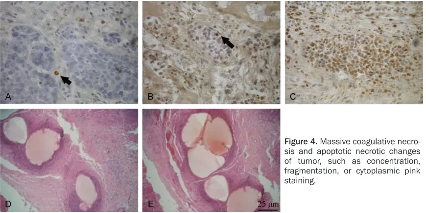
characterized by peritoneal thickening in enhanced CT, as well as massive ascites in the perihepatic and abdominal cavity (Figure 3). After embolization, the tumor cells in the cen-tral part of the tumor showed massive coagula-tive necrosis, and a large number of tumor cells were found apoptotic necrotic changes such as concentration, fragmentation, or cytoplasmic pink staining in the part close to the periphery (Figure 4).
VEGF immunohistochemistry results and MVD count
Immunohistochemistry results: normal liver tis-sue and peritumoral liver tistis-sue have no stain-ing or only very weak stainstain-ing. The positive rates of VEGF expression in group A, B, C, and D were 66.7%, 50%, 100%, and 83.3%, respec-tively. There was no significant difference in the comparison among the four groups (P>0.05).
CD31 staining and MVD count: the endothelial cells showed different degrees of brown-yellow to brown staining in the tumor tissue. In this experiment, the MVD counts were 55.36 ± 7.02, 41.27 ± 8.45, 82.42 ± 6.23, and 67.81 ± 11.42, respectively (Table 1). The MVD value in group D was higher than group B and group C, and the MVD value in group D was lower than group E (P<0.05). The Spearman rank correla-tion analysis between VEGF and MVD showed a correlation coefficient of 0.726, P<0.01 (Table 2).
Three weeks after TACE, the gross specimen showed liquefaction necrosis in the central part of the tumor, but residual tumor parenchyma still remained certain white fish-like tissue in the periphery of the tumor. In the specimens with positive TUNEL staining, more nuclear enlargement, cytoplasm with deeply-stained brown granules, and fragmentation and con-centration of nucleoli can be observed at the junction of irregular necrotic center/surround-ing area of the tumor with the boundary of residual tissue, suggesting apoptosis of a large number of tumor cells (Figure 5). The apoptotic indexes in the five groups were: 0.3%, 11.4%, 14.3%, 1.7%, and 5.1%, respectively (P<0.01). There were scattered apoptotic cells in group A, and apoptotic cells in each embolization group increased, mainly locating in the periphery of the tumor, and the apoptotic cells in group B also increased significantly. The comparison among groups B-D showed that the apoptotic cells in group C and D significantly increased. In the normal liver tissue of group E, a large
num-Table 2. VEGF staining and MVD counts in
differ-ent groups
Group n VEGF positive+ ++ +++ Positive rate (%) MVD count
A 6 1 1 2 66.7 55.36±7.02
B 6 1 0 2 50 41.27±8.45a
C 6 3 1 2 100 82.42±6.23a,b
D 6 1 3 1 83.3 67.81±11.42b,c
[image:4.612.90.525.71.289.2]Note: Compared with group A, aP<0.05; compare with group B, bP<0.05; compare with group C, cP<0.05. Spearman rank correlation analysis between VEGF and MVD shows a correla-tion coefficient of 0.726, P<0.01.
[image:4.612.90.303.336.415.2]ber of apoptotic normal hepatocytes with posi-tive TNNEL staining were found.
Discussion
Anti-tumor mechanism of sodium alginate
There is now more and more evidence indicat-ing that [15, 16], in addition to causindicat-ing emboli-zation by itself, TACE may cause recurrence of residual foci, and different embolic agents also affect the long-term efficacy of TACE. The tradi-tional embolic agent lipiodol has a short resi-dence time in tumor blood vessels, and because of its liquid state, it is difficult to achieve complete embolization due to siphon action and blood flow in tumor blood vessels [17], so chemotherapy drugs can achieve effec-tive distribution. In this study, ADM-alginate microspheres were used as the embolic agent. Due to their good and rapid expansion charac-teristics, after the microspheres reached the target blood vessels, the volume can rapidly expand to twice as much as the original, so the tumor blood supply artery can be rapidly occluded. The effects of tumor necrosis were more complete than iodized oil embolization. The rapid necrosis of the tumor preceded the establishment of residual blood supply, which greatly reduced the recurrence of residual tumor. At the end of Week 5, the growth of liver VX2 xenografts in group C was more inhibited than group B and group D, and the tumor necrotic rate was greater. Although simple lipi-odol embolization and conventional chemoem-bolization had a certain inhibitory effect on the
growth of orthotopic transplantation tumors, their inhibitory effects against intrahepatic metastasis and distant metastasis were not significant when compared with group A. The application of ADM in TACE can not only further inhibit the growth of orthotopic liver xenografts but also significantly lower the intrahepatic metastasis rate and distant metastasis rate than group A, B, and D.
Role of angiogenic factors such as VEGF in tumor angiogenesis
[image:5.612.90.523.71.289.2]Tumor angiogenesis is particularly important in tumor recurrence and metastasis. Due to the incomplete vascular structure of the tumor, the intercellular connections are relaxed, so the tumor cells can directly enter the blood without invasion. During invasion, the tumor cells can release various factors and proteolytic enzymes to promote the metastasis of liver tumor cells. In patients with liver cancer, the VEGF concen-tration is statistically high in patients with tumor diameter >5 cm, tumor thrombus, or microscopic tumor thrombus (P<0.05). Tumor angiogenesis is essential for tumor growth and metastasis. Studies have reported that basic fibroblast growth factor (bFGF) is the strongest vascular endothelial growth factor, followed by the vascular endothelial growth factor (VEGF) and the platelet-derived endothelial cell growth factor (PD-ECGF) [18]. VEGF is a well-known cytokine that promotes the vascular endotheli-al growth and has the functions of increasing the microvascular permeability, promoting the
8577 Int J Clin Exp Med 2019;12(7):8572-8578 Sodium alginate microspheres have good bio-compatibility, and the final degradation prod-ucts in the body are non-toxic polysaccharides, mannose, and gulose, which are mainly excret-ed by the kidneys. Therefore, the apoptosis-inducing effect of ADM-alginate microspheres is mainly related to the effects of the chemo-therapeutic drugs embedded inside the embol-ic agents, such as damaging the DNA of tumor cells, increasing the expression of pro-apoptot-ic genes, inactivating the apoptosis-inhibiting genes, and causing ischemia and hypoxia in the tumor area.
Conclusion
In conclusion, ADM-alginate microspheres have many advantages such as rapid blood supply cutoff and continuous induction of apoptosis of tumor cells, can significantly inhibit the growth and metastasis of orthotopic transplanted tumors. However, it should also be noted that at present, ADM-alginate microspheres are mainly characterized by strong antitumor effects in TACE. With deepening of research, sodium alginate microspheres containing dif-ferent drugs may provide better pharmaceuti-cal dosage forms and play important roles in anti-tumor growth and metastasis.
Disclosure of conflict of interest
None.
Address correspondence to: Dandan Xiao, De- partment of Outpatient, Shenzhen Hospital, Sou- thern Medical University, Shenzhen 518010, China. Tel: +86 10 2336 0604; Fax: +86 10 23323777; E-mail: [email protected]
References
[1] Glantzounis GK, Tokidis E, Basourakos SP, Nt-zani EE, Lianos GD and Pentheroudakis G. The role of portal vein embolization in the surgical management of primary hepatobiliary can-cers. a systematic review. Eur J Surg Oncol 2017; 43: 32-41.
[2] Petrowsky H and Busuttil RW. Resection or ab-lation of small hepatocellular carcinoma: what is the better treatment? J Hepatol 2008; 49: 502-504.
[3] Shindoh J, Tzeng CW, Aloia TA, Curley SA, Huang SY, Mahvash A, Gupta S, Wallace MJ endothelial cell division and vascular
construc-tion from different sources, promoting the endothelial cell migration, and the inducing tumor angiogenesis. It is closely related to the growth of solid tumors and the metastasis of malignant tumors [19, 20].
Tumor angiogenesis after embolization.
In this study, expression of VEGF and MVD in group D was significantly higher than group B (P<0.05), but the expression showed a decrease in group C compared with group B, indicating that VEGF expression increase and angiogenesis are in the process of tumor growth. The expression of VEGF increased sig-nificantly after embolization with simple lipi-odol, and the angiogenesis of tumor was more obvious. The application of ADM in the process of ADM microsphere embolization reduced expression of VEGF in tumors and inhibited the tumor angiogenesis to a certain extent. Mor- eover, rapid and effective expansion of the sodium alginate microspheres made the embo-lization more thorough, reduced retention of residual foci, and inhibited tumor recurrence for a period of time.
Evaluation of ADM-alginate microsphere em-bolization
and Vauthey JN. Safety and efficacy of portal vein embolization before planned major or ex-tended hepatectomy: an institutional experi-ence of 358 patients. J Gastrointest Surg 2014; 18: 45-51.
[4] Li D, Kang J and Madoff DC. Locally ablative therapies for primary and metastatic liver can-cer. Expert Rev Anticancer Ther 2014; 14: 931-945.
[5] Simon CJ, Dupuy DE and Mayo-Smith WW. Mi-crowave ablation: principles and applications. Radiographics 2005; 25: S69-83.
[6] Maruyama H, Yoshikawa M and Yokosuka O. Current role of ultrasound for the management of hepatocellular carcinoma. World J Gastroen-terol 2008; 14: 1710-1719.
[7] de Graaf W, van den Esschert JW, van Lienden KP, van Gulik TM. Induction of tumor growth after preoperative portal vein embolization: is it a real problem? Ann Surg Oncol 2009; 16: 423-430.
[8] Chung JW. Transcatheter arterial chemoembo-lization of hepatocellular carcinoma. Hepato-gastroenterology 1998; 45: 1236-1241. [9] Katayama N, Sugimoto K, Okada T, Ueha T,
Sakai Y, Akiyoshi H, Mie K, Ueshima E, Sofue K, Koide Y, Tani R, Gentsu T and Yamaguchi M. Intra-arterially infused carbon dioxide-saturat-ed solution for sensitizing the anticancer effect of cisplatin in a rabbit VX2 liver tumor model. Int J Oncol 2017; 51: 695-701.
[10] Tomozawa Y, Nitta N, Ohta S, Watanabe S, So-noda A, Nitta-Seko A, Tsuchiya K and Murata K. Anti-tumor effects of sorafenib administered at different time points in combination with transarterial embolization in a rabbit VX2 liver tumor model. Cardiovasc Intervent Radiol 2017; 40: 1763-1768.
[11] Kim GM, Kim MD, Kim do Y, Kim SH, Won JY, Park SI, Lee do Y, Shin W and Shin M. Transar -terial chemoembolization using sorafenib in a rabbit VX2 liver tumor model: pharmacokinet-ics and antitumor effect. J Vasc Interv Radiol 2016; 27: 1086-1092.
[12] Akinwande O, Kim D, Edwards J, Brown R, Phil-ips P, Scoggins C and Martin RC 2nd. Is radio-embolization ((90)Y) better than doxorubicin drug eluting beads (DEBDOX) for hepatocellu-lar carcinoma with portal vein thrombosis? a retrospective analysis. Surg Oncol 2015; 24: 270-275.
[13] Chen J, Qian T, Zhang H, Wei C, Meng F and Yin H. Combining dynamic contrast enhanced magnetic resonance imaging and microvessel density to assess the angiogenesis after PEI in a rabbit VX2 liver tumor model. Magn Reson Imaging 2016; 34: 177-182.
[14] Kim DY, Choi MS, Lee JH, Koh KC, Paik SW, Yoo BC, Shin SW, Choo SW, Do YS and Rhee JC. Milan criteria are useful predictors for favor-able outcomes in hepatocellular carcinoma patients undergoing liver transplantation after transarterial chemoembolization. World J Gas-troenterol 2006; 12: 6992-6997.
[15] Poon RT, Ngan H, Lo CM, Liu CL, Fan ST and Wong J. Transarterial chemoembolization for inoperable hepatocellular carcinoma and postresection intrahepatic recurrence. J Surg Oncol 2000; 73: 109-114.
[16] Trinchet JC, Ganne-Carrie N and Beaugrand M. Intra-arterial chemoembolization in patients with hepatocellular carcinoma. Hepatogastro-enterology 1998; 45: 1242-1247.
[17] Iwai K, Maeda H and Konno T. Use of oily con -trast medium for selective drug targeting to tumor: enhanced therapeutic effect and x-ray image. Cancer Res 1984; 44: 2115-2121. [18] Kumar R, Yoneda J, Bucana CD and Fidler IJ.
Regulation of distinct steps of angiogenesis by different angiogenic molecules. Int J Oncol 1998; 12: 749-757.
[19] Kobayashi N, Ishii M, Ueno Y, Kisara N, Chida N, Iwasaki T and Toyota T. Co-expression of bcl-2 protein and vascular endothelial growth fac-tor in hepatocellular carcinomas treated by chemoembolization. Liver 1999; 19: 25-31. [20] An FQ, Matsuda M, Fujii H and Matsumoto Y.
Expression of vascular endothelial growth fac-tor in surgical specimens of hepatocellular car-cinoma. J Cancer Res Clin Oncol 2000; 126: 153-160.
[21] Kerr JF, Wyllie AH and Currie AR. Apoptosis: a basic biological phenomenon with wide rang-ing implication in tissue kinetics. Br J Cancer 1972; 26: 239-257.