Case Report
Type II old odontoid fractures in the
ankylosing spondylitis patient: successful
surgical treatment by O-arm navigation: a case report
Kai Tang*, Xiao-Qiang Cheng*, Hui-Lin Yang, Feng Zhou
Department of Orthopedic Surgery, The First Affiliated Hospital of Soochow University, Suzhou 215006, Jiangsu, China. *Equal contributors.
Received September 28, 2018; Accepted February 11, 2019; Epub June 15, 2019; Published June 30, 2019
Abstract: Background: Ankylosing spondylitis (AS) is a chronic inflammatory disease that mainly affects the axial bone. Because of the poor blood supply, conservative treatment for type II old odontoid fractures is not effective. It often requires surgery. However, cases of odontoid fracture complicated with ankylosing spondylitis are very rare in the clinic. Case presentation: A case of a 60-year-old male patient is described who felt neck pain after he fell down on the ground two months before. The patient had a history of ankylosing spondylitis and did not undergo regular treatment. The X-ray and computed tomography (CT) showed a type II odontoid fracture and fusion of all spinal segments below C2. MR found included interruption of bony continuity of odontoid process basilar part. This patient was diagnosed with an old type II odontoid fracture complicated with ankylosing spondylitis. Posterior bone
graft fusion screw-rod fixation assisted by O-arm navigation surgery was performed for the patient. After surgery, the
patient’s neck pain completely disappeared. Conclusions: Cases of odontoid fracture complicated with ankylosing
spondylitis are very rare in clinic. This is a very important case of providing spine surgeons with a treatment plan for treating patients with old type II odontoid fractures complicated with AS. It also requires us to discuss that what treatments are more suitable for patients.
Keywords: Type II old odontoid fractures, ankylosing spondylitis, old fracture, posterior fusion screw-rod fixation, O-arm navigation
Introduction
Ankylosing spondylitis (AS) is a systemic inflam
-matory disease that mainly affects the spine,
ankle joints, and ambient joints [1, 2]. It
eventu-ally leads to severe spinal deformity, spinal fusion, and ankylosis. The patients are at high risk of spine fractures. The odontoid fracture is very common in cervical vertebrae fracture, accounting for 10% to 15% of cervical fractures [2, 3]. Most odontoid fractures are not compli -cated with spinal cord injury, namely without
obvious symptoms. As a result, the rate of missed diagnosis is high. After more than three weeks, fresh fractures evolved into old frac
-tures. In the literature, the probability of type II odontoid fracture is 26%-76% [4]. However, cases of odontoid fracture complicated with AS
are rare in clinic. According to our review there
have been few reports on type II old odontoid
fractures complicated with AS. In this article, a rare case of a patient with type II old odontoid fractures complicated with AS is reported. Posterior bone graft fusion and screw-rod fixa
-tion assisted by O-arm naviga-tion surgery was chosen for the patient, and the patient recov
-ered well after the surgery.
Case presentation
The 60-year-old patient man came to our
hospi-tal because he fell down on the ground and he felt neck pain for two months. The patient was
diagnosed as ankylosing spondylitis when he was 30 and he did not undergo regular
treat-ment. The patient was given cervical fixation
treatment. Physical examination showed ten-derness on the cervical spine. The patient’s thoracic and lumbar vertebrae were supported
the anterior and posterior longitudinal ligaments, and
formation of the ossify
bri-dge. Typical bamboo appe-
arance of the spine from the CT scan was found. MR
(Figure 3A-C) revealed that
the interruption of bony co-ntinuity of the base of odon -toid process basilar part. From the MR, the posterior
displacement of the frac
-ture site and soft tissue swelling of the cervical
sp-ine could be seen. At the
fracture site, there was a
stripy hypo-intense on T1- W1, and a patchy hyper-in- tense was seen on the sh- ort time inversion recovery
(STIR). The spinal cord of
C1-2 spinal cord was com-pressed. There was no obvi-ous abnormal signal in the spinal cord. According to the patient’s medical histo-ry and radiology examina-tions, the patient was diag-nosed as type II old
odon-toid fractures with AS.
Due to the history of AS,
the cervical vertebra activi-ty degree was basically lo-
st, and the anterior flexion and posterior extension of
the cervical vertebrae was
very poor. Therefore, ante -rior cervical operative posi-tion was not an opposi-tion. Co- nsidering various aspects (activity, surgical position, anesthesia intubation, etc.) tension are normal, and the patient had normal
sensation. The pathological reflex was
negati-ve.
Cervical X-ray (Figure 1A, 1B) revealed change
of physiological curvature of cervical spine, old type II odontoid fracture, and fusion of all spinal segments below C2. Calcified cervical anterior
longitudinal ligament was also observed. Com- puterized tomography (Figure 2A, 2B) showed
that old type II odontoid fracture, ossification of
factors, it was decided to perform posterior bone graft fusion and screw-rod fixation as-sisted by O-arm navigation surgery (C1-2).
Af-ter the operation, the patient got pain relie- ve. The postoperative X-ray (Figure 4A, 4B)
showed the internal fixation was in good po-sition. The patient was able to get up out of bed 7 days and was discharged 10 days
af-ter operation and continued to be protected
by neck collar for four weeks more. Five mon-ths after operation, X-rays of the cervical
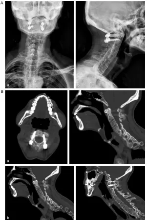
[image:2.612.90.389.71.279.2]sp-Figure 1. Cervical X-ray (A, B) showed the change of physiological curvature of cervical spine, old type II odontoid fracture and fusion of all spinal segments below C2. Calcified cervical anterior longitudinal ligament can also be seen.
Figure 2. CT of the cervical spine (A, B) showed that old type II dens fracture,
the right cortical bone destruction was discontinuous, the CT appearances were
hyperostosis of the anterior and posterior margins of vertebral body, ossification of the anterior and posterior longitudinal ligaments, and formation of the ossify bridge. The classic bamboo appearance of the spine from the CT scan can be
[image:2.612.89.387.345.497.2]valence in China is about
0.26% [5, 6]. During the de-velopment of the disease, spontaneous bone fusion
occurs in patients with AS, and the patients usually have osteoporosis. The
bio-mechanical properties of
the spine have changed,
and spinal fractures can occur at any time if the
pa-tients with AS are not
care-ful. It is reported that the probability of spinal frac -tures in AS patients is near-ly 5 times higher than that in the normal population,
and the probability of com
-bining with other types of fr-acture is increased by 35%
almost [3]. Cervical
verte-brae is a common site of fracture in patients with AS. ine (Figure 5Aa, 5Ab) and CT (Figure 5Ba,
5Bb) showed good internal fixation and bo-ne callus was observed at the bobo-ne graft
si-te. The patient’s neck pain completely dis-
appeared and the patient was still followed
up.
Discussion and conclusion
Ankylosing spondylitis (AS) is an unexplained
systemic inflammatory disease that mainly affects the spine and sacroiliac joint. The
pre-Due to the biomechanical property changes of AS, there are some characteristics of cervical vertebrae fractures in the patients with AS.
First, there can be low-energy injuries even
when there is no definite trauma that could result in fracture. Second, the fracture site is unstable, accompanied by fracture dislocation, which can easily lead to spinal cord injury. Old odontoid fractures are not common in clinical
practice and are easily missed diagnosed. Th-
ere are many reasons for missed diagnosis [1,
[image:3.612.92.523.71.237.2]5, 6].
Figure 3. MR (A-C) showed that the change of physiological curvature of cervical spine, and the interruption of bony continuity of the base of odontoid process basilar part. From the MR, the posterior displacement of the fracture site and soft tissue swelling of the cervical spine can be seen. At the fracture site, there was a stripy low signal on T 1W1, and a patchy hyper-intense signal was seen on the short time inversion recovery (STIR). The spinal cord of
C1-2 spinal cord was under compression. There was no obvious abnormal signal in the spinal cord.
[image:3.612.90.388.320.538.2]not be treated at the same time. Various reasons lead to unreasonable treatment and patients’ condition will
probably be concealed
fr-om multiple injury. In this case, the patient was diag-nosed with a type II old
odontoid fracture associat -ed with AS. Studies have shown that the odontoid blood supply mainly comes
from the anterior ascend -ing artery, the posterior ascending artery, and the
cleft perforators artery. Ty-pe II odontoid fractures can
easily damage blood supply
and lead to odontoid frac -tures nonunion.
According to investigation,
the odontoid fracture can occur in all age groups.
Ol-der people may have this
type of fracture because of a low-energy fall, while
younger patients’ odontoid
are more vulnerable to frac
-ture after motor vehicle
ac-cidents [6-8]. Dr. Anderson
and Dr. D’Alonzo put for
-ward a classification sys -tem in 1974 and there are
3 types of the odontoid fractures. Type I fractures, described asavulsion frac
-ture, near the tip of the
od-ontoid process, accounting
for 1% to 5% and were con
-sidered stable. Type II frac
-tures, accounting for 38% to 80%, are usually trans
-First of all, due to insufficient understanding of the disease, there is a large space for buff-ering because of a wide diameter of cervical spinal canal. Therefore, some fresh odontoid fractures may be missed because there are no symptoms and signs of spinal cord injury. Second, because of complicated with other body parts injury, with insufficient experience, some doctors are focused on the major injuries that affect vital signs and ignore the odontoid fracture. Moreover, some serious trauma can
-verse fractures that occur at the base of the
odontoid and above the axis [3, 9]. At the same
time, this type of fractures usually occur
be-tween the transverse ligament and the
verte-bral body. Type III fractures, extend into the ver -tebral body, involving the superior articular
pro-cess of the axis, one side articular propro-cess or bilateral facet fractures, accounting for 15% to 40%. The type II fractures are the most com
-mon type, and the probability of nonunion of type II odontoid fractures is 26%-76% accord -Figure 5. (A, B) Five months after operation, the X-rays of the cervical spine (Aa,
[image:4.612.90.391.70.520.2]ing to other literatures due to the unstable
structure [4]. Most of the type I and type III odontoid fractures can be cured by conserva
-tive treatment. However, the fracture line of type II odontoid fracture is located at the odon -toid waist, and the blood supply is very poor,
and thus conservative treatment is not effec
-tive and most patients require surgery [3, 10, 11]. At the same time, according to the fracture time, it is divided into fresh fracture and old fracture. The former refers to fracture within 3 weeks and the latter refers to fracture over 3 weeks. Obviously, old odontoid fractures are
not common in clinical practice.
Treatment options for a type II OF remain con -troversial. Conservative treatment, such as prolonged cervical immobilization may be a risk
for the patient, resulting in bone nonunion.
Fu-rthermore, operation intervention may pose a
risk for the patient. For the fresh type II odon
-toid fractures, conservative treatment has poor effect. The anterior cancellous bone screw fixa
-tion is the first choice in clinical. The operative method can retain the rotation function of
atlanto-axial vertebral. Considering the patient
has a long history of ankylosing spondylitis, activity degree of cervical vertebra is very sm-all, the significance of anterior surgery for the patient is not great. Furthermore, for the old type II odontoid fractures, the fracture inter
-space may be partially filled with soft tissue scars. If the anterior dentate lag screw fixation or posterior non-fusion screw fixation is cho
-sen, there is still a risk of bone nonunion. Therefore, the posterior atlanto-axial fixation [8, 12-14] assisted by O-arm navigation was
more inclined to be chosen.
According to the literature, it has been revealed that a higher median placement accuracy with a CT-based navigation system, and the CT- based navigation system may provide higher
accuracy of pedicle screw placement than con
-ventional non-navigated techniques. Our initial experience with the O-arm navigated spinal
surgery allowed us to anticipate that this
sys-tem may significantly increase the precision of
spinal instrumented procedures and shorten the operation time [6, 10-12, 15]. It allows intra-operative control and immediate
correc-tion of misplaced spinal implants that can eventually result in lower incidence of redo sur
-gery. Therefore the patient was received surgi
-cal assisted by O-arm navigation [3, 16].
In conclusion, for the old type II odontoid frac -tures, the appropriate surgical treatment sh-
ould be selected based on different individual conditions, such as the type of odontoid frac -ture, displacement degree, reduction status, as well as the technical skills in clinical. In this way the best clinical treatment can be chosen.
The patient gained good recovery after the sur -gery. Three days later, the X-rays were reviewed (Figure 4A, 4B). The fracture internal fixation was good and the fracture site was completely
repositioned. The X-ray (Figure 5Aa, 5Ab) and
CT scan of the cervical vertebrae (Figure 5Ba,
5Bb) showed good internal fixation after 5
months. Bone callus can be seen at the bone
graft site.
Disclosure of conflict of interest
None.
Address correspondence to: Dr. Feng Zhou, Depart-
ment of Orthopaedic Surgery, The First Affiliated Hospital of Soochow University, 188 Pinghai Road,
Suzhou 215006, Jiangsu, China. Tel: +86-512-67- 780111; Fax: +86-512-67780999; E-mail: liw72@ 126.com
References
[1] Josten C, Jarvers JS, Glasmacher S, Heyde CE,
Spiegl UJ. Anterior transarticular atlantoaxial screw fixation in combination with dens screw fixation for type II odontoid fractures with as -sociated atlanto-odontoid osteoarthritis. Eur Spine J 2016; 25: 2210-2217.
[2] Ozgocmen S, Ardicoglu O. Odontoid fracture
complicating ankylosing spondylitis. Spinal Cord 2000; 38: 117-119.
[3] Bhattacharyya S, Kim M. Cervical spine frac -ture associated with ankylosing spondylitis. Neurology 2014; 83: 1297.
[4] Vaccaro AR, Kepler CK, Kopjar B, Chapman J,
Shaffrey C, Arnold P, Gokaslan Z, Brodke D, France J, Dekutoski M. Functional and quality-of-life outcomes in geriatric patients with type-II dens fracture. J Bone Joint Surg Am 2013;
95: 729-735.
[5] Meyer C, Oppermann J, Meermeyer I, Eysel P,
Muller LP, Stein G. [Management and outcome
of type II fractures of the odontoid process]. Der Unfallchirurg 2018; 121: 397-402.
[6] Pal D, Sell P, Grevitt M. Type II odontoid frac -tures in the elderly: an evidence-based
narra-tive review of management. Eur Spine J 2011;
[7] Clark S, Nash A, Shasti M, Brown L, Jauregui JJ, Mistretta K, Koh E, Banagan K, Ludwig S, Gelb
D. Mortality rates after posterior C1-2 fusion for displaced type II odontoid fractures in octo -genarians. Spine 2018; 43: e1077-e1081. [8] Hadjicostas PT, Tsirogianni AK, Soucacos PN,
Thielemann FW. Odontoid fracture in severe
ankylosing spondylitic patient. Injury 2010; 41: 231-234.
[9] Nugent M, Berney MJ, Morris S. Clinical
out-comes following spinal fracture in patients
with ankylosing spondylitis. Ir J Med Sci 2017; 186: 677-681.
[10] Vosse D, van der Heijde D, Landewe R, Geu- sens P, Mielants H, Dougados M, van der Lin-
den S. Determinants of hyperkyphosis in pa -tients with ankylosing spondylitis. Ann Rheum Dis 2006; 65: 770-774.
[11] Alaranta H, Luoto S, Konttinen YT. Traumatic spinal cord injury as a complication to ankylos-ing spondylitis. An extended report. Clin Exp Dermatol 2002; 20: 66-68.
[12] Konieczny MR, Gstrein A, Muller EJ. Treatment
algorithm for dens fractures: non-halo immobi
-lization, anterior screw fixation, or posterior transarticular C1-C2 fixation. J Bone Joint Surg
Am 2012; 94: e144, 141-146.
[13] Thumbikat P, Hariharan RP, Ravichandran G, McClelland MR, Mathew KM. Spinal cord injury in patients with ankylosing spondylitis: a 10-year review. Spine 2007; 32: 2989-2995. [14] Pavlu J, Jan-Mohamed R, Kaczmarski R. Path-
ological odontoid fracture. Lancet Oncol 2006;
7: 96.
[15] Liu H, Chen W, Liu T, Meng B, Yang H. Accuracy
of pedicle screw placement based on preoper -ative computed tomography versus
intraopera-tive data set acquisition for spinal navigation system. J Orthop Surg (Hong Kong) 2017; 25:
2309499017718901.
[16] Fu Z, Zhang X, Shi Y, Dong Q. Comparison of
surgical outcomes between short-segment
open and percutaneous pedicle screw fixation techniques for thoracolumbar fractures. Med