Original Article
Maxillary sinus floor elevation without bone
grafting using an animal model
Zhen Luo, Andi Zhu, Di Zhang, Huilan Zhong, Bo Yang, Jiansheng Huang, Pingping Xu
Stomatological Hospital, Southern Medical University, S366, Jiangnan Boulevard, Guangzhou 510280, China
Received January 9, 2019; Accepted February 13, 2019; Epub May 15, 2019; Published May 30, 2019
Abstract: Objectives: The aim of this study was to evaluate the amount and source of new bone formation after max-illary sinus floor elevation without bone grafting, investigating the effects of maxmax-illary sinus membrane perforation using this technique. Methods: Six beagles were used, establishing an animal model of insufficient alveolar bone volume in the maxillary region. Bilateral maxillary sinus floor elevations were performed. A space-maintaining device was placed on one side with the membrane intact, while another device was placed on the contralateral side with the membrane perforated. The animals were sacrificed after 3 or 6 months. Results: Results showed newly formed bone inside the device. The heights of newly formed bones, 3 months after surgery, were 3.60 ± 0.63 mm and 2.81 ± 1.23 mm, with intact membranes and perforated membranes, respectively. The heights of the bones, 6 months after surgery, were 3.47 ± 0.29 mm and 2.91 ± 1.98 mm, with intact membranes and perforated membranes, re-spectively. Osteoid sedimentary mineralization was observed in the pores of the device, with a percentage of 48%. Conclusion: Maxillary sinus floor elevation without bone grafting induced new bone formation. Smaller membrane perforations did not affect new bone formation.
Keywords: No bone grafting, maxillary sinus floor elevation, membrane perforation, space maintenance, top de-sign
Introduction
Classic lateral antrostomy in the maxillary sinus fills the space between raised maxillary sinus membranes and sinus floor bones with bone grafts, creating a scaffold for subsequent blood vessel and cell growth and providing space for new bone formation [1]. Clinical findings have shown new bone formation after maxillary sinus floor elevation without bone grafting [2], confirmed by other researchers via animal experiments [3-5]. Maxillary sinus floor eleva-tion, alone, can provide a relatively closed space filled with blood clots, enabling bone tis-sue regeneration [6]. Maxillary sinus elevation without bone grafting is theoretically feasible. However, a method of maintaining the voided space after maxillary sinus floor elevation with-out bone grafting must be determined. Two main methods are currently available. One plac-es the implants concurrently with the sinus floor elevation procedure to maintain the void [7]. However, in many cases, the available bone graft is too tall to fit the space. The implant will not stay in place. Thus, maxillary sinus floor elevation cannot be performed concurrently
with implant placement. However, a specific space-maintaining device can be used to sup-port the lifted membrane [8, 9].
Animal experiments for maxillary sinus floor elevation with an implant, without bone graft-ing, have shown that, in the early stages of heal-ing, the void often collapses due to blood clot shrinkage and air pressure in the maxillary sinus. The membrane may attach to the implant surface or even be perforated. Thus, the im- plant protrudes into the sinus cavity. New bones are often confined to the lateral implant wall [10]. A space-maintaining device may partially maintain the void, a prerequisite for new bone formation. However, whether the new bone is derived from sinus floor bones or membranes and whether the void has a height limit for new bone formation remains unclear.
mucus and bacteria penetrating the void through the perforation. If the void with the per-forated membrane is filled with bone graft material, the antigenicity and movability of the bone grafts may delay new bone formation. If no bone grafting is performed, membrane per-foration effects on new bone formation after maxillary sinus elevation must be determined. The current study established an animal model with insufficient bone volume in the posterior maxilla after loss of the molar teeth. A space-maintaining device was used to maintain the void in the maxillary sinus membrane elevation. This model was used to evaluate the amount and source of new bone formation after maxil-lary sinus floor elevation without bone grafting, investigating the effects of maxillary sinus membrane perforation using this technique. Materials and methods
Design of the space-maintaining device
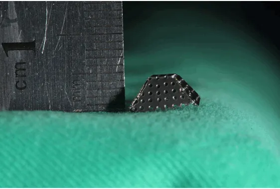
The space-maintaining device was constructed of pure titanium (TA2, Baoji, Shanxi Province, China), forming a cap-like structure. Surface pores were 0.5 mm in diameter and each pore was 0.5 mm apart from the others (porosity: 1.34/mm2). The top support surface was blunt
[image:2.612.90.371.71.261.2]and the device was approximately 4 mm high (Figure 1).
Preparation of the animal experimental model
The current study was approved by the Ethics Committee of the Guangdong Laboratory Ani-
mals were anaesthetized with atropine (0.05 mg/kg, Tianjin Pharmaceutical Group Xinzheng Co., Ltd., Tianjin, China) and ketamine (15 mg/ kg, Tianjin Pharmaceutical Group Xinzheng Co., Ltd., Tianjin, China). The operative field was dis-infected with 0.5% iodophor disinfectant and draped. Anaesthesia was infiltrated locally using a 1.7 mL dose of articaine hydrochloride (Primacaine, France) containing 1:10,000 epi-nephrine. Surgical procedures were performed in a strictly sterile environment.
Surgical procedure
The third premolar and first and second molars in the bilateral maxilla were removed three months before surgery, establishing an animal model with insufficient bone volume in the maxilla. Cone-beam computed tomography (CBCT) (NewTom, Italy) was used to exclude maxillary sinus inflammation and anatomical variation. A near-midline-incision was made in the alveolar ridge crest to flip the mucoperi-osteal flap and expose the lateral wall of the infraorbital nerve. The bone wall was removed to expose the infraorbital nerve and vascular bundle. These were ligated and divided. The lat-eral wall of the maxillary sinus was exposed. A bone chisel was used to create a bone window of 10 mm × 7 mm ± 2 mm. A sinus membrane-lifting tool was used to peel off and lift the max-illary sinus membrane. One side was randomly selected to be the model with a 2-mm mem-brane perforation, while the other side served as the model with an intact membrane. A pure titanium space-maintaining device was placed on each side (Figure 2). All experimental
ani-Figure 1. Space-maintaining device design.
ani-mals were given ampicillin sodium (80 mg/kg, Harbin Pharmaceutical Group, Harbin, China) and gentamicin (0.5 million units/kg, Chongqing Pioneer Pharmaceutical Co., Ltd., Chongqing, China) for a week and fed a liquified diet for 2 weeks. Three experimental animals were sacri-ficed using air embolization under general anaesthesia, 3 months after surgery. The re- maining experimental animals were sacrificed 6 months after surgery. Specimens were har-vested and soft tissues were removed and fixed in a 10% formalin solution.
and fine aluminium powder mixed with distilled water. Section surfaces were examined micro-scopically, with no scratches observed. The sections were then stained with toluidine blue and sealed. Heights of the new bones were measured using an optical microscope (Ol- ympus, Olympus Co., Japan) and Image-Pro-Plus software. The percentage of pores with sedimentary mineralization was calculated from the total pores. Each specimen was meas-ured three times and the average was re- corded.
Results
Micro-CT observation
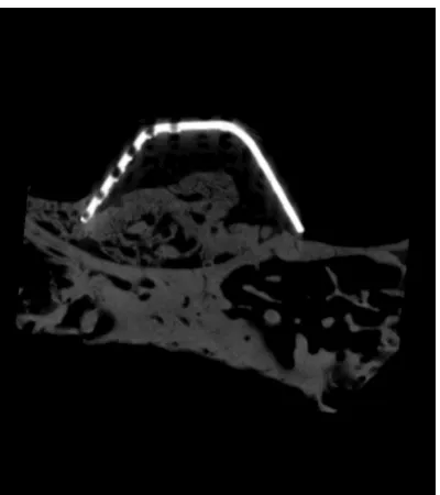
The space-maintaining device was located ab- ove the maxillary sinus floor, with no obvious changes in axial direction. No obvious imaging artifacts were seen around the new bones. The device’s internal space was occupied by bone tissue that was continuous with the sinus floor bone. New bone tissues showed an irregular shape and bone intensity remained relatively low (Figure 3).
Histological observation
[image:3.612.91.371.73.259.2]Histological changes did not significantly differ between the two experimental groups. The maxillary sinus membrane was attached to the surface of the device, which did not protrude directly into the sinus cavity.
Figure 2. Surgical procedure for maxillary sinus floor elevation after ligating the infraorbital neurovascular bundle.
Figure 3. Effects diagram of the Micro-CT.
Radiography and histological preparation
[image:3.612.90.290.309.534.2]New bone was visible inside the device and was similarly to the void. However, it did not occupy the entire voided space and was located mainly in the center. A small amount of new bone “climbing” occurred on the lateral wall of the device near the sinus floor. No obvious newly formed bone was observed in the space between the device and the membrane, oppo-site the sinus floor or in the area surrounding the device.
[image:4.612.90.525.72.234.2]Three months after surgery, the boundary between sinus floor bone and new bone was obvious. The new bone was an immature woven bone, in which loosely trabecular bones were arranged in a meshwork. Obvious spaces were visible between the trabecular bone. Cartilage lacuna were visible in the trabecular bone. Six months after surgery, the boundary between the sinus floor bone and new bone was unclear. The new bone was a mature bone structure, continuous with the sinus floor. Obvious spaces were observed between trabecular bones (Figure 4).
Osteoid sedimentary mineralization was seen in the pores at the side and top of the device. Sedimentary mineralization was composed of many scattered, long fusiform, and small min-eralization units, arranged in a regular pattern with clear boundaries. More oval mineralization was located near the membrane, while most of the long fusiform mineralization was located inside the device. Sedimentary mineralization originated from the maxillary sinus membrane and protruded into the pores. Sedimentary
min-eralization occurred in 48% of the pores (Figure 5).
Three months after surgery, newly formed bones were 3.60 ± 0.63 mm and 2.81 ± 1.23 mm high in dogs with intact membranes and perforated membranes, respectively. Six mon- ths after surgery, newly formed bones were 3.47 ± 0.29 mm and 2.91 ± 1.98 mm high in dogs with intact membranes and perforated membranes, respectively (Figure 6).
Discussion
The current study shows that new bone was formed in the space below the pure titanium space-maintaining device after maxillary sinus floor elevation. Therefore, without bone grafting material, the effects of maxillary sinus floor elevation can be achieved via the body’s own vascularization repair. During blood aggrega-tion, coagulation is activated by red blood cells and platelets. Blood clots begin to form. Blood clots contain many active factors and growth factors that promote capillary formation and proliferation and osteoblast differentiation [13-15]. Wang [16] showed that thrombin promotes new bone formation. This new bone formation has the same effects as fracture repair by regu-lating growth factors and angiogenesis. Al- though blood clots, alone, can promote new bone formation, blood clots do not maintain the void after maxillary membrane floor elevation, due to instability and shrinkage. Over time, the membranes gradually collapse and new bone formation is limited or even absorbed. Asai [17]
reported that air pressure in the maxillary sinus causes the membrane to collapse. This, in turn, affects the amount of new bone formed. Therefore, a specific space-maintaining device is used to maintain the void after sinus eleva-tion, providing a relatively closed environment and ensuring enough blood clot formation required for maxillary sinus floor elevation with-out bone grafting.
Schweikert used a titanium plate to maintain the void after sinus floor elevation. Due to a lack of three-dimensional support in the space, the membrane partially collapsed and the de- vice protruded into the sinus cavity. Johansson [18] placed a circular, hollow, and absorbable device with a 12-mm diameter in the lower part of the void. These authors endoscopically observed the membrane attached to the device surface, confirming that new bone was formed inside the device after 6 months. In this study,
the device’s design was in accord with an ideal space-maintaining device, proposed by Cricchio [8, 9] after maxillary sinus floor elevation. The device, like a tent, is stable and enhances the intactness of the void. It maintains the space three-dimensionally to prevent possible mem-brane collapse. Moreover, the space-maintain-ing device is made of pure titanium, with good tissue compatibility and flexibility. It easily fits different anatomical variations. The device has many 0.5-mm pores on its surface. Blood accu-mulates inside the device through the pores and is unaffected by atmospheric pressure dur-ing agglutination.
[image:5.612.91.524.72.399.2]Maxillary sinus membrane perforation reduces bone graft stability, displacing the bone grafts. This causes maxillary sinus inflammation, affecting bone graft maturation and calcifica-tion [19]. Furthermore, the sealed internal envi-ronment is lost due to membrane perforation
and stable blood clots cannot form. This hin-ders vascularization and new bone formation. No bone grafts exist in maxillary sinus elevation without bone grafting. Perforation factors only affect blood clot aggregation. In this study, the maxillary sinus membrane adhered to the sur-face of the device in both groups, the intact membrane group and the membrane perfora-tion group. The porous surface supported the perforation area, allowing blood permeation. Therefore, the device did not protrude into the sinus cavity and new bone formation was undis-turbed. Kaneko [20] performed imaging stud-ies of maxillary sinus elevation without bone grafting, finding no differences in new bone for-mation between perforated and unperforated areas of the membrane. This may have been because the perforated membrane was att- ached to the device’s surface, distanced from the blood clotting area. In addition, continuous oozing of soft tissue after surgery can fill the void.
The height of new bones after maxillary sinus elevation without bone grafting may be closely related to the void’s height. In 2007, Thor [21] reported results from 4-year follow-ups of 20 patients undergoing maxillary sinus elevation without bone graft material, but with 44 im- plants. Mean bone formation volume in the
requires new bone remodelling, including blood clot aggregation, mechanization, woven bone formation, and maturation [23]. Schmitz and Hollinger [24] first proposed the concept of criti-cal size defects, referring to the smallest bone defects that cannot heal on their own during the life cycle of an animal’s specific bone tis-sue. Rudert [25, 26] defined critical size defects as bone defects of 3 mm in diameter that can be completely repaired by self-formation of new bone. Although bone formation was observed in the bone defect area (4 mm or over), the cal-cification degree was low and the cortical bone was discontinuous. Bone graft materials and collagen membranes may be required to pro-mote bone repair [27]. Although the height of the space-maintaining device was designed to be 4 mm in this study, the concave shape of the maxillary sinus floor and blood clot aggregation may have caused device floating. Therefore, the height of the void may be greater than 4 mm, exceeding the limit of the body’s ossifica-tion potential. New bone volume was insuffi-cient to completely fill the void.
[image:6.612.90.368.70.319.2]The origin of new bone after maxillary sinus elevation has been a controversial issue. In this study, sedimentary mineralization was observ- ed in the pores of the device opposite the sinus floor. This may indicate that new bone was de-
Figure 6. Double-bar graph of new bone heights 3 months and 6 months after surgery.
rived from the membrane. In addition, this study showed that new bone was mainly locat-ed in the center of the void, with no obvious new bone formation surrounding the device. New bone morphology was compatible with the elevated membrane morphology. New bone for-mation did not occur at the same mineraliza-tion deposimineraliza-tion rate and the highest point of the elevated membrane was also the highest point of new bone formation. These factors suggest possible membrane induction for new bone for-mation. Srouj [28, 29] isolated and cultured human maxillary sinus membranes, analyzing the culture using flow cytometry. Expression of various osteogenic markers, such as alkaline phosphatase, BMP-2 and osteonectin, in the maxillary sinus membrane culture was signifi-cantly increased. Amplified maxillary sinus membrane cells were transplanted subcutane-ously into mice and calcified nodules were observed at the transplantation site after eight weeks. Kim [30] suggested that mesenchymal stem cells in the maxillary sinus membrane can differentiate into osteoblasts after osteogenic induction. Studies [31] have shown that a par-acrine signalling pathway may exist between the periosteum and new bone. After stimula-tion, the signalling pathway is activated. Spe- cific growth factors are stimulated to promote
[image:7.612.89.375.70.297.2]formation was observed at the top of the implant. Hatano [34] showed that bone resorp-tion at the top of the implant gradually increased over the observation period. This study demon-strated osteoid sedimentary mineralization in the pores of the device’s lateral and top walls, with no obvious sedimentary mineralization observed between the membrane and device. This may be because the membrane was tightly attached to the device because of sinus pres-sure. Thus, the space required for new bone formation was compressed. However, the pores provide a space for new bone formation. Therefore, new bone can be observed in some pores. This gave present researchers the idea to design the top of an implant as a mesh-like stud, with a diameter of 0.5 mm and interval of 0.5 mm between the studs (Figure 7). When the maxillary sinus membrane is attached to the top of the implant, the protruding studs pro-vide support. The space between protruding studs does not disappear. New bone can be deposited in the space between the protruding studs by bone formation. Bone formation at the top of the implant allows the entire implant to be embedded in bone tissue. This increases rates of bone-implant contact, thereby improv-ing success rates of maxillary sinus floor eleva-tion without bone grafting.
Figure 7. Effects diagram of the top of the implant designed specifically for maxillary sinus floor elevation. A. Effects diagram of the specialized implant installed in the maxillary sinus. B. The top includes studs that induce osteo-genesis in the pores.
undifferentiated mesenchymal stem cells to the cartilage pre-cursor, gradually forming new bones. The maxillary sinus membrane elevation may be considered a similar stimulus in the sinus floor.
Conclusion
In summary, maxillary sinus elevation without bone grafting induces new bone formation. Maxillary sinus membranes may be the source. Moreover, a special implant for maxillary sinus floor elevation could be designed based on the principle of osteogenesis in the pores.
Acknowledgements
This work was supported by a grant from Sto- matological Hospital Southern Medical Uni- versity Projects (no. PY2017010).
Address correspondence to: Jiansheng Huang and Pingping Xu, Stomatological Hospital, Southern Medical University, S366, Jiangnan Boulevard, Guangzhou 510280, China. Tel: (+86) 020-34811937; Fax: (+86) 020-020-34811937; E-mail: 515725901@qq.com (JSH); Tel: (+86) 020-348125- 26; Fax: (+86) 020-34812526; E-mail: zzxf0504@ 163.com (PPX)
References
[1] Schweikert M, Botticelli D, de Oliveira JA, Scala A, Salata LA, Lang NP. Use of a titanium device in lateral sinus floor elevation: an experimental study in monkeys. Clin Oral Implants Res 2012; 23: 100-105.
[2] Chen TW, Chang HS, Leung KW, Lai YL, Kao SY. Implant placement immediately after the lat-eral approach of the trap door window proce-dure to create a maxillary sinus lift without bone grafting: a 2-year retrospective evalua-tion of 47 implants in 33 patients. J Oral Maxillofac Surg 2007; 65: 2324-2328. [3] Ahmed M, Abu SA, Hamdy RM, Ezz M.
Bioresorbable versus titanium space-main-taining mesh in maxillary sinus floor elevation: a split-mouth study. Int J Oral Maxillofac Surg 2017; 46: 1178-1187.
[4] Fouad W, Osman A, Atef M, Hakam M. Guided maxillary sinus floor elevation using deprotein-ized bovine bone versus graftless schneideri-an membrschneideri-ane elevation with simultschneideri-aneous implant placement: randomized clinical trial. Clin Implant Dent Relat Res 2018; 20: 424-433.
[5] Palma VC, Magro-Filho O, de Oliveria JA, Lundgren S, Salata LA, Sennerby L. Bone refor-mation and implant integration following maxil-lary sinus membrane elevation: an experimen-tal study in primates. Clin Implant Dent Relat Res 2006; 8: 11-24.
[6] Pinchasov G, Juodzbalys G. Graft-free sinus augmentation procedure: a literature review. J Oral Maxillofac Res 2014: 5: e1.
[7] Balleri P, Veltri M, Nuti N, Ferrari M. Implant placement in combination with sinus mem-brane elevation without biomaterials: a 1-year study on 15 patients. Clin Implant Dent Relat Res 2012; 14: 682-689.
[8] Cricchio G, Palma VC, Faria PE, de Oliveira JA, Lundgren S, Sennerby L, Salata LA. Histological findings following the use of a space-making device for bone reformation and implant inte-gration in the maxillary sinus of primates. Clin Implant Dent Relat Res 2009; 11 Suppl 1: e14-e22.
[9] Cricchio G, Palma VC, Faria PE, de Olivera JA, Lundgren S, Sennerby L, Salata LA. Histological outcomes on the development of new space-making devices for maxillary sinus floor aug-mentation. Clin Implant Dent Relat Res 2011; 13: 224-230.
[10] Scala A, Botticelli D, Faeda RS, Garcia RI, de Oliveira JA, Lang NP. Lack of influence of the schneiderian membrane in forming new bone apical to implants simultaneously installed with sinus floor elevation: an experimental study in monkeys. Clin Oral Implants Res 2012; 23: 175-181.
[11] Nolan PJ, Freeman K, Kraut RA. Correlation be-tween schneiderian membrane perforation and sinus lift graft outcome: a retrospective evaluation of 359 augmented sinus. J Oral Maxillofac Surg 2014; 72: 47-52.
[12] Clementini M, Ottria L, Pandolfi C, Bollero P. A novel technique to close large perforation of sinus membrane. Oral Implantol 2013; 6: 11-14.
[13] Pagel CN, de Niese MR, Abraham LA, Chinni C, Song SJ, Pike RN, Mackie EJ. Inhibition of os-teoblast apoptosis by thrombin. Bone 2003; 33: 733-743.
[14] Bluteau G, Pilet P, Bourges X, Bilban M, Spa-ethe R, Daculsi G, Guicheux J. The modulation of gene expression in osteoblasts by thrombin coated on biphasic calcium phosphate ceram-ic. Biomaterials 2006; 27: 2934-2943. [15] Frost A, Jonsson KB, Ridefelt P, Nilsson O,
Ljunghall S, Ljunggren O. Thrombin, but not bradykinin, stimulates proliferation in isolated human osteoblasts, via a mechanism not de-pendent on endogenous prostaglandin forma-tion. Acta Orthop Scand 1999; 70: 497-503. [16] Wang H, Li X, Tomin E, Doty SB, Lane JM,
Carney DH, Ryaby JT. Thrombin peptide (TP508) promotes fracture repair by up-regu-lating inflammatory mediators, early growth factors, and increasing angiogenesis. J Orthop Res 2005; 23: 671-679.
[18] Johansson LA, Isaksson S, Adolfsson E, Lindh C, Sennerby L. Bone regeneration using a hol-low hydroxyapatite space-maintaining device for maxillary sinus floor augmentation--a clini-cal pilot study. Clin Implant Dent Relat Res 2012; 14: 575-584.
[19] Artzi Z, Weinreb M, Carmeli G, Lev-Dor R, Dard M, Nemcovsky CE. Histomorphometric assess-ment of bone formation in sinus augassess-mentation utilizing a combination of autogenous and hy-droxyapatite/biphasic tricalcium phosphate graft materials: at 6 and 9 months in humans. Clin Oral Implants Res 2008; 19: 686-692. [20] Kaneko T, Masuda I, Horie N, Shimoyama T.
New bone formation in nongrafted sinus lifting with space-maintaining management: a novel technique using a titanium bone fixation de-vice. J Oral Maxillofac Surg 2012; 70: e217-e224.
[21] Thor A, Sennerby L, Hirsch JM, Rasmusson L. Bone formation at the maxillary sinus floor fol-lowing simultaneous elevation of the mucosal lining and implant installation without graft material: an evaluation of 20 patients treated with 44 Astra tech implants. J Oral Maxillofac Surg 2007; 65: 64-72.
[22] Tatum H. Maxillary and sinus implant recon-structions. Dent Clin North Am 1986; 30: 207-229.
[23] Scala A, Botticelli D, Rangel IG, de Oliveira JA, Okamoto R, Lang NP. Early healing after eleva-tion of the maxillary sinus floor applying a lat-eral access: a histological study in monkeys. Clin Oral Implants Res 2010; 21: 1320-1326. [24] Schmitz JP, Hollinger JO. The critical size defect
as an experimental model for craniomandibu-lofacial nonunions. Clin Orthop Relat Res 1986: 299-308.
[25] Rudert M. Histological evaluation of osteo-chondral defects: consideration of animal models with emphasis on the rabbit, experi-mental setup, follow-up and applied methods. Cells Tissues Organs 2002; 171: 229-240. [26] Brittberg M, Nilsson A, Lindahl A, Ohlsson C,
Peterson L. Rabbit articular cartilage defects treated with autologous cultured chondro-cytes. Clin Orthop Relat Res 1996: 270-283.
[27] Caudill R, Lancaster D. Histologic analysis of the osseointegration of endosseous implants in simulated extraction sockets with and with-out e-PTFE barriers. Part II: histomorphometric findings. J Oral Implantol 1993; 19: 209-215. [28] Srouji S, Ben-David D, Lotan R, Riminucci M,
Livne E, Bianco P. The innate osteogenic poten-tial of the maxillary sinus (schneiderian) mem-brane: an ectopic tissue transplant model simulating sinus lifting. Int J Oral Maxillofac Surg 2010; 39: 793-801.
[29] Srouji S, Kizhner T, Ben David D, Riminucci M, Bianco P, Livne E. The schneiderian membrane contains osteoprogenitor cells: in vivo and in vitro study. Calcif Tissue Int 2009; 84: 138-145.
[30] Kim SW, Lee IK, Yun KI, Kim CH, Park JU. Adult stem cells derived from human maxillary sinus membrane and their osteogenic differentia-tion. Int J Oral Maxillofac Implants 2009; 24: 991-998.
[31] Saris DB, Sanyal A, An KN, Fitzsimmons JS, O’Driscoll SW. Periosteum responds to dynam-ic fluid pressure by proliferating in vitro. J Orthop Res 1999; 17: 668-677.
[32] Li X, Chen SL, Zhu SX, Zha GQ. Guided bone regeneration using collagen membranes for sinus augmentation. BR J Oral Maxillofac Surg 2012; 1: 69-73.
[33] Hurzeler MB, Quinones CR, Kirsch A, Schup-bach P, Krausse A, Strub JR, Caffesse RG. Max-illary sinus augmentation using different graft-ing materials and dental implants in monkeys. Part III. Evaluation of autogenous bone com-bined with porous hydroxyapatite. Clin Oral Im-plants Res 1997; 8: 401-411.