This paper is made available online in accordance with
publisher policies. Please scroll down to view the document
itself. Please refer to the repository record for this item and our
policy information available from the repository home page for
further information.
To see the final version of this paper please visit the publisher’s website.
Access to the published version may require a subscription.
Author(s): M Ahearne, Y Yang, K Y Then, K-K Liu
Article Title: Non-destructive mechanical characterisation of
UVA/riboflavin crosslinked collagen hydrogels
Year of publication: 2007
Link to published article:
http://dx.doi.org/10.1136/bjo.2007.130104
Non-destructive mechanical characterisation of UVA/
riboflavin crosslinked collagen hydrogels
M Ahearne,
1Y Yang,
1K Y Then,
2K-K Liu
11Institute of Science and
Technology in Medicine, School of Medicine, Keele University, Stoke-on-Trent, UK;
2
Birmingham and Midland Eye Centre, City Hospital, Birmingham, UK
Correspondence to:
K-K Liu, Institute of Science and Technology in Medicine, School of Medicine, Keele University, Stoke-on-Trent ST4 7QB, UK; i.k.liu@pmed.keele.ac.uk
Accepted 20 October 2007 Published Online First 16 November 2007
ABSTRACT
Aims:To establish a non-destructive method of characterising the mechanical properties of collagen hydrogels to model corneal tissue and to examine the effect of photochemical crosslinking on their mechanical properties.
Methods:Collagen hydrogels were manufactured, sub-merged in 0.1% riboflavin solution and crosslinked using two UVA tube bulbs with an intensity of between 2.8 and 3.2 mW/cm2. The hydrogels were clamped around their
outer edge and deformed using a sphere. The deformation was measured in situ using a long-working-distance microscope connected to a CCD camera, and the deformation displacement was used with a theoretical model to calculate the Young modulus of the hydrogels. Collagen hydrogels seeded with human corneal fibroblasts were used to examine cell viability after UVA irradiation. Results:There was an increase in Young modulus of the collagen hydrogels after UVA/riboflavin treatment that was dependent on the exposure time. UVA irradiation without riboflavin showed decreased mechanical integrity and strength. Cell viability was reduced with increased UVA exposure time.
Conclusion:The non-destructive technique demon-strated a new methodology comparable with strip extensiometry for cornea or corneal model specimens but with more convenient features. This approach could be used as an initial step in developing new crosslinking treatments for patients with keratoconus.
Keratoconus is a non-inflammatory disease that causes thinning of the corneal stroma which can lead to bulging, reduction of vision and discomfort. Several treatment options are under investigation to stabilise the progression of the disease. One of the most promising approaches involves the cross-linking of collagen fibrils in the cornea using ultraviolet light in the presence of a photosensitiser to improve its mechanical strength.1–3 Extensiometry has been used to characterise the mechanical properties of the corneas under inves-tigation. However, extensiometry has several limitations including destructive measurement, using strips of cornea not whole cornea, the corneal curvature not being taken into considera-tion, and tension primarily on fibrils that are parallel to the direction of strain.4 5
In addition, the method used to crosslink cornea for extensiometry differs from the method used to treat patients in vivo where only the centre of cornea is irradiated.3 For these reasons, an alternative approach to mechanical characterisation is desirable.
We have developed a long-working-distance microscope spherical microindentation system cap-able of measuring the mechanical properties of
hydrogel materials under cell culture conditions.6 This system can be used to obtain mechanical properties of biological materials on-line, non-destructively and in situ. Previous studies have shown that type 1 collagen can be used to model corneal tissue.7–9This is particularly useful, since a viable human cornea is difficult to obtain. In this paper, a spherical indentation technique was used to measure the Young modulus of collagen hydro-gels as a model for corneal tissue in order to demonstrate the capability of this technique to detect changes in the mechanical properties after photochemical crosslinking. A UVA/riboflavin crosslinking treatment similar to that previously used to crosslink cornea was used.1 Collagen hydrogels embedded with human corneal fibro-blasts were used to examine the cell viability after crosslinking.
MATERIALS AND METHODS Sample preparation and treatment
Rat-tail collagen type-1 (BD Bioscience, Mountain View, CA) was used as the source of collagen for the hydrogels. The hydrogels were formed follow-ing the manufacturer’s protocol except that a concentration 10 times that of standard Dulbecco
modified eagles medium (DMEM, ICN
Biomedicals, Aurora, OH) was used instead of PBS. Collagen concentrations of 2.5 mg/ml and 3.5 mg/ml were used. Five hundred microlitres of the hydrogel solution was poured inside a filter paper ring of inner diameter 20 mm. The solution was allowed to set for 1 h in an incubator at 37uC, 5% CO2. Once set, the newly formed hydrogel was cultured overnight with 5 ml of culture media supplemented by 10% fetal calf serum (Sigma, St Louis, MO), 1% L-glutamine (Sigma) and 1% antibiotic-antimycotic solution (Sigma).
0.1% riboflavin solution was made by dissolving 2 g of dextran (Sigma) in 10 ml of distilled water and adding 10 mg of riboflavin-5-phosphate (Sigma). Prior to UVA exposure, the collagen hydrogels were submerged in 3 ml of riboflavin solution for 5 min to allow infiltration. Some hydrogels, which were not placed into the ribo-flavin solution, were used as controls to examine if riboflavin influences the crosslinking process.
were found to have provided an irradiance of between 2.8 and 3.2 mW/cm2 across the whole collagen hydrogel surface.
Different exposure times up to 60 min were examined. The Young modulus of the hydrogels was measured before UV exposure by the technique described below. After the exposure, they were placed back into culture media and incubated at 37uC for 20 min to compensate for any dehydration during cross-linking. The modulus was then remeasured to examine the change in mechanical properties of the hydrogels. A number of hydrogels were placed back into culture media and cultured at 37uC for 1 week, after which their modulus was re-measured.
To examine the effect of UVA exposure on the viability of cells within the collagen hydrogels, human corneal fibroblasts were used. The cultivation of fibroblasts from human corneal tissue has been described previously.6
Cells at passage number 3 were used. The cells were suspended throughout the hydrogel solution at a concentration of 1 million cells/ml. Cell viability was assessed using a live/dead cell double staining kit (L-Fluka, Buchs, Switzerland). Five hundred microlitres of the staining solution was applied over the surface of the hydrogel and left at 37uC. After 20 min, the hydrogel was washed in PBS and examined using a confocal microscope (FV300, Olympus, Tokyo). The cell viability was calculated by dividing the number of live cells by the total number of cells.
Measurement instrumentation
The instrument, which has previously been reported,6consisted of two parts; a sample holder with a spherical indenter and an image acquisition system. Each hydrogel was clamped around its outer circumference by two transparent plastic circular hoops of inner diameter 20 mm. The hoops were held together using two thin stainless steel plates and a number of stainless steel screws as shown in fig 1. The whole assembly was submerged in PBS within a large rectangular Petri dish in an incubator at 37uC. The hydrogel was deformed (indented) by using a PTFE sphere (Spheric-Trafalgar, London) of diameter 4 mm. A sphere was placed at the centre of the hydrogel, and its weight caused the hydrogel to deform. In this study, the diameter ratio of the hydrogel to the sphere was kept constant at 5.0, which was identical to the value used in the previous analyses.6 10 11
The image acquisition system consisted of a long-working-distance objective microscope (Edmund Industrial Optics, Barrington, NJ) with a computer-linked CCD camera (XC-ST50CE, Sony, Tokyo) as shown in fig 2. The system allowed a high magnification up to 120 times for acquiring the side-view images of the deformation profile from outside the incubator through a glass window. The magnification of the system was
calibrated with the computer-acquired images of a stage micrometer. As well as the deformation, the thickness of the samples was measured using the same approach. The samples were placed onto a flat surface and their thickness was measured laterally based on the distance between the two edges of the hydrogel image that was acquired from the long-working-distance CCD microscope system.
Theoretical modelling
The mechanical behaviours of swollen hydrogels are believed to have similar behaviours to rubber-like materials.12 Based on rubber-like Mooney constitutive equations, Yang and Hsu developed a model that describes the deformation of a membrane due to the weight of a ball.11These equations have previously been applied to find the Young modulus of polyurethane films10 and hydrogel membranes.6 The central deformation (d) caused by the weight of the sphere on the hydrogel was measured by the image-acquisition system. This information was used to determine the Young modulus (E) from the following equation:
where w is the weight of the sphere, h is the hydrogel thickness and R is the radius of the sphere.6 10
When a sphere and hydrogel sample system satisfies the dimensional characteristics of a/ R = 5 andd/R(1.7, where a is the radius of the hydrogel, this model assumes that the ratio of thickness to the radius is low, and the deformation is large; hence stretching of the hydrogel dominates over bending.13 14
RESULTS
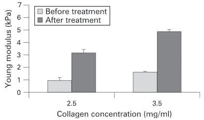
The effects of UVA/riboflavin crosslinking treatment on the Young modulus of the collagen hydrogels with a concentration of 3.5 mg/ml are displayed in fig 3. There was an increase in the modulus after UVA/riboflavin treatment that was dependent on the UVA exposure time. The increase in modulus was statistically significant after 15 minutes’ exposure comparable with non-treated samples. In addition, there was a significant difference between the hydrogels exposed for 15, 30 and 45 min but no statistically significant difference after 45 min, as determined using the ANOVA–Tukey test with a 95% confidence interval (Minitab). This suggests that the majority of crosslinking occurred in the first 30–45 min, and a longer exposure time might be unnecessary. Hydrogels with a collagen concentration of both 2.5 mg/ml and 3.5 mg/ml showed an increase in modulus of approximately 240% and 200%, respectively, after UVA exposure for 30 min (fig 4). The increased modulus was maintained over 1 week in culture (fig 5).
Four of the hydrogels were exposed to UVA without treatment with riboflavin. After 15 minutes’ irradiance, these hydrogels showed a decrease in modulus, demonstrated by an increase in indentation displacement depth. The indentation depth exceeded the maximum depth allowable to calculate the Young modulus from our theoretical model. After 30 minutes’ exposure to UVA radiation, the hydrogels reverted to a liquid state. This suggests that without the presence of the riboflavin, UVA irradiation caused the collagen fibres to degrade rather than crosslink.
[image:3.624.42.287.603.728.2]The effect of UVA/riboflavin treatment on the cells viability within the collagen hydrogels was examined. It was found that
UVA had a negative effect on the cells, as there was a reduction in cell viability after UVA exposure with only some cells still remaining viable. The percentage of viable cells was 83% without exposure to UVA, 60% after 15 minutes’ exposure and 37% after 30 minutes’ exposure.
DISCUSSION
The long-working-distance microscope spherical microindenta-tion technique has been used to examine the effect of photochemical crosslinking on the mechanical properties of collagen hydrogels. Unlike extensiometry, which has been previously used to examine mechanical properties of corneal strips after crosslinking,1 3
our non-destructive mechanical characterisation technique allows hydrogels and tissue engi-neered corneas to model corneal tissue, thus enabling examina-tion of crosslinking treatments. This is particularly useful, as viable human corneal tissue for experimentation is difficult to obtain due to its high demand for use in transplantations. In addition, our mechanical characterisation technique utilises a long-working-distance microscope and spherical microindenta-tion system that can be used on the same sample both before and after treatment. The stability of the crosslinking reactions could be demonstrated because of the non-destructive nature of the mechanical tests. This approach allowed a direct compar-ison and monitoring in the change in modulus of each hydrogel. In addition to examining the effect of photochemical cross-linking treatment on the mechanical properties of collagen hydrogels, the spherical indentation technique could be used for several other applications. The effect of other crosslinking agents such as glutaraldehyde on the mechanical properties of the hydrogel could be investigated. The long-term effects of the cells within the hydrogel on the overall mechanical properties of the hydrogel membrane could be investigated. The effect of
multiple treatments on the mechanical properties of the corneal constructs over a time frame could be measured. This technique could also be adopted to examine biomaterials used as a keratoprosthesis or tissue-engineered cornea.
A significant increase in Young modulus was demonstrated after UVA/riboflavin treatment (increase factor of 3.6 after 45 minutes’ UVA exposure). This increase in modulus was of a similar magnitude to that found with strips of human cornea.3 The results also suggest that the optimum UVA exposure time to increase the modulus of the hydrogels was between 30 and 45 min. This increase in modulus was maintained over time. These results demonstrate how the spherical microindentation technique can be used to optimise crosslinking techniques.
Hydrogels exposed to UVA without riboflavin showed a reduced mechanical integrity, while those with riboflavin showed an increased modulus. Menter et al15 observed that riboflavin added to collagen had a significant effect on fibrillogenesis kinetics under UVA radiation. Our spherical microindentation technique could be used to examine the effect of different concentrations of riboflavin solution or other photosensitising agents, which would allow an optimum concentration for crosslinking to be determined.
UVA exposure appeared to have a negative affect on cell viability in hydrogels. This was expected as UVA treatments on real corneal tissue have induced cytotoxicity and apoptosis of keratocytes.16 17Despite these findings, a populations of viable cells remain after UVA exposure. The long-term affect of UVA exposure on the cell behaviour has not yet been examined. Finding the optimum crosslinking conditions that allow increased modulus while maintaining high levels of cell viability is important in developing and optimising photochemical crosslinking treatments.
[image:4.624.325.556.79.163.2]One limitation with the spherical microindentation techni-que is that at present, it can only be used on hydrogels and tissue-engineered corneas but not real corneal tissue. We have recently developed an alternative indentation technique using a
Figure 2 Schematic of the instrument system used to measure the deformation and creep behaviour of membranes. (A) Sample holder and sphere; (B) incubator at 37uC, 5% CO2; (C) long-working-distance microscope; (D) CCD camera; (E) precision X–Y translation stage; (F) image-analysis system.
[image:4.624.41.288.576.719.2]Figure 3 Young modulus of collagen hydrogels of concentration 3.5 mg/ml before and after UVA/riboflavin treatment using different exposure times (n = 4,¡SD).
[image:4.624.327.533.599.720.2]fine-tipped indenter connected to a force transducer, which has been used to measure the mechanical properties of whole human cornea.5Thus, this technique could be used to examine the change in mechanical properties of corneas after photo-chemical crosslinking in vitro. The advantage of measuring the Young modulus of intact corneas is that it would represent the real value more accurately, since the damage of epithelia or endothelia layer can lead to inflammation and swelling.18 19
The hydrogels used in this project have a substantially lower mechanical strength than real corneal tissue. However, the use of hydrogels to model tissue provides an attractive option due to the lack of available corneal tissues. The development of more sophisticated hydrogels and tissue-engineered corneas to model corneal tissue will reduce the need for using real tissue for laboratory experiments. Our indentation characterisation tool can provide a convenient and reliable monitoring for further investigations.
CONCLUSIONS
A non-destructive technique for characterising the mechanical properties of UVA/riboflavin-treated collagen hydrogels has been demonstrated. UVA/riboflavin treatment has been shown to provide a dramatic improvement in the mechanical properties of the hydrogels tested. The non-destructive mechanical
characterisation technique will provide a useful first step in developing and optimising crosslinking treatments for patients suffering from keratoconus.
Funding:The authors are grateful for the partial financial support provided from North Staffordshire R&D Consortium.
Competing interests:None.
Ethics approval:This research has received approval from Birmingham NHS Health Authority Local Research Ethics Committee.
REFERENCES
1. Spoerl E,Huhle M, Seiler T. Induction of cross-links in corneal tissue.Exp Eye Res 1998;66:97–103.
2. Wollensak G,Spoerl E, Seiler T. Riboflavin/ultraviolet-A-induced collagen crosslinking for the treatment of keratoconus.Am J Ophthalmol2003;135:620–7. 3. Wollensak G,Spoerl E, Seiler T. Stress-strain measurements of human and porcine
corneas after riboflavin-ultraviolet-A-induced cross-linking.J Cataract Refract Surg 2003;29:1780–5.
4. Maurice DM.Mechanics of the cornea. In: Cavanagh HD, ed,The cornea: Transactions of the World Congress on the Cornea III. New York: Raven 1988:187–93. 5. Ahearne M,Yang Y, Then KY,et al. An indentation technique to characterize the
mechanical and viscoelastic properties of human and porcine cornea.Ann Biomed Eng2007;35:1608–16.
6. Ahearne M,Yang Y, El Haj AJ,et al. Characterizing the viscoelastic properties of thin hydrogel-based constructs for tissue engineering applications.J R Soc Interf 2005;2:455–63.
7. Minami Y,Sugihara H, Oono S. Reconstruction of cornea in three-dimensional collagen gel matrix culture.Invest Ophthalmol Vis Sci1993;34:2316–24. 8. Orwin EJ,Hubel A. In vitro culture characteristics of corneal epithelial, endothelial
and keratocyte cells in a native collagen matrix.Tissue Eng2000;6:307–19. 9. Reichl S,Bednarz J, Muller-Goymann CC. Human corneal equivalent as cell culture
model for in vitro drug permeation studies.Br J Ophthalmol2004;88:560–5. 10. Liu KK,Ju BF. A novel technique for mechanical characterization of thin elastomeric
membrane.J Phys D Appl Phys2001;34:L91–4.
11. Yang WH,Hsu KH. Indentation of a circular membrane.J Appl Mech1971;38:227– 30.
12. Anseth KS,Bowman CN, Brannon-Peppas L. Mechanical properties of hydrogels and their experimental determination.Biomaterials1996;17:1647–57.
13. Begley M,Mackin TJ. Spherical indentation of freestanding circular thin films in the membrane regime.J Mech Phys Solids2004;52:2005–23.
14. Wan KT.Fracture mechanics of a shaft-loaded blister test—transition from a bending plate to a stretching membrane.J Adhesion1999;70:209–19.
15. Menter JM,Patta AM, Sayre RM,et al. Effect of UV irradiation on type I collagen fibril formation in neutral collagen solutions.Photodermatol Photoimmunol Photomed 2001;17:114–20.
16. Wollensak G,Spoerl E, Reber F,et al. Keratocyte cytotoxicity of riboflavin/UVA-treatment in vitro.Eye2004;18:718–22.
17. Wollensak G,Spoerl E, Wilsch M,et al. Keratocyte apoptosis after corneal collagen cross-linking using riboflavin/UVA treatment.Cornea2004;23:43–9.
18. Lundberg B,Jonsson M, Behndig A. Postoperative corneal swelling correlates strongly to corneal endothelial cell loss after phacoemulsification cataract surgery. Am J Ophthalmol2005;139:1035–41.
[image:5.624.65.265.56.235.2]19. Ozturk E,Ergun MA, Ozturk Z,et al. Chitosan-coated alginate membranes for cultivation of limbal epithelial cells to use in the restoration of damaged corneal surfaces.Int J Artif Organs2006;29:228–38.
doi: 10.1136/bjo.2007.130104
November 30, 2007
2008 92: 268-271 originally published online
Br J Ophthalmol
M Ahearne, Y Yang, K Y Then, et al.
crosslinked collagen hydrogels
characterisation of UVA/riboflavin
Non-destructive mechanical
http://bjo.bmj.com/content/92/2/268.full.html
Updated information and services can be found at:
These include:
References
http://bjo.bmj.com/content/92/2/268.full.html#related-urls
Article cited in:
http://bjo.bmj.com/content/92/2/268.full.html#ref-list-1
This article cites 18 articles, 2 of which can be accessed free at:
service
Email alerting
the box at the top right corner of the online article.
Receive free email alerts when new articles cite this article. Sign up in
Collections
Topic
(447 articles)
Ocular surface
(375 articles)
Cornea
Articles on similar topics can be found in the following collections
Notes
http://group.bmj.com/group/rights-licensing/permissions
To request permissions go to:
http://journals.bmj.com/cgi/reprintform
To order reprints go to:
http://group.bmj.com/subscribe/