Orphenadrinium picrate
Jerry P. Jasinski,a* Ray J. Butcher,bB. P. Siddaraju,c H. S. Yathirajancand B. Narayanad
a
Department of Chemistry, Keene State College, 229 Main Street, Keene, NH 03435-2001, USA,bDepartment of Chemistry, Howard University, 525 College Street NW, Washington, DC 20059, USA,cDepartment of Studies in Chemistry,
University of Mysore, Manasagangotri, Mysore 570 006, India, anddDepartment of
Studies in Chemistry, Mangalore University, Mangalagangotri, 574 199, India Correspondence e-mail: jjasinski@keene.edu
Received 28 November 2010; accepted 29 November 2010
Key indicators: single-crystal X-ray study;T= 123 K; mean(C–C) = 0.003 A˚; disorder in main residue;Rfactor = 0.065;wRfactor = 0.188; data-to-parameter ratio = 11.5.
In the title molecular salt {systematic name: N,N -dimethyl-2-[(2-methylphenyl)(phenyl)methoxy]ethanaminium 2,4,6-tri-nitrophenolate}, C18H24NO
+
C6H2N3O7
, the phenyl rings of the orphenadrinum cation are disordered [occupancies = 0.662 (4) and 0.338 (4)]. The N atom in the orphenadrinum cation is protonated. The picrate anion interacts with the protonated N atom through a bifurcated N—H O hydrogen bond, forming anR1
2
(6) ring motif with an adjacent cation. The mean planes of the twoo-NO2and singlep-NO2groups in the
picrate anion are twisted by 23.0 (6), 31.3 (3) and 6.3 (2)with
respect to the mean planes of the six-membered ring. Weak intermolecular C—H O hydrogen bonds, C—H inter-molecular interactions and weak – stacking interactions [centroid–centroid distances = 3.677 (2) and 3.515 (3) A˚ } stabilize the crystal packing, creating a three-dimensional network.
Related literature
For the pharmacological activity of the title compound, see: Hunskaar & Donnel (1991). For related structures, see: Funet al.(2010); Glaseret al.(1992).
Experimental
Crystal data
C18H24NO+C6H2N3O7
Mr= 498.49 Triclinic,P1
a= 9.9434 (10) A˚
b= 11.2216 (8) A˚
c= 11.3523 (12) A˚
= 78.658 (7) = 76.342 (9)
= 87.660 (7)
V= 1206.82 (19) A˚3
Z= 2
CuKradiation
= 0.88 mm1
T= 123 K
0.520.430.16 mm
Data collection
Oxford Diffraction Xcalibur Ruby Gemini diffractometer Absorption correction: multi-scan
(CrysAlis RED; Oxford Diffraction, 2007)
Tmin= 0.635,Tmax= 1.000
7402 measured reflections 4677 independent reflections 3760 reflections withI> 2(I)
Rint= 0.031
Refinement
R[F2> 2(F2)] = 0.065
wR(F2) = 0.188
S= 1.09 4677 reflections
407 parameters
H-atom parameters constrained
max= 0.45 e A˚3
[image:1.610.313.566.383.521.2]min=0.40 e A˚3
Table 1
Hydrogen-bond geometry (A˚ ,).
Cg2 and Cg3 are the centroids of the C9A–C7A and C2C–C7C rings, respectively.
D—H A D—H H A D A D—H A
N1A—H1AB O1B 0.93 1.85 2.661 (2) 144 N1A—H1AB O7B 0.93 2.36 3.031 (3) 129 C4A—H4AA O4Bi
0.95 2.46 3.346 (4) 155 C16A—H16A O3Bii
0.99 2.57 3.519 (3) 160 C17A—H17A O2Bii
0.98 2.57 3.470 (4) 153 C18A—H18A O6Biii 0.98 2.41 3.167 (3) 133 C18A—H18C O4Biv
0.98 2.36 3.317 (3) 166 C8C—H8CB O6B 0.96 2.48 3.239 (9) 136 C6A—H6AA Cg2v
0.93 2.88 3.643 (2) 138 C6A—H6AA Cg3v
0.93 3.00 3.836 (4) 148 C12C—H12B Cg2v
0.93 2.62 3.492 (4) 153 C12C—H12B Cg3v
0.93 2.83 3.704 (4) 153
Symmetry codes: (i) x;y;z1; (ii) xþ1;yþ1;zþ2; (iii)
xþ2;yþ1;zþ1; (iv)xþ2;yþ1;zþ2; (v)xþ1;yþ2;zþ1.
Data collection: CrysAlis PRO(Oxford Diffraction, 2007); cell refinement: CrysAlis PRO; data reduction: CrysAlis RED (Oxford Diffraction, 2007); program(s) used to solve structure:SHELXS97 (Sheldrick, 2008); program(s) used to refine structure:SHELXL97 (Sheldrick, 2008); molecular graphics:SHELXTL(Sheldrick, 2008); software used to prepare material for publication:SHELXTL.
BPS thanks the University of Mysore (UOM) for research facilities and HSY thanks UOM for sabbatical leave. RJB acknowledges the NSF MRI program (grant No. CHE-0619278) for funds to purchase an X-ray diffractometer.
Supplementary data and figures for this paper are available from the IUCr electronic archives (Reference: BT5424).
Acta Crystallographica Section E
Structure Reports
Online
References
Fun, H.-K., Hemamalini, M., Siddaraju, B. P., Yathirajan, H. S. & Narayana, B. (2010).Acta Cryst.E66, o682–o683.
Glaser, R., Donnel, D. & Maartmann-Moe, K. (1992).J. Pharm. Sci.81, 858– 862.
Hunskaar, S. & Donnel, D. (1991).J. Int. Med. Res.19, 71–87.
Oxford Diffraction (2007). CrysAlis PRO and CrysAlis RED Oxford Diffraction Ltd, Abingdon, Oxfordshire, England.
supporting information
Acta Cryst. (2011). E67, o190–o191 [https://doi.org/10.1107/S1600536810049937]
Orphenadrinium picrate
Jerry P. Jasinski, Ray J. Butcher, B. P. Siddaraju, H. S. Yathirajan and B. Narayana
S1. Comment
Orphenadrine (systematic IUPAC name: N, N-dimethyl-2-[(2-methylphenyl) phenyl-methoxy]ethanamine) is an
anticholinergic drug of the ethanolamine antihistamine class with prominent CNS and peripheral actions used to treat
painful muscle spasm and other symptoms and conditions as well as some aspects of Parkinson's disease. It is closely
related to diphenhydramine and therefore related to other drugs used for Parkinson's like benztropine and trihexyphenidyl
and is also structurally related to nefopam, a centrally acting yet non-opioid analgesic. Clinical and pharmacological
review of the efficacy of orphenadrine and its combination with paracetamol has been described (Hunskaar & Donnel,
1991).
The solid-state structure of orphenadrine hydrochloride and conformational comparisons with diphenhydramine
hydro-chloride and nefopam hydrohydro-chloride was reported (Glaser et al., 1992). The crystal structure of orphenadrinium picrate
picric acid is recently reported (Fun et al., 2010). The present work reports the crystal structure of the title compound, (I),
which was obtained by the interaction between orphenadrine hydrochloride and 2,4,6-trinitrophenol in aqueous medium.
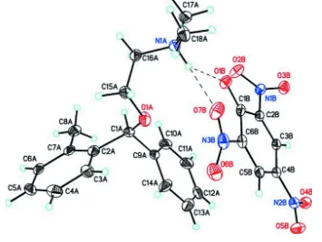
In the crystal structure of the title compound, C18H24NO+. C6H2N3O7-, there is one cation-anion pair in the asymmetric
unit (Fig. 1). The two phenyl rings in the orphenadrinum cation are disordered [occupancy C1A–C14A = 0.662 (4); C1C–
C13C = 0.338 (4)] with a protonated N atom in the N-dimethylethanamine group (Fig. 2). The dihedral angle between the
mean planes of the two cation phenyl rings [occupancy C1A–C14A = 0.662 (4)] is 73.2 (1)°. The picrate anion interacts
with the protonated N atom through a bifurcated N—H···O hydrogen bond forming a R12(6) ring motif with an adjacent
cation. The dihedral angle between the mean planes of the anion benzene and two cation phenyl rings [occupancy C1A–
C14A = 0.662 (4)] is 77.2 (6)° and 9.7 (0)°, respectively. The mean planes of the two o-NO2 and single p-NO2 groups in
the picrate anion are twisted by 23.0 (6)°, 31.3 (3)° and 6.3 (2)° with respect to the mean planes of the 6-membered
benzene ring. Weak Intermolecular C—H···O hydrogen bonds, C—H···Cg intermolecular interactions (Table 1), and weak
π–π stacking interactions (Table 2) dominate the crystal packing creating a 3-D supramolecular structure (Fig. 3).
S2. Experimental
Orphenadrine hydrochloride (3.05 g, 0.01 mol) was dissolved in 25 ml of water and picric acid (2.4 g, 0.01 mol) was also
dissolved in 25 ml of water. Both solutions were mixed and stirred in a beaker at room temperature for 1 h. The mixture
was warmed at 323 K for 10 min & kept aside for 2 days at room temperature. The formed product was filtered and dried
in vaccum desiccator over phosphorous pentoxide. The product was recrystallized from dimethyl sulphoxide by slow
evaporation (m.p. 341–344 K).
S3. Refinement
The two o-phenyl rings in the orphenadrinium cation are disordered [occupancy C1A–C14A = 0.662 (4); C1C–C13C =
—H lengths of 0.95Å (CH), 0.96 & 0.99Å (CH2), 0.98Å (CH3) or 0.93Å (NH). Isotropic displacement parameters for
[image:4.610.130.485.111.378.2]these atoms were set to 1.19 times (NH), 1.19–1.21 (CH, CH2) or 1.49–1.50 (CH3) times Ueq of the parent atom.
Figure 1
Molecular structure of the title compound, (I), showing the atom labeling scheme and 50% probability displacement
ellipsoids. Only the highest occupied atoms in the disordered phenyl rings [occupancy C1A–C14A = 0.662 (4)] are
shown. Dashed lines indicate weak C—H···O intermolecular hydrogen bonds between the cation and anion and R12(6)
Figure 2
Molecular structure of the title compound, (I), showing the disordered atoms in the two phenyl rings [occupancy C1A–
Figure 3
Packing diagram of the title compound viewed down the a axis. Only the highest occupied atoms in the disordered
phenyl rings [occupancy C1A–C14A = 0.662 (4)] are shown. Dashed lines indicate weak intermolecular C—H···O
hydrogen bond interactions creating a 3-D supramolecular structure.
N,N-dimethyl-2-[(2-methylphenyl)(phenyl)methoxy]ethanaminium 2,4,6-trinitrophenolate
Crystal data
C18H24NO+·C6H2N3O7−
Mr = 498.49
Triclinic, P1 Hall symbol: -P 1
a = 9.9434 (10) Å
b = 11.2216 (8) Å
c = 11.3523 (12) Å
α = 78.658 (7)°
β = 76.342 (9)°
γ = 87.660 (7)°
V = 1206.82 (19) Å3
Z = 2
F(000) = 524
Dx = 1.372 Mg m−3
Cu Kα radiation, λ = 1.54184 Å Cell parameters from 3744 reflections
θ = 4.6–74.4°
µ = 0.88 mm−1
T = 123 K
Triangular plate, yellow 0.52 × 0.43 × 0.16 mm
Data collection
Oxford Diffraction Xcalibur Ruby Gemini diffractometer
Radiation source: Enhance (Cu) X-ray Source Graphite monochromator
Detector resolution: 10.5081 pixels mm-1
ω scans
Absorption correction: multi-scan
(CrysAlis RED; Oxford Diffraction, 2007)
Tmin = 0.635, Tmax = 1.000
7402 measured reflections 4677 independent reflections 3760 reflections with I > 2σ(I)
Rint = 0.031
θmax = 74.6°, θmin = 4.6°
h = −12→12
k = −13→13
Refinement
Refinement on F2 Least-squares matrix: full
R[F2 > 2σ(F2)] = 0.065
wR(F2) = 0.188
S = 1.09 4677 reflections 407 parameters 0 restraints
Primary atom site location: structure-invariant direct methods
Secondary atom site location: difference Fourier map
Hydrogen site location: inferred from neighbouring sites
H-atom parameters constrained
w = 1/[σ2(F
o2) + (0.093P)2 + 0.5659P] where P = (Fo2 + 2Fc2)/3
(Δ/σ)max < 0.001 Δρmax = 0.45 e Å−3 Δρmin = −0.40 e Å−3
Extinction correction: SHELXL97 (Sheldrick, 2008), Fc*=kFc[1+0.001xFc2λ3/sin(2θ)]-1/4 Extinction coefficient: 0.0038 (11)
Special details
Geometry. All e.s.d.'s (except the e.s.d. in the dihedral angle between two l.s. planes) are estimated using the full
covariance matrix. The cell e.s.d.'s are taken into account individually in the estimation of e.s.d.'s in distances, angles and torsion angles; correlations between e.s.d.'s in cell parameters are only used when they are defined by crystal symmetry. An approximate (isotropic) treatment of cell e.s.d.'s is used for estimating e.s.d.'s involving l.s. planes.
Refinement. Refinement of F2 against ALL reflections. The weighted R-factor wR and goodness of fit S are based on F2,
conventional R-factors R are based on F, with F set to zero for negative F2. The threshold expression of F2 > σ(F2) is used only for calculating R-factors(gt) etc. and is not relevant to the choice of reflections for refinement. R-factors based on F2 are statistically about twice as large as those based on F, and R- factors based on ALL data will be even larger.
Fractional atomic coordinates and isotropic or equivalent isotropic displacement parameters (Å2)
x y z Uiso*/Ueq Occ. (<1)
O1A 0.7336 (3) 0.61869 (16) 0.6143 (2) 0.0829 (7) N1A 0.7148 (2) 0.35671 (16) 0.72631 (17) 0.0460 (5) H1AB 0.7569 0.4148 0.7541 0.055*
H11A 0.5629 0.8594 0.9801 0.104* 0.662 (4) C12A 0.7428 (6) 0.9283 (4) 0.8627 (4) 0.075 (3) 0.662 (4) H12A 0.7656 0.9769 0.9150 0.091* 0.662 (4) C13A 0.8299 (5) 0.9269 (4) 0.7475 (5) 0.081 (2) 0.662 (4) H13A 0.9121 0.9747 0.7210 0.097* 0.662 (4) C14A 0.7966 (4) 0.8558 (4) 0.6709 (4) 0.0721 (18) 0.662 (4) H14A 0.8561 0.8549 0.5922 0.087* 0.662 (4) C15A 0.7104 (3) 0.5276 (2) 0.5497 (2) 0.0519 (6)
H15A 0.8002 0.4997 0.5049 0.062* H15B 0.6562 0.5621 0.4884 0.062* C16A 0.6334 (3) 0.4220 (2) 0.6388 (2) 0.0473 (5) H16A 0.5464 0.4517 0.6867 0.057* H16B 0.6085 0.3642 0.5917 0.057* C17A 0.6223 (3) 0.2806 (2) 0.8360 (2) 0.0589 (7) H17A 0.5585 0.3332 0.8822 0.088* H17B 0.6785 0.2345 0.8895 0.088* H17C 0.5694 0.2241 0.8084 0.088* C18A 0.8262 (3) 0.2799 (2) 0.6658 (2) 0.0598 (7) H18A 0.8846 0.3300 0.5925 0.090* H18B 0.7841 0.2140 0.6414 0.090* H18C 0.8828 0.2454 0.7240 0.090*
O1B 0.78560 (18) 0.46162 (16) 0.89387 (15) 0.0545 (4) O2B 0.6057 (2) 0.4845 (2) 1.10418 (19) 0.0708 (6) O3B 0.71427 (19) 0.50961 (19) 1.23996 (16) 0.0624 (5) O4B 1.0321 (2) 0.85253 (18) 1.1050 (2) 0.0723 (6) O5B 1.1774 (2) 0.8672 (2) 0.9274 (2) 0.0818 (7) O6B 1.0764 (2) 0.6666 (2) 0.62610 (18) 0.0766 (6) O7B 0.98789 (19) 0.4863 (2) 0.68726 (18) 0.0680 (6) N1B 0.70630 (19) 0.52022 (17) 1.13249 (17) 0.0461 (5) N2B 1.0733 (3) 0.8227 (2) 1.0032 (2) 0.0606 (6) N3B 1.0114 (2) 0.5869 (2) 0.70603 (19) 0.0574 (6) C1B 0.8494 (2) 0.5445 (2) 0.9157 (2) 0.0429 (5) C2B 0.8186 (2) 0.58148 (19) 1.03509 (19) 0.0406 (5) C3B 0.8897 (2) 0.6690 (2) 1.0645 (2) 0.0434 (5) H3BA 0.8658 0.6876 1.1449 0.052* C4B 0.9972 (2) 0.7300 (2) 0.9748 (2) 0.0474 (5) C5B 1.0340 (2) 0.7033 (2) 0.8576 (2) 0.0491 (6) H5BA 1.1069 0.7469 0.7967 0.059* C6B 0.9642 (2) 0.6133 (2) 0.8304 (2) 0.0466 (5)
Atomic displacement parameters (Å2)
U11 U22 U33 U12 U13 U23
C7C 0.058 (5) 0.029 (4) 0.063 (5) −0.001 (3) −0.027 (4) −0.007 (3) C8C 0.066 (5) 0.050 (4) 0.080 (6) −0.009 (4) −0.006 (4) −0.010 (4) C9C 0.070 (7) 0.029 (5) 0.047 (7) 0.001 (4) −0.032 (6) −0.012 (4) C10C 0.072 (6) 0.043 (6) 0.066 (7) 0.005 (4) −0.045 (6) 0.001 (5) C11C 0.138 (14) 0.036 (6) 0.092 (9) −0.004 (7) −0.079 (10) −0.001 (5) C12C 0.102 (11) 0.029 (5) 0.083 (8) 0.002 (5) −0.056 (8) −0.019 (5) C13C 0.096 (8) 0.050 (5) 0.100 (10) −0.004 (6) −0.047 (8) −0.038 (6) C14C 0.110 (10) 0.048 (6) 0.075 (7) 0.018 (5) −0.054 (7) −0.039 (5) O1B 0.0585 (10) 0.0651 (11) 0.0472 (9) −0.0057 (8) −0.0170 (7) −0.0212 (8) O2B 0.0593 (11) 0.0916 (15) 0.0678 (12) −0.0224 (10) −0.0065 (9) −0.0355 (11) O3B 0.0582 (10) 0.0834 (13) 0.0450 (9) −0.0066 (9) −0.0144 (8) −0.0062 (9) O4B 0.0985 (15) 0.0594 (11) 0.0697 (13) −0.0230 (10) −0.0421 (11) −0.0059 (10) O5B 0.0861 (15) 0.0765 (14) 0.0795 (14) −0.0389 (12) −0.0260 (12) 0.0090 (11) O6B 0.0816 (14) 0.0921 (16) 0.0471 (11) 0.0039 (12) −0.0065 (10) −0.0027 (10) O7B 0.0557 (11) 0.0980 (16) 0.0585 (11) 0.0003 (10) −0.0114 (8) −0.0363 (11) N1B 0.0460 (10) 0.0497 (11) 0.0460 (10) −0.0003 (8) −0.0126 (8) −0.0148 (8) N2B 0.0753 (15) 0.0520 (12) 0.0589 (13) −0.0151 (11) −0.0351 (12) 0.0054 (10) N3B 0.0517 (11) 0.0799 (16) 0.0392 (11) 0.0083 (10) −0.0123 (9) −0.0079 (11) C1B 0.0445 (11) 0.0504 (12) 0.0400 (11) 0.0042 (9) −0.0205 (9) −0.0115 (9) C2B 0.0436 (11) 0.0416 (11) 0.0403 (11) 0.0020 (8) −0.0177 (9) −0.0069 (9) C3B 0.0520 (12) 0.0425 (11) 0.0412 (11) 0.0013 (9) −0.0236 (10) −0.0056 (9) C4B 0.0529 (13) 0.0437 (11) 0.0501 (13) −0.0050 (9) −0.0282 (10) 0.0009 (10) C5B 0.0473 (12) 0.0542 (13) 0.0440 (12) −0.0009 (10) −0.0195 (10) 0.0059 (10) C6B 0.0460 (12) 0.0581 (13) 0.0390 (11) 0.0088 (10) −0.0190 (9) −0.0078 (10)
Geometric parameters (Å, º)
O1A—C1C 1.208 (7) C1C—C2C 1.548 (8)
O1A—C1A 1.346 (4) C1C—H1CA 1.0000
O1A—C15A 1.425 (3) C2C—C3C 1.3900
N1A—C16A 1.491 (3) C2C—C7C 1.3900
N1A—C17A 1.494 (3) C3C—C4C 1.3900
N1A—C18A 1.495 (3) C3C—H3CA 0.9500
N1A—H1AB 0.9300 C4C—C5C 1.3900
C1A—C9A 1.522 (4) C4C—H4CA 0.9500
C1A—C2A 1.549 (5) C5C—C6C 1.3900
C1A—H1AA 1.0000 C5C—H5CA 0.9500
C2A—C3A 1.3900 C6C—C7C 1.3900
C2A—C7A 1.3900 C6C—H6CA 0.9500
C3A—C4A 1.3900 C7C—C8C 1.478 (10)
C3A—H3AA 0.9500 C8C—C8Ci 1.789 (15)
C4A—C5A 1.3900 C8C—H8CA 0.9600
C4A—H4AA 0.9500 C8C—H8CB 0.9600
C5A—C6A 1.3900 C8C—H8CC 0.9601
C5A—H5AA 0.9500 C9C—C10C 1.3900
C6A—C7A 1.3900 C9C—C14C 1.3900
C6A—H6AA 0.9500 C10C—C11C 1.3900
C8A—H8AA 0.9600 C11C—C12C 1.3900
C8A—H8AB 0.9600 C11C—H11B 0.9500
C8A—H8AC 0.9601 C12C—C13C 1.3900
C9A—C10A 1.3900 C12C—H12B 0.9500
C9A—C14A 1.3900 C13C—C14C 1.3900
C10A—C11A 1.3900 C13C—H8AA 1.5316
C10A—H10A 0.9500 C13C—H13B 0.9500
C11A—C12A 1.3900 C14C—H14B 0.9500
C11A—H11A 0.9500 O1B—C1B 1.242 (3)
C12A—C13A 1.3900 O2B—N1B 1.225 (3)
C12A—H12A 0.9500 O3B—N1B 1.223 (3)
C13A—C14A 1.3900 O4B—N2B 1.238 (3)
C13A—H13A 0.9500 O5B—N2B 1.231 (3)
C14A—H14A 0.9500 O6B—N3B 1.221 (3)
C15A—C16A 1.501 (3) O7B—N3B 1.230 (3)
C15A—H15A 0.9900 N1B—C2B 1.457 (3)
C15A—H15B 0.9900 N2B—C4B 1.438 (3)
C16A—H16A 0.9900 N3B—C6B 1.463 (3)
C16A—H16B 0.9900 C1B—C6B 1.447 (3)
C17A—H17A 0.9800 C1B—C2B 1.455 (3)
C17A—H17B 0.9800 C2B—C3B 1.369 (3)
C17A—H17C 0.9800 C3B—C4B 1.385 (3)
C18A—H18A 0.9800 C3B—H3BA 0.9500
C18A—H18B 0.9800 C4B—C5B 1.383 (3)
C18A—H18C 0.9800 C5B—C6B 1.371 (3)
C1C—C9C 1.528 (8) C5B—H5BA 0.9500
N1A—C17A—H17A 109.5 C2B—C3B—H3BA 120.6 N1A—C17A—H17B 109.5 C4B—C3B—H3BA 120.6 H17A—C17A—H17B 109.5 C5B—C4B—C3B 121.3 (2) N1A—C17A—H17C 109.5 C5B—C4B—N2B 118.8 (2) H17A—C17A—H17C 109.5 C3B—C4B—N2B 119.9 (2) H17B—C17A—H17C 109.5 C6B—C5B—C4B 119.3 (2) N1A—C18A—H18A 109.5 C6B—C5B—H5BA 120.4 N1A—C18A—H18B 109.5 C4B—C5B—H5BA 120.4 H18A—C18A—H18B 109.5 C5B—C6B—C1B 124.4 (2) N1A—C18A—H18C 109.5 C5B—C6B—N3B 116.5 (2) H18A—C18A—H18C 109.5 C1B—C6B—N3B 119.1 (2) H18B—C18A—H18C 109.5
O1A—C15A—C16A—N1A −65.3 (3) O4B—N2B—C4B—C3B −6.2 (3) C1A—O1A—C1C—C9C −53.1 (6) C3B—C4B—C5B—C6B −1.1 (3) C15A—O1A—C1C—C9C 43.5 (9) N2B—C4B—C5B—C6B 178.5 (2) C1A—O1A—C1C—C2C 77.7 (6) C4B—C5B—C6B—C1B 1.9 (4) C15A—O1A—C1C—C2C 174.4 (4) C4B—C5B—C6B—N3B −178.1 (2) O1A—C1C—C2C—C3C −43.8 (7) O1B—C1B—C6B—C5B −179.0 (2) C9C—C1C—C2C—C3C 90.7 (6) C2B—C1B—C6B—C5B −1.0 (3) O1A—C1C—C2C—C7C 135.6 (5) O1B—C1B—C6B—N3B 0.9 (3) C9C—C1C—C2C—C7C −89.8 (7) C2B—C1B—C6B—N3B 178.98 (19) C7C—C2C—C3C—C4C 0.0 O6B—N3B—C6B—C5B −21.7 (3) C1C—C2C—C3C—C4C 179.5 (6) O7B—N3B—C6B—C5B 156.2 (2) C2C—C3C—C4C—C5C 0.0 O6B—N3B—C6B—C1B 158.3 (2) C3C—C4C—C5C—C6C 0.0 O7B—N3B—C6B—C1B −23.8 (3) C4C—C5C—C6C—C7C 0.0
Symmetry code: (i) −x+2, −y+2, −z+1.
Hydrogen-bond geometry (Å, º)
Cg2 and Cg3 are the centroids of the C9A–C7A and C2C–C7C rings, respectively.
D—H···A D—H H···A D···A D—H···A
N1A—H1AB···O1B 0.93 1.85 2.661 (2) 144 N1A—H1AB···O7B 0.93 2.36 3.031 (3) 129 C4A—H4AA···O4Bii 0.95 2.46 3.346 (4) 155 C16A—H16A···O3Biii 0.99 2.57 3.519 (3) 160 C17A—H17A···O2Biii 0.98 2.57 3.470 (4) 153 C18A—H18A···O6Biv 0.98 2.41 3.167 (3) 133 C18A—H18C···O4Bv 0.98 2.36 3.317 (3) 166 C8C—H8CB···O6B 0.96 2.48 3.239 (9) 136 C6A—H6AA···Cg2vi 0.93 2.88 3.643 (2) 138 C6A—H6AA···Cg3vi 0.93 3.00 3.836 (4) 148 C12C—H12B···Cg2vi 0.93 2.62 3.492 (4) 153 C12C—H12B···Cg3vi 0.93 2.83 3.704 (4) 153