1
H
-Pyrrole-2-carboxylic acid
Gui Hong Tang, Dong Dong Li, Gang Huang, Xing Yan Xu and Xiang Chao Zeng*
Department of Chemistry, Jinan University, Guangzhou, Guangdong 510632, People’s Republic of China
Correspondence e-mail: xczeng@126.com
Received 27 March 2009; accepted 15 April 2009
Key indicators: single-crystal X-ray study;T= 173 K; mean(C–C) = 0.004 A˚;
Rfactor = 0.063;wRfactor = 0.191; data-to-parameter ratio = 13.6.
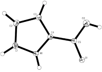
In the title compound, C5H5NO2, the pyrrole ring and its carboxyl substituent are close to coplanar, with a dihedral angle of 11.7 (3)between the planes. In the crystal structure,
adjacent molecules are linked by pairs of O—H O hydrogen bonds to form inversion dimers. Additional N—H O hydrogen bonds link these dimers into chains extending along theaaxis.
Related literature
For pyrroles sourced from marine organisms, see: Faulkner (2002). For the bioactivity of pyrrole derivatives, see: Banwell
et al.(2006); Sosaet al.(2002). For related structures, see: Zeng (2006); Zenget al.(2007). For graph-set motifs, see: Bernstein
et al.(1995).
Experimental
Crystal data
C5H5NO2
Mr= 111.10 Monoclinic,C2=c a= 14.080 (3) A˚
b= 5.0364 (10) A˚
c= 14.613 (3) A˚
= 98.969 (3)
V= 1023.6 (3) A˚3
MoKradiation
= 0.11 mm1
0.420.400.37 mm
Data collection
Bruker SMART 1K CCD area-detector diffractometer Absorption correction: multi-scan
(SADABS; Sheldrick, 1996)
Tmin= 0.954,Tmax= 0.959
2277 measured reflections 1006 independent reflections 875 reflections withI> 2(I)
Rint= 0.015
Refinement
R[F2> 2(F2)] = 0.063
wR(F2) = 0.191
S= 1.06 1006 reflections
74 parameters
H-atom parameters constrained
max= 0.74 e A˚3
min=0.73 e A˚3
Table 1
Hydrogen-bond geometry (A˚ ,).
D—H A D—H H A D A D—H A
N1—H1A O1i
0.88 2.22 2.951 (3) 141 O2—H2A O1ii
0.84 2.16 2.986 (3) 166
Symmetry codes: (i)xþ1 2;yþ
5
2;zþ1; (ii)x;yþ2;zþ1.
Data collection:SMART(Bruker,1999); cell refinement: SAINT-Plus(Bruker, 1999); data reduction:SAINT-Plus; program(s) used to solve structure: SHELXS97(Sheldrick, 2008); program(s) used to refine structure:SHELXL97(Sheldrick, 2008); molecular graphics: SHELXTL(Sheldrick, 2008); software used to prepare material for publication:SHELXTL.
We thank the Natural Science Foundation of Guangdong Province, China (grant No. 06300581), for generously supporting this study.
Supplementary data and figures for this paper are available from the IUCr electronic archives (Reference: SJ2604).
References
Banwell, M. G., Hamel, E., Hockless, D. C. R., Verdier-Pinard, P., Willis, A. C. & Wong, D. J. (2006).Bioorg. Med. Chem.14, 4627–4638.
Bernstein, J., Davis, R. E., Shimoni, L. & Chang, N.-L. (1995).Angew. Chem. Int. Ed. Engl.34, 1555–1573.
Bruker (1999). SMART and SAINT-Plus. Bruker AXS Inc., Madison, Wisconsin, USA.
Faulkner, D. J. (2002).Nat. Prod. Rep.18, 1–48.
Sheldrick, G. M. (1996).SADABS. University of Go¨ttingen, Germany. Sheldrick, G. M. (2008).Acta Cryst.A64, 112–122.
Sosa, A. C. B., Yakushijin, K. & Horne, D. A. (2002).J. Org. Chem.67, 4498– 4500.
Zeng, X.-C. (2006).Acta Cryst.E62, o5505–o5507.
Zeng, X.-C., Zeng, J., Li, X. & Ling, X. (2007).Acta Cryst.E63, o3424. Structure Reports
Online
supporting information
Acta Cryst. (2009). E65, o1121 [doi:10.1107/S1600536809014044]
1
H
-Pyrrole-2-carboxylic acid
Gui Hong Tang, Dong Dong Li, Gang Huang, Xing Yan Xu and Xiang Chao Zeng
S1. Comment
Pyrrole derivatives are well known in many marine organisms (Faulkner, 2002), some show important bioactivities, such
as antitumor activity (Banwell et al., 2006) and protein kinase inhibiting activity (Sosa et al., 2002). This is the reason
they have attracted our interest. This study is related to our previous structural investigations of methyl
2-(4,5-di-bromo-1H-pyrrole-2-carboxamido)propionate (Zeng et al., 2007) and
3-bromo-1-methyl-6,7-dihydropyrrolo[2,3-c]azepine- 4,8(1H,5H)-dione (Zeng, 2006). In the crystal structure, molecules of the title compound are linked through
N1—H1···O1i hydrogen bonds to form centrosymmetric dimers (Fig. 2) of graph-set motif R
22(10) (Bernstein et al.,
1995), which are linked by O2—H2···O1ii hydrogen bonds (another kind of centrosymmetric dimers of graph-set motif
R22(8) are formed), generating chains extending to the a axis (also shown in Fig. 2).
S2. Experimental
The commercially available 1H-pyrrole-2-carboxylic acid was dissolved in the mixture of EtOH (80%) and ethyl acetate
(20%). Colorless monoclinic crystals suitable for X-ray analysis were obtained when the solution was exposed to the air
at room temperature for about 5 d.
S3. Refinement
All non-H atoms were refined with anisotropic displacement parameters. The H atoms were positioned geometrically [C
—H = 0.95Å for CH, O—H = 0.84Å for OH, and N—H = 0.88 Å] and refined using a riding model, with Uiso = 1.2Ueq
(1.5Ueq for the methyl group) of the parent atom. In the final difference Fourier map the highest peak (0.74 eÅ-3) is 1.01Å
Figure 1
The molecular structure of the title compound, with the atom-numbering scheme. Displacement ellipsoids are drawn at
Figure 2
Crystal packing of (I) showing the chains formed by hydrogen bonds (dashed lines).
1H-Pyrrole-2-carboxylic acid
Crystal data
C5H5NO2 Mr = 111.10 Monoclinic, C2/c Hall symbol: -C 2yc a = 14.080 (3) Å b = 5.0364 (10) Å c = 14.613 (3) Å β = 98.969 (3)° V = 1023.6 (3) Å3 Z = 8
F(000) = 464 Dx = 1.442 Mg m−3
Melting point: 480 K
Mo Kα radiation, λ = 0.71073 Å Cell parameters from 1751 reflections θ = 2.8–27.0°
µ = 0.11 mm−1 T = 173 K Block, colorless 0.42 × 0.40 × 0.37 mm
Data collection
Bruker SMART 1K CCD area-detector diffractometer
Radiation source: fine-focus sealed tube Graphite monochromator
φ and ω scans
Absorption correction: multi-scan (SADABS; Sheldrick, 1996) Tmin = 0.954, Tmax = 0.959
θmax = 26.0°, θmin = 2.8° h = −17→13
l = −14→18
Refinement
Refinement on F2
Least-squares matrix: full R[F2 > 2σ(F2)] = 0.063 wR(F2) = 0.191 S = 1.06 1006 reflections 74 parameters 0 restraints
Primary atom site location: structure-invariant direct methods
Secondary atom site location: difference Fourier map
Hydrogen site location: inferred from neighbouring sites
H-atom parameters constrained w = 1/[σ2(F
o2) + (0.1108P)2 + 3.3345P]
where P = (Fo2 + 2Fc2)/3
(Δ/σ)max = 0.001
Δρmax = 0.74 e Å−3
Δρmin = −0.73 e Å−3
Special details
Geometry. All e.s.d.'s (except the e.s.d. in the dihedral angle between two l.s. planes) are estimated using the full covariance matrix. The cell e.s.d.'s are taken into account individually in the estimation of e.s.d.'s in distances, angles and torsion angles; correlations between e.s.d.'s in cell parameters are only used when they are defined by crystal symmetry. An approximate (isotropic) treatment of cell e.s.d.'s is used for estimating e.s.d.'s involving l.s. planes.
Refinement. Refinement of F2 against ALL reflections. The weighted R-factor wR and goodness of fit S are based on F2,
conventional R-factors R are based on F, with F set to zero for negative F2. The threshold expression of F2 > σ(F2) is used
only for calculating R-factors(gt) etc. and is not relevant to the choice of reflections for refinement. R-factors based on F2
are statistically about twice as large as those based on F, and R- factors based on ALL data will be even larger.
Fractional atomic coordinates and isotropic or equivalent isotropic displacement parameters (Å2)
x y z Uiso*/Ueq
O1 0.12435 (12) 1.1503 (3) 0.53422 (12) 0.0223 (5)
C4 0.23786 (16) 0.8483 (5) 0.61313 (15) 0.0176 (6)
O2 0.07382 (14) 0.7350 (4) 0.56343 (15) 0.0373 (6)
H2A 0.0220 0.7923 0.5336 0.056*
N1 0.31542 (14) 1.0100 (4) 0.61094 (15) 0.0216 (6)
H1A 0.3144 1.1614 0.5808 0.026*
C3 0.26837 (17) 0.6325 (5) 0.66849 (17) 0.0208 (6)
H3 0.2299 0.4879 0.6828 0.025*
C5 0.14189 (16) 0.9228 (5) 0.56657 (15) 0.0173 (6)
C2 0.36767 (18) 0.6681 (5) 0.69974 (17) 0.0245 (6)
H2 0.4085 0.5521 0.7393 0.029*
C1 0.39405 (17) 0.9010 (6) 0.66242 (18) 0.0251 (6)
H1 0.4570 0.9740 0.6712 0.030*
Atomic displacement parameters (Å2)
U11 U22 U33 U12 U13 U23
C5 0.0192 (12) 0.0164 (11) 0.0167 (11) −0.0002 (9) 0.0042 (9) −0.0008 (9) C2 0.0220 (13) 0.0291 (14) 0.0215 (12) 0.0052 (10) 0.0009 (9) 0.0038 (10) C1 0.0174 (12) 0.0318 (14) 0.0256 (13) −0.0013 (10) 0.0019 (9) 0.0030 (11)
Geometric parameters (Å, º)
O1—C5 1.250 (3) N1—H1A 0.8800
C4—N1 1.367 (3) C3—C2 1.413 (3)
C4—C3 1.383 (3) C3—H3 0.9500
C4—C5 1.464 (3) C2—C1 1.369 (4)
O2—C5 1.342 (3) C2—H2 0.9500
O2—H2A 0.8400 C1—H1 0.9500
N1—C1 1.354 (3)
N1—C4—C3 107.8 (2) O1—C5—O2 122.4 (2)
N1—C4—C5 121.3 (2) O1—C5—C4 121.6 (2)
C3—C4—C5 130.8 (2) O2—C5—C4 116.0 (2)
C5—O2—H2A 109.5 C1—C2—C3 107.2 (2)
C1—N1—C4 109.4 (2) C1—C2—H2 126.4
C1—N1—H1A 125.3 C3—C2—H2 126.4
C4—N1—H1A 125.3 N1—C1—C2 108.6 (2)
C4—C3—C2 106.9 (2) N1—C1—H1 125.7
C4—C3—H3 126.5 C2—C1—H1 125.7
C2—C3—H3 126.5
C3—C4—N1—C1 0.7 (3) N1—C4—C5—O2 171.9 (2)
C5—C4—N1—C1 177.3 (2) C3—C4—C5—O2 −12.3 (4)
N1—C4—C3—C2 −0.2 (3) C4—C3—C2—C1 −0.3 (3)
C5—C4—C3—C2 −176.4 (2) C4—N1—C1—C2 −0.9 (3)
N1—C4—C5—O1 −10.0 (3) C3—C2—C1—N1 0.7 (3)
C3—C4—C5—O1 165.7 (2)
Hydrogen-bond geometry (Å, º)
D—H···A D—H H···A D···A D—H···A
N1—H1A···O1i 0.88 2.22 2.951 (3) 141
O2—H2A···O1ii 0.84 2.16 2.986 (3) 166