Methyl eucomate
Linglin Li,aGuang-Xiong Zhouband Ren-Wang Jiangb*
aNutrition and Metabolism Laboratory, Beth Israel Deaconess Medical Center, Boston, MA 02215, USA, andbInstitute of Traditional Chinese Medicine and Natural Products, College of Pharmacy, Jinan University, Guangzhou 510632, People’s Republic of China
Correspondence e-mail: [email protected]
Received 27 April 2008; accepted 20 June 2008
Key indicators: single-crystal X-ray study;T= 293 K; mean(C–C) = 0.003 A˚; Rfactor = 0.029;wRfactor = 0.067; data-to-parameter ratio = 7.6.
The crystal structure of the title compound [systematic name: methyl 3-carboxy-3-hydroxy-3-(4-hydroxybenzyl)propanoate], C12H14O6, is stabilized by intermolecular O—H O and C—
H O hydrogen bonds. The molecules are arranged in layers, parallel to (001), which are interconnected by the O—H O hydrogen bonds.
Related literature
For related literature, see: Heller & Tamm (1974); Jianget al. (2002, 2006).
Experimental
Crystal data
C12H14O6
Mr= 254.23
Orthorhombic,P212121 a= 5.9109 (6) A˚ b= 7.0348 (7) A˚ c= 29.109 (3) A˚
V= 1210.4 (2) A˚3
Z= 4
MoKradiation = 0.11 mm1
T= 293 (2) K 0.400.320.25 mm
Data collection
Bruker SMART CCD diffractometer
Absorption correction: none 6709 measured reflections
1279 independent reflections 1047 reflections withI> 2(I) Rint= 0.045
Refinement
R[F2> 2(F2)] = 0.029 wR(F2) = 0.066
S= 1.05 1278 reflections
168 parameters
H-atom parameters constrained
max= 0.14 e A˚
3
min=0.12 e A˚
3
Table 1
Hydrogen-bond geometry (A˚ ,).
D—H A D—H H A D A D—H A
O1—H1 O3i
0.82 1.96 2.775 (2) 172 O2—H2 O1ii
0.82 2.33 2.888 (2) 125 O4—H4 O2iii
0.82 1.85 2.639 (2) 161 C12—H12B O5iv
0.96 2.42 3.268 (4) 148
Symmetry codes: (i)x;y1 2;zþ
1
2; (ii) xþ1;yþ 1 2;zþ
1
2; (iii)x1;y;z; (iv) xþ1
2;yþ 3 2;z.
Data collection:SMART(Bruker, 1998); cell refinement:SAINT (Bruker, 1998); data reduction:SAINTandXPREPin SHELXTL (Sheldrick, 2008); program(s) used to solve structure: SHELXS97 (Sheldrick, 2008); program(s) used to refine structure:SHELXL97 (Sheldrick, 2008); molecular graphics:XP(Siemens, 1998); software used to prepare material for publication:SHELXTL.
This work was supported by the Starting Fund for Excellent Talents of Jinan University.
Supplementary data and figures for this paper are available from the IUCr electronic archives (Reference: FB2096).
References
Bruker (1998).SMARTandSAINT. Bruker AXS Inc., Madison, Wisconsin, USA.
Heller, W. & Tamm, C. (1974).Helv. Chim. Acta,57, 1766–1784.
Jiang, J. Q., Li, Y. F., Chen, Z., Min, Z. D. & Lou, F. C. (2006).Steroids,71, 1073–1077.
Jiang, J. Q., Ye, W. C., Chen, Z., Lou, F. C. & Min, Z. D. (2002).J. Chin. Pharm. Sci.11, 1–3.
Sheldrick, G. M. (2008).Acta Cryst.A64, 112–122.
Siemens (1998).XP. Siemens Analytical X-ray Instruments Inc., Madison, Wisconsin, USA.
Acta Crystallographica Section E
Structure Reports
Online
supporting information
Acta Cryst. (2008). E64, o1354 [doi:10.1107/S1600536808018734]
Methyl eucomate
Linglin Li, Guang-Xiong Zhou and Ren-Wang Jiang
S1. Comment
Methyl eucomate is the methyl ester of the eucomic acid. The title compound has been isolated from several edible
plants, e. g. Opuntia dillenii (Jiang et al., 2006) or Opuntia vulgaris (Jiang et al., 2002). However, the stereochemistry of
the ester has not been established yet. In the present paper, we report its crystal structure.
The molecule contains phenol, carboxyl, ester and hydroxyl functional groups (Fig. 1). The mean deviation of the
benzene ring from planarity is 0.0004 Å and its dihedral angle with the plane of the carboxylic group at C8 is 50.3 (3)°,
while it is roughly perpendicular to the ester group at C10 with a dihedral angle of 87.3 (3)°.
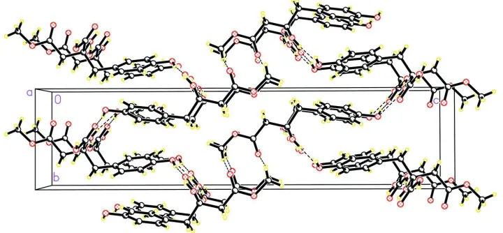
The intermolecular hydrogen bonds O1—H···O3, O2—H···O1 and O4—H···O2 (Tab. 1) link the molecules into layers
that are parallel to (001) (Fig. 2).
There is no heavy atom with a significant anomalous dispersion contribution, so the absolute configuration from the
diffraction pattern itself could not be determined. However, the absolute configuration of the eucomic acid has been
established by synthesis (Heller & Tamm, 1974) though its crystal structure has not been determined. Therefore the title
compound is expected to share the same R configuration at the chiral centre C8.
S2. Experimental
The title compound was purified from the stems of Opuntia vulgaris according to the reported procedures (Jiang et al.,
2002). Briefly, the stems of Opuntia vulgaris (1 kg) was extracted with 95% ethanol under room temperature. The
extracted solution was concentrated with rotary evaporator to afford a crude extract, which was suspended in distilled
water and partitioned with petroleum ether, ethyl acetate and n-butanol. Then the n-butanol fraction was subjected to
silica gel column chromatography eluted with methanol-chloroform gradient solvent system to afford the title compound
(16 mg). The transparent rectangular crystals of the title compound with average size of 0.50 × 0.40 × 0.30 mm were
obtained by slow evaporation of the methanol solution at room temperature.
S3. Refinement
Though all the hydrogens were discernible in the difference electron density maps. Neverheless, the hydrogens were
situated into the idealized position and constrained during the refinement. Hydroxyl hydrogens: O-H equalled to 0.82 Å,
Uiso(H)=1.5 UeqO; Caryl-H equalled to 0.93 Å, Uiso(H)=1.2 UeqCaryl; Cmethylene-H equalled to 0.97 Å, Uiso(H)=1.2 UeqCmethylene;
Cmethyl-H equalled to 0.96 Å, Uiso(H)=1.5 UeqCmethyl.
There is no heavy atom with significant anomalous dispersion contribution in the structure for the used wavelength, so
the absolute configuration from the diffraction pattern itself was not determined. 836 Friedel reflections were merged
before the refinement. However, the absolute configuration of the related eucomic acid has been established previously
(Heller & Tamm, 1974) and therefore the title compound has been expected to share the same R configuration at the
Reflection (0 0 2) was omitted.
Figure 1
The molecular structure of the title structure showing 30% probability displacement ellipsoids and the atom-numbering
scheme.
Figure 2
The packing diagram of the title structure viewed down the a axis.
methyl 3-carboxy-3-hydroxy-3-(4-hydroxybenzyl)propanoate
Crystal data
C12H14O6 Mr = 254.23
Orthorhombic, P212121 Hall symbol: P 2ac 2ab a = 5.9109 (6) Å b = 7.0348 (7) Å c = 29.109 (3) Å V = 1210.4 (2) Å3 Z = 4
F(000) = 536 Dx = 1.395 Mg m−3
Mo Kα radiation, λ = 0.71073 Å Cell parameters from 6709 reflections θ = 1.4–25.0°
µ = 0.11 mm−1 T = 293 K
[image:3.610.126.486.292.459.2]Data collection
Bruker SMART/CCD diffractometer
Radiation source: fine-focus sealed tube Graphite monochromator
ω scans
6709 measured reflections 1279 independent reflections
1047 reflections with I > 2σ(I) Rint = 0.046
θmax = 25.0°, θmin = 1.4° h = −6→7
k = −8→7 l = −27→34
Refinement
Refinement on F2 Least-squares matrix: full R[F2 > 2σ(F2)] = 0.029 wR(F2) = 0.066 S = 1.05 1278 reflections 168 parameters 0 restraints 42 constraints
Primary atom site location: structure-invariant direct methods
Secondary atom site location: difference Fourier map
Hydrogen site location: difference Fourier map H-atom parameters constrained
w = 1/[σ2(F
o2) + (0.0328P)2] where P = (Fo2 + 2Fc2)/3 (Δ/σ)max < 0.001
Δρmax = 0.14 e Å−3 Δρmin = −0.12 e Å−3
Extinction correction: SHELXL97 (Sheldrick, 2008), Fc*=kFc[1+0.001xFc2λ3/sin(2θ)]-1/4 Extinction coefficient: 0.0070 (19)
Special details
Geometry. All e.s.d.'s (except the e.s.d. in the dihedral angle between two l.s. planes) are estimated using the full covariance matrix. The cell e.s.d.'s are taken into account individually in the estimation of e.s.d.'s in distances, angles and torsion angles; correlations between e.s.d.'s in cell parameters are only used when they are defined by crystal symmetry. An approximate (isotropic) treatment of cell e.s.d.'s is used for estimating e.s.d.'s involving l.s. planes.
Refinement. Refinement of F2 against ALL reflections. The weighted R-factor wR and goodness of fit S are based on F2, conventional R-factors R are based on F, with F set to zero for negative F2. The threshold expression of F2 > σ(F2) is used only for calculating R-factors(gt) etc. and is not relevant to the choice of reflections for refinement. R-factors based on F2 are statistically about twice as large as those based on F, and R- factors based on ALL data will be even larger.
Fractional atomic coordinates and isotropic or equivalent isotropic displacement parameters (Å2)
x y z Uiso*/Ueq
O1 0.2505 (3) 0.2830 (2) 0.32392 (5) 0.0466 (5)
H1 0.1313 0.2346 0.3323 0.070*
O2 0.5510 (2) 0.5081 (2) 0.11441 (5) 0.0400 (4)
H2 0.5099 0.5865 0.1335 0.060*
O3 0.1405 (3) 0.6278 (2) 0.13901 (5) 0.0425 (4)
O4 −0.0301 (3) 0.3948 (3) 0.09911 (6) 0.0499 (5)
H4 −0.1460 0.4500 0.1070 0.075*
O5 0.2745 (4) 0.6569 (3) 0.03265 (6) 0.0640 (6)
O6 0.3709 (3) 0.4543 (2) −0.02286 (5) 0.0543 (5)
C1 0.2722 (4) 0.2631 (3) 0.27718 (8) 0.0344 (6)
C2 0.4773 (4) 0.3133 (3) 0.25799 (8) 0.0367 (6)
H2B 0.5932 0.3598 0.2764 0.044*
C3 0.5089 (4) 0.2940 (3) 0.21133 (8) 0.0370 (6)
H3A 0.6475 0.3286 0.1987 0.044*
C4 0.3420 (4) 0.2253 (3) 0.18265 (8) 0.0346 (6)
H5A 0.0209 0.1285 0.1843 0.047*
C6 0.1010 (4) 0.1933 (3) 0.24947 (9) 0.0387 (6)
H6A −0.0373 0.1588 0.2622 0.046*
C7 0.3804 (4) 0.2011 (3) 0.13179 (8) 0.0400 (6)
H7A 0.2693 0.1119 0.1201 0.048*
H7B 0.5286 0.1451 0.1272 0.048*
C8 0.3660 (4) 0.3855 (3) 0.10324 (7) 0.0336 (5)
C9 0.3831 (4) 0.3384 (3) 0.05217 (8) 0.0403 (6)
H9A 0.5339 0.2904 0.0459 0.048*
H9B 0.2767 0.2375 0.0452 0.048*
C10 0.3374 (4) 0.5023 (4) 0.02090 (8) 0.0424 (6)
C11 0.1476 (4) 0.4865 (4) 0.11534 (7) 0.0347 (5)
C12 0.3196 (5) 0.5985 (4) −0.05667 (9) 0.0681 (9)
H12A 0.3552 0.5515 −0.0868 0.102*
H12B 0.4079 0.7102 −0.0505 0.102*
H12C 0.1617 0.6297 −0.0552 0.102*
Atomic displacement parameters (Å2)
U11 U22 U33 U12 U13 U23
O1 0.0418 (10) 0.0604 (11) 0.0377 (10) −0.0079 (10) 0.0050 (8) 0.0063 (9)
O2 0.0255 (9) 0.0527 (11) 0.0418 (11) −0.0021 (8) 0.0013 (7) −0.0105 (9)
O3 0.0368 (9) 0.0474 (10) 0.0432 (10) 0.0061 (9) 0.0051 (8) −0.0103 (8)
O4 0.0229 (9) 0.0617 (12) 0.0651 (12) 0.0047 (9) −0.0011 (9) −0.0157 (11)
O5 0.0819 (15) 0.0646 (12) 0.0454 (11) 0.0208 (12) 0.0057 (11) −0.0017 (10)
O6 0.0678 (12) 0.0652 (11) 0.0298 (9) −0.0045 (11) 0.0042 (10) −0.0054 (9)
C1 0.0334 (13) 0.0335 (13) 0.0363 (14) 0.0024 (11) 0.0021 (11) 0.0060 (11)
C2 0.0315 (12) 0.0395 (14) 0.0391 (15) −0.0086 (12) −0.0029 (11) 0.0020 (12) C3 0.0277 (12) 0.0404 (13) 0.0429 (15) −0.0040 (12) 0.0035 (11) 0.0041 (12) C4 0.0294 (12) 0.0326 (12) 0.0417 (14) 0.0046 (11) −0.0010 (11) 0.0021 (11) C5 0.0293 (12) 0.0412 (13) 0.0463 (15) −0.0009 (12) −0.0045 (12) 0.0000 (12) C6 0.0263 (12) 0.0430 (13) 0.0470 (15) −0.0029 (11) 0.0030 (11) 0.0068 (13) C7 0.0336 (12) 0.0431 (13) 0.0431 (14) 0.0062 (12) −0.0021 (12) −0.0074 (12) C8 0.0223 (11) 0.0441 (14) 0.0344 (13) 0.0009 (12) −0.0012 (11) −0.0054 (12)
C9 0.0334 (13) 0.0521 (14) 0.0356 (14) 0.0060 (12) 0.0028 (11) −0.0071 (12)
C10 0.0315 (13) 0.0572 (16) 0.0385 (14) −0.0011 (15) 0.0029 (12) −0.0075 (14) C11 0.0277 (12) 0.0460 (14) 0.0303 (12) 0.0012 (13) −0.0001 (11) 0.0007 (12)
C12 0.085 (2) 0.080 (2) 0.0393 (15) −0.023 (2) −0.0078 (16) 0.0111 (16)
Geometric parameters (Å, º)
O1—C1 1.374 (3) C4—C5 1.392 (3)
O1—H1 0.8200 C4—C7 1.507 (3)
O2—C8 1.430 (3) C5—C6 1.382 (3)
O2—H2 0.8200 C5—H5A 0.9300
O3—C11 1.210 (3) C6—H6A 0.9300
O4—C11 1.320 (3) C7—C8 1.543 (3)
O5—C10 1.200 (3) C7—H7B 0.9700
O6—C10 1.333 (3) C8—C11 1.515 (3)
O6—C12 1.446 (3) C8—C9 1.526 (3)
C1—C2 1.380 (3) C9—C10 1.494 (3)
C1—C6 1.384 (3) C9—H9A 0.9700
C2—C3 1.378 (3) C9—H9B 0.9700
C2—H2B 0.9300 C12—H12A 0.9600
C3—C4 1.380 (3) C12—H12B 0.9600
C3—H3A 0.9300 C12—H12C 0.9600
C1—O1—H1 109.5 C8—C7—H7B 108.5
C8—O2—H2 109.5 H7A—C7—H7B 107.5
C11—O4—H4 109.5 O2—C8—C11 108.40 (17)
C10—O6—C12 116.2 (2) O2—C8—C9 107.55 (19)
O1—C1—C2 117.2 (2) C11—C8—C9 112.63 (19)
O1—C1—C6 123.0 (2) O2—C8—C7 110.03 (18)
C2—C1—C6 119.8 (2) C11—C8—C7 108.44 (18)
C3—C2—C1 119.5 (2) C9—C8—C7 109.77 (19)
C3—C2—H2B 120.2 C10—C9—C8 114.4 (2)
C1—C2—H2B 120.2 C10—C9—H9A 108.6
C2—C3—C4 122.3 (2) C8—C9—H9A 108.6
C2—C3—H3A 118.9 C10—C9—H9B 108.6
C4—C3—H3A 118.9 C8—C9—H9B 108.6
C3—C4—C5 117.2 (2) H9A—C9—H9B 107.6
C3—C4—C7 121.8 (2) O5—C10—O6 123.3 (2)
C5—C4—C7 121.0 (2) O5—C10—C9 125.6 (2)
C6—C5—C4 121.5 (2) O6—C10—C9 111.1 (2)
C6—C5—H5A 119.2 O3—C11—O4 125.3 (2)
C4—C5—H5A 119.2 O3—C11—C8 123.2 (2)
C5—C6—C1 119.6 (2) O4—C11—C8 111.43 (19)
C5—C6—H6A 120.2 O6—C12—H12A 109.5
C1—C6—H6A 120.2 O6—C12—H12B 109.5
C4—C7—C8 115.19 (19) H12A—C12—H12B 109.5
C4—C7—H7A 108.5 O6—C12—H12C 109.5
C8—C7—H7A 108.5 H12A—C12—H12C 109.5
C4—C7—H7B 108.5 H12B—C12—H12C 109.5
O1—C1—C2—C3 179.3 (2) C4—C7—C8—C9 −174.1 (2)
C6—C1—C2—C3 0.3 (4) O2—C8—C9—C10 −68.8 (2)
C1—C2—C3—C4 −0.2 (4) C11—C8—C9—C10 50.5 (3)
C2—C3—C4—C5 0.1 (3) C7—C8—C9—C10 171.5 (2)
C2—C3—C4—C7 −178.7 (2) C12—O6—C10—O5 −2.1 (4)
C3—C4—C5—C6 0.0 (3) C12—O6—C10—C9 176.6 (2)
C7—C4—C5—C6 178.7 (2) C8—C9—C10—O5 −5.7 (4)
C4—C5—C6—C1 0.1 (4) C8—C9—C10—O6 175.6 (2)
O1—C1—C6—C5 −179.2 (2) O2—C8—C11—O3 −13.9 (3)
C2—C1—C6—C5 −0.2 (4) C9—C8—C11—O3 −132.8 (2)
C5—C4—C7—C8 102.8 (3) O2—C8—C11—O4 169.74 (19)
C4—C7—C8—O2 67.7 (3) C9—C8—C11—O4 50.9 (3)
C4—C7—C8—C11 −50.7 (3) C7—C8—C11—O4 −70.8 (2)
Hydrogen-bond geometry (Å, º)
D—H···A D—H H···A D···A D—H···A
O1—H1···O3i 0.82 1.96 2.775 (2) 172
O2—H2···O1ii 0.82 2.34 2.888 (2) 125
O4—H4···O2iii 0.82 1.85 2.639 (2) 161
C12—H12B···O5iv 0.96 2.42 3.268 (4) 148