N
,
N
000-Dimethoxy-
N
,
N
000-dimethyl-succinamide
Sumei Yao
Medical College of Henan University, Henan University, Kaifeng 475004, People’s Republic of China
Correspondence e-mail: ysum@yahoo.cn
Received 16 June 2008; accepted 17 June 2008
Key indicators: single-crystal X-ray study;T= 296 K; mean(C–C) = 0.002 A˚; Rfactor = 0.055;wRfactor = 0.174; data-to-parameter ratio = 14.2.
The title compound, C8H16N2O4, is a Weinreb amide that is also an important intermediate for the preparation of ketones and aldehydes. The molecule possesses a centre of symmetry.
Related literature
For related literature, see: Nahm & Weinreb (1981).
Experimental
Crystal data
C8H16N2O4 Mr= 204.23 Monoclinic,P21=c a= 4.2645 (15) A˚ b= 11.152 (4) A˚ c= 11.165 (4) A˚ = 98.485 (5)
V= 525.2 (3) A˚3 Z= 2
MoKradiation = 0.10 mm1 T= 296 (2) K 0.200.160.13 mm
Data collection
Bruker SMART APEX CCD area-detector diffractometer Absorption correction: multi-scan
(SADABS; Sheldrick, 2001) Tmin= 0.980,Tmax= 0.987
2116 measured reflections 909 independent reflections 776 reflections withI> 2(I) Rint= 0.015
Refinement
R[F2> 2(F2)] = 0.055 wR(F2) = 0.174 S= 1.01 909 reflections
64 parameters
H-atom parameters constrained
max= 0.25 e A˚
3
min=0.24 e A˚
3
Data collection:SMART(Bruker, 2001); cell refinement:SMART; data reduction:SAINT-Plus(Bruker, 2001); program(s) used to solve structure:SHELXS97(Sheldrick, 2008); program(s) used to refine structure: SHELXL97 (Sheldrick, 2008); molecular graphics:
PLATON (Spek, 2003); software used to prepare material for publication:PLATON.
Supplementary data and figures for this paper are available from the IUCr electronic archives (Reference: AT2577).
References
Bruker (2001). SAINT-Plus and SMART. Bruker AXS Inc., Madison, Wisconsin, USA.
Nahm, S. & Weinreb, S. M. (1981).Tetrahedron Lett.22, 3815–3818. Sheldrick, G. M. (2001).SADABS. Bruker AXS Inc., Madison, Wisconsin,
USA.
Sheldrick, G. M. (2008).Acta Cryst.A64, 112–122. Spek, A. L. (2003).J. Appl. Cryst.36, 7–13. Acta Crystallographica Section E
Structure Reports Online
supporting information
Acta Cryst. (2008). E64, o1315 [doi:10.1107/S1600536808018369]
N,N
′
-Dimethoxy-N,N
′
-dimethylsuccinamide
Sumei Yao
S1. Comment
The Weinreb amides are widely recognized as effective acylating agents since they react with organometallics (RM, M = MgBr, Li) to produce ketones without side products in organic synthesis, including the total synthesis of complex natural
products (Nahm & Weinreb, 1981). We here reported the structure of the Weinreb amides related title compound, (I).
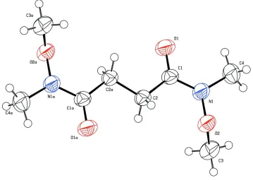
Compound (I), is the synthetic intermediate, whose molecule is the centrosymmetric structure (Fig.1). In the symmetric
unit, the C1—O1 bond distance is 1.224 (2) Å, which displays a typical double-bond of ketone carbonyl. Whereas, the
N1—C1 bond distance of 1.342 (2) Å is obviously shorter than N1—C4 of 1.445 (2) Å, indicates that amide bond N1—
C1 has some proporties of double-bond.
S2. Experimental
Triethylamine (25 ml, 180 mmol) was added slowly by cannulation to a stirred suspension of N,O -dimethylhydroxyl-amine (9.0 g, 92.25 mmol) and succinyl chloride (100 ml) in dichloromethane at 273 K under N2. After stirring for 2 h the
solution was allowed to warm to room temperature and quenched with saturated aqueous sodium bicarbonate solution
(50 ml). The layers were separated and the aqueous layer was extracted with dichloromethane (2×25 ml). The combined
organic extracts were washed with brine (18.5 ml), dried (MgSO4) and evaporated under reduced pressure to give the
compound (I) (7.365 g, 83%) as light brown needles. The molecule formula, C8H16N2O4 was established by EIMS
m/z:144(M+ –N(CH3)OCH3). Spectroscopic analysis, 1H NMR (400 MHz; CDCl3-d6) δ:3.75 (6H, s, OCH3), 3.19 (6H, s,
NCH3) and 2.78 (4H, s, CH2); 13C NMR (400 MHz; CDCl3-d6) δ:173.8 (C=O), 61.6 (OCH3), 32.6 and 26.8.
S3. Refinement
H atoms were treated as riding, with C—H distances in the range of 0.96–0.97 Å, and were refined as riding with Uiso(H)
Figure 1
The molecular structure of (I), showing the atom-labelling scheme. Displacement ellipsoids are drawn at the 50%
probability level.
N,N′-Dimethoxy-N,N′-dimethylsuccinamide
Crystal data
C8H16N2O4
Mr = 204.23 Monoclinic, P21/c
Hall symbol: -P 2ybc
a = 4.2645 (15) Å
b = 11.152 (4) Å
c = 11.165 (4) Å
β = 98.485 (5)°
V = 525.2 (3) Å3
Z = 2
F(000) = 220
Dx = 1.291 Mg m−3
Mo Kα radiation, λ = 0.71073 Å Cell parameters from 925 reflections
θ = 2.6–26.6°
µ = 0.10 mm−1
T = 296 K Block, yellow
0.20 × 0.16 × 0.13 mm
Data collection
Bruker SMART APEX CCD area-detector diffractometer
Radiation source: fine-focus sealed tube Graphite monochromator
φ and ω scans
Absorption correction: multi-scan (SADABS; Sheldrick, 2001)
Tmin = 0.980, Tmax = 0.987
2116 measured reflections 909 independent reflections 776 reflections with I > 2σ(I)
Rint = 0.015
θmax = 25.0°, θmin = 2.6°
h = −5→5
k = −9→13
Refinement
Refinement on F2
Least-squares matrix: full
R[F2 > 2σ(F2)] = 0.055
wR(F2) = 0.174
S = 1.01 909 reflections 64 parameters 0 restraints
Primary atom site location: structure-invariant direct methods
Secondary atom site location: difference Fourier map
Hydrogen site location: inferred from neighbouring sites
H-atom parameters constrained
w = 1/[σ2(F
o2) + (0.117P)2 + 0.1547P]
where P = (Fo2 + 2Fc2)/3
(Δ/σ)max < 0.001
Δρmax = 0.25 e Å−3
Δρmin = −0.24 e Å−3
Special details
Geometry. All e.s.d.'s (except the e.s.d. in the dihedral angle between two l.s. planes) are estimated using the full covariance matrix. The cell e.s.d.'s are taken into account individually in the estimation of e.s.d.'s in distances, angles and torsion angles; correlations between e.s.d.'s in cell parameters are only used when they are defined by crystal symmetry. An approximate (isotropic) treatment of cell e.s.d.'s is used for estimating e.s.d.'s involving l.s. planes.
Refinement. Refinement of F2 against ALL reflections. The weighted R-factor wR and goodness of fit S are based on F2,
conventional R-factors R are based on F, with F set to zero for negative F2. The threshold expression of F2 > σ(F2) is used
only for calculating R-factors(gt) etc. and is not relevant to the choice of reflections for refinement. R-factors based on F2
are statistically about twice as large as those based on F, and R- factors based on ALL data will be even larger.
Fractional atomic coordinates and isotropic or equivalent isotropic displacement parameters (Å2)
x y z Uiso*/Ueq
O1 0.2198 (3) 0.51856 (11) 0.21773 (11) 0.0633 (4)
O2 0.4066 (3) 0.76550 (10) 0.05213 (10) 0.0498 (3)
N1 0.3748 (4) 0.69395 (13) 0.15260 (12) 0.0525 (4)
C1 0.2269 (4) 0.58794 (14) 0.13325 (15) 0.0434 (4)
C2 0.0721 (4) 0.56191 (14) 0.00580 (15) 0.0452 (4)
H2A 0.2289 0.5689 −0.0485 0.054*
H2B −0.0920 0.6210 −0.0183 0.054*
C3 0.2116 (5) 0.87038 (16) 0.0512 (2) 0.0643 (6)
H3A 0.2351 0.9186 −0.0181 0.096*
H3B 0.2754 0.9159 0.1237 0.096*
H3C −0.0061 0.8468 0.0475 0.096*
C4 0.5720 (5) 0.72712 (18) 0.26424 (17) 0.0616 (5)
H4A 0.5347 0.6728 0.3274 0.092*
H4B 0.5214 0.8073 0.2861 0.092*
H4C 0.7910 0.7232 0.2535 0.092*
Atomic displacement parameters (Å2)
U11 U22 U33 U12 U13 U23
C4 0.0767 (12) 0.0605 (11) 0.0445 (10) −0.0137 (9) −0.0015 (9) −0.0093 (9)
Geometric parameters (Å, º)
O1—C1 1.2238 (19) C2—H2B 0.9700
O2—N1 1.3994 (18) C3—H3A 0.9600
O2—C3 1.434 (2) C3—H3B 0.9600
N1—C1 1.342 (2) C3—H3C 0.9600
N1—C4 1.445 (2) C4—H4A 0.9600
C1—C2 1.506 (2) C4—H4B 0.9600
C2—C2i 1.510 (3) C4—H4C 0.9600
C2—H2A 0.9700
N1—O2—C3 110.25 (13) O2—C3—H3A 109.5
C1—N1—O2 118.16 (13) O2—C3—H3B 109.5
C1—N1—C4 124.34 (15) H3A—C3—H3B 109.5
O2—N1—C4 115.63 (14) O2—C3—H3C 109.5
O1—C1—N1 119.82 (15) H3A—C3—H3C 109.5
O1—C1—C2 123.31 (15) H3B—C3—H3C 109.5
N1—C1—C2 116.87 (14) N1—C4—H4A 109.5
C1—C2—C2i 111.95 (17) N1—C4—H4B 109.5
C1—C2—H2A 109.2 H4A—C4—H4B 109.5
C2i—C2—H2A 109.2 N1—C4—H4C 109.5
C1—C2—H2B 109.2 H4A—C4—H4C 109.5
C2i—C2—H2B 109.2 H4B—C4—H4C 109.5
H2A—C2—H2B 107.9
C3—O2—N1—C1 110.95 (17) O2—N1—C1—C2 −7.3 (2)
C3—O2—N1—C4 −83.94 (19) C4—N1—C1—C2 −171.05 (17)
O2—N1—C1—O1 173.52 (15) O1—C1—C2—C2i −3.6 (3)
C4—N1—C1—O1 9.8 (3) N1—C1—C2—C2i 177.24 (18)
![7,7 [Ethane 1,2 diylbis(oxy)] 2 [hydroxy(phenyl)methyl]bicyclo[3 3 1]nonan 3 one](data:image/gif;base64,R0lGODlhAQABAIAAAP///wAAACH5BAEAAAAALAAAAAABAAEAAAICRAEAOw==)