Original Article
Gene Expression Profiles Associated with Advanced
Pancreatic Cancer
Domenico Campagna1, Leslie Cope2, Sindhu S. Lakkur1, 3, Clark Henderson1, Daniel Laheru2, 3 and Christine A. Iacobuzio-Donahue1, 2, 3
Departments of Pathology1, Oncology2, and The Sol Goldman Pancreatic Cancer Research Center3, The Johns
Hopkins Hospital, Baltimore, Maryland, USA and The Meyerhoff Scholar Program4, University of Maryland,
Baltimore, Maryland, USA
Received 8 July 2007; Accepted 15 July 2007; Available online 1 January 2008
Abstract: Few studies have addressed the expression profiles associated with progression of pancreatic cancer to advanced disease. Towards this end, we performed expression profiling of a series of normal pancreas, pancreatitis and cancer tissues representing early stage resected pancreatic cancers (stages pT2/T3), late stage unresectable cancers (stage pT4) and matched metastases to a variety of organ sites. Microarray data was analyzed using linear modeling of microarray data (LIMMA), and differentially expressed genes were subjected to Gene Set Enrichment Analysis (GSEA). While robust differences were found in primary cancers as compared to normal pancreatic tissues, no differences were found between primary cancers and metastases, whether using matched or unmatched samples. When resected pancreatic cancers were specifically compared to advanced pancreatic cancers, significant differences in gene expression were found associated with growth at the primary site. These differentially expressed genes were most prominent in gene classes that related to MAPK and Wnt pathway, metabolism, immune regulation, cell-cell and cell-matrix interactions within the infiltrating carcinoma. One candidate upregulated gene (MXI1) was validated as having increased expression in advanced stage (T4) carcinomas by real-time PCR (p<0.05) and immunolabeling (p<0.003). We conclude that in addition to the robust changes in expression that accompany pancreatic carcinogenesis additional specific changes occur in association with growth at the primary site. By contrast, metastatic spread is not accompanied by reproducible changes in gene expression. These findings add to our understanding of pancreatic cancer and offer new topics for investigation into the aggressive nature of this deadly tumor type.
Key Words: MAX-interacting protein 1, c-Myc, MAP kinase, microarray, metastasis, pancreas, carcinoma, oncogene, autopsy
Introduction
Despite several advances in our basic understanding and the clinical management of pancreatic cancer, most patients diagnosed with pancreatic cancer in the United Stages will die from this disease [1]. The late stage at which this neoplasm is usually detected, its aggressiveness, and the lack of effective systemic therapies all contribute to the poor survival rate. Approximately 80% of patients with pancreatic cancer are not candidates for surgical resection because they have either locally advanced or metastatic disease at the time of diagnosis; of those patients who undergo surgical resection, 70% will develop recurrent or metastatic disease within 1 year
[2]. Despite this sobering reality, few studies are designed to understand advanced pancreatic cancers and their metastases in a comprehensive manner.
signaling [11], glycoprotein expression [12, 13], decreased cell adhesion and enhanced motility [14-16] in increased metastatic ability of pancreatic cancer. By contrast, fewer studies have used unbiased methods such as whole genome expression profiling to understand the features associated with the metastatic phenotype of pancreatic cancer. Nonetheless, evidence for a gene signature associated with lymph node metastasis of pancreatic cancer has been shown [17].
An improved understanding of pancreatic cancer metastasis may afford novel strategies for intervention and treatment. Towards this goal, we have previously reported the utility of a rapid autopsy approach to the collection of high quality tissues from patients who have succumbed to metastatic pancreatic cancer [18]. We now report our analyses of the expression profiles of a series of resected pancreatic cancers and advanced stage unresectable pancreatic adenocarcinoma in an effort to begin characterization of the gene signatures associated with progression of this tumor type.
Materials and Methods
Clinical Specimens and Cell lines
Samples of pancreatic carcinoma or chronic pancreatitis were collected from resection specimens at Johns Hopkins Hospital. All samples were harvested within 1 hour from areas free of gross hemorrhage or necrosis, and were immediately snap frozen in liquid nitrogen. Samples of advanced stage and/or metastatic pancreatic carcinoma were collected from participants of the Johns Hopkins Gastrointestinal Cancer Rapid Medical Donation Program (GICRMDP) as previously described [18]. For all tissue samples used, a frozen section was prepared to confirm the diagnosis of normal tissue, chronic pancreatitis or pancreatic cancer. For cancer tissue samples, all non-neoplastic tissue (i.e. adjacent normal liver, normal lung, normal pancreas) was removed from the specimen before macrodissection of the neoplastic cells from frozen sections prepared for each sample. Only cancer tissues in which we achieved a neoplastic cellularity of at least 50% were used. Archival samples of primary and metastatic pancreatic cancer from an additional 34 patients were also obtained from the Pathology Files of The Johns Hopkins
Hospital as previously described [19]. The collection and use of all fresh frozen and paraffin-embedded surgical and autopsy samples for use in this project was approved by the Johns Hopkins Institutional Review Board. The two immortalized normal pancreatic ductal epithelium cell lines HPDE and HPNE were also used and prepared as previously described [20].
RNA Extractions and Oligonucleotide Array Hybridization
Total RNA was isolated from cell lines or frozen tissues using the RNeasy mini kit (Qiagen, Valencia, CA), according to the manufacturer’s instructions and quantified using a NanoDrop Spectrophotometer (NanoDrop Technologies, Wilmington, Delaware). Only samples with yields ≥100 μg/uL total RNA and 260/230 absorbance ratios ≥ 1.9 were subjected to a second screen of RNA integrity with an Agilent 2100 Bioanalyzer (Agilent Technologies, Palo Alto, CA). High quality samples of RNA were prepared for hybridization to U133plus2.0 microarrays according to the protocol described in the Affymetrix GeneChip® Expression Analysis Manual (Santa Clara, CA), using 3-10 μg of high quality total RNA as starting material. Microarray images were processed with Microarray Analysis Suite 5.0 (Affymetrix). All samples that demonstrated characteristics of high-quality cRNA (3′/5′ ratio of probe sets for glyceraldehyde-3-phosphate dehydrogenase <4.0) were subjected to subsequent statistical analysis.
Statistical Analyses of Microarray Data
The raw data for microarray results were normalized using the methods described by Irizarry et al [21]. Genes with expression levels below the detection limits of the Affymetrix platform and that therefore generated an absent call based on a proprietary algorithm developed by Affymetrix in all experiments were eliminated from analysis. Inter-array comparisons and determinations of false discovery rates (FDR) for each comparison were performed using the Bioconductor package ‘Limma’ [22]. Genes with p values ≤
were tested using a variation on Gene Set Enrichment analysis (GSEA) [23] that is implemented in 'Limma' by use of a Wilcoxon test to examine whether genes in a gene category are more differentially expressed than the remaining genes. GSEA was performed using the March 2005 build of gene set collections.
Quantitative Real-Time PCR Amplification
Total RNA was extracted from tissue samples and aliquots of 1μg were reverse-transcribed to cDNA in a 20 μL final volume using the SuperScript II Reverse Transcriptase kit (Invitrogen Inc, CA). For quantitative PCR of differentially expressed genes, Taqman Gene Expression Assays were obtained from Applied Biosystems (Foster City, CA). Details of all assays used are available upon request. All reactions were performed in triplicate in the same run according to the manufacturers’ instructions using 1μL of total cDNA per reaction. Negative controls in which cDNA was replaced with an equal volume of water were included in each PCR reaction to rule out contamination. Real-time quantitative RT-PCR analysis was performed using an automated sequence detection instrument (7300 Real Time PCR System, Applied Biosystems). Relative expression of mRNA in each sample was calculated using the comparative CT method as compared to the endogenous reference gene beta-GUS [24].
Immunohistochemistry and Analysis of Data
Immunolabeling was performed as described in detail in previous publications [25, 26]. The primary antibody used was goat polyclonal anti-human Mxi1 (Calbiochem, San Diego, CA #PC725) at a 1:25 dilution that was incubated at room temperature for 2 hours. Scoring of immunolabeling patterns were performed by two of the authors (D.C. and C.I.D.) at a two-headed microscope. Scoring was accom-plished by independent evaluation of labeling intensity and labeling percentage within the tissue. For labeling intensity, 0 corresponded to no labeling, 1+ to weak positive labeling (labeling most convincingly seen at 10x or greater), 2+ to unequivocal positive labeling (labeling convincingly seen at 4x) and 3+ to intense positive labeling. The intensity value and the percent positive cells were multiplied to generate a Histology Score (H-score) with H = % positive cells X intensity for each tissue
that was used for subsequent statistical analysis.
Statistics
All summary values are expressed as a mean ± standard deviation (SD) unless otherwise indicated. For parametric distributions a Student’s T test was used, and for frequency distributions a Chi-squared test was used with modification by the Fishers exact test to account for frequency values less than 5. P values ≤0.05 were considered statistically significant.
Results
Samples and RNA Integrity
A total of 60 neoplastic samples were collected corresponding to 19 primary carcinomas and 41 samples of metastatic carcinoma to liver, lung, peritoneum or lymph node. Seven of 19 primary carcinomas were obtained from surgical resection specimens, and twelve of 19 primary carcinomas and all 41 metastases were obtained from rapid autopsy participants of the Johns Hopkins GICRMDP [18]. In addition, eight non-neoplastic tissues were collected to include three samples of chronic pancreatitis and five samples of normal bulk pancreas. Two immortalized normal pancreatic ductal epithelium cell lines (HPDE, HPNE) were also used.
Table 1 Patient and sample characteristics used for microarray analysis
a The primary carcinoma of these two patients was previously resected, thus detailed information was not available. b The RNA quality of the primary
carcinoma in these two patients was suboptimal for microarray evaluation. c PR, primary carcinoma; LV, liver metastasis; LU, lung metastasis;
LN, lymph node metastasis; PE, peritoneal metastasis.
Samples used for Microarray Analysisc
Patient Age/Gender Sample Origin Tumor Location Tumor Differentiation
Pathologic Stage at Time of Sampling
Primary Carcinoma Diameter (cm)
Gross Metastatic Disease
PR LV LU LN PE
S8 57F Resection Head Poor pT2N1bM0 3.5 No +
S3 72M Resection Head Poor pT3N1bM0 1.7 No +
S4 59M Resection Head Poor pT3N1bM0 2.5 No +
S5 74F Resection Tail Poor pT3N0M0 4.6 No +
S9 71F Resection Head Moderate/Poor pT2N1bM0 2.5 No +
A2 63M Autopsy Body Poor pT4 7.0b Yes + +
A3 68F Autopsy Body Moderate pT4 naa Yes + +
A6 57M Autopsy Head Poor pT4 5.0 Yes + + + +
A10 60M Autopsy Head Poor pT4 10.0 Yes + + + +
A13 60M Autopsy Body Poor pT4 15.0 Yes + + +
A17 50F Autopsy Head Moderate/Poor pT4 4.0 Yes + +
A21 69M Autopsy naa Moderate pT4 3.0b Yes + +
A22 52M Autopsy Body Moderate pT4 4.5 Yes + + +
A26 50M Autopsy Tail Moderate/Poor pT4 naa Yes + +
peritoneal metastases and three lymph node metastases, and two samples each of chronic pancreatitis, normal pancreas, and normal duct epithelium. Among these samples, no significant difference was found for the source of tissue (surgery versus autopsy) and GAPDH 3’/5’ ratios (1.46±0.42 versus 1.73±0.27, p=0.19). The clinicopathologic features of these patients whose RNA samples were ultimately used for microarray analysis are shown in Table 1.
Expression Profiles Associated with Pancreatic Carcinogenesis
To determine the changes in gene expression associated with pancreatic carcinogenesis in our sample set, we compared the 10 primary pancreatic cancers (both surgically resected and autopsy) to six non-neoplastic samples represented by two samples of bulk normal pancreas, two samples of chronic pancreatitis and two samples of normal duct epithelium. This analysis identified 8352 probesets with an estimated FDR of ≤0.30 indicating those genes whose expression was significantly different in cancer versus normal samples and with a false discovery rate of 30%. Of these, 171 probesets had an estimated FDR of ≤0.05 and 825 probesets had an estimated FDR of
≤0.10. The subset of known genes with a FDR of ≤0.05 within the larger group of 8352 probesets encoded proteins known to play a role in pancreatic carcinogenesis and behavior, including members of the Notch signaling pathway (NOTCH1, HES1) [27, 28], the WNT signaling pathway (WNT5A and WNT5B) [29], the MAPK signaling pathway (DUSP6) [30], angiogenesis (VEGF) [31, 32], and cell-cell contacts (CLDN4, MUC1) [12, 26, 33] among others (Supplementary Table 1). These genes not only confirm the many gene expression profiling studies that have implicating these gene products in pancreatic cancer, but also validate our sample set and methodological approach as well in identifying gene expression of biologic significance.
Expression Profiles Associated with Metastatic Disease
As the ultimate purpose of our study was to understand the expression profiles associated with pancreatic cancer progression, we next compared the 10 primary carcinomas to the 20 metastatic carcinomas from a variety of target sites. No robust differences in gene
expression were found, with an estimated false discovery rate of 1.0 for all genes in this comparison, indicating the observed differences are no greater than expected by chance. To determine if the lack of detectable changes in gene expression was due to the use of unmatched samples, we repeated this comparison using only the five primary carcinomas for which their 11 matched distant metastases were available. Similar to our finding using the entire subset of primary and metastatic carcinoma samples, no remarkable differences in gene expression were found for these matched primary and metastatic carcinomas, with all estimated false discovery rates ≥0.81. It is important to note that these findings do not rule out the presence of gene expression of biologic significance in pancreatic cancer metastasis; rather, it indicates that only a few genes with potential differences in expression exist within the range of false discovery. Finally, we determined if changes in gene expression associated with progression could best be detected when each patient’s primary carcinoma was individually compared to their own matched metastases. Several genes showed fold-change differences in each matched primary and metastatic carcinoma, although each gene list was largely unique to each matched pair (Supplementary Table 2). Nonetheless, two genes were present in four of five matched pairs (PCK1 and SFRP2) and four genes were present in three of five matched pairs (COL10A1, FBXO32, MFAP5, and PDGFD). In addition, 31 genes were present in two of five matched pairs that included MMP9, a gene well described as playing a role in metastatic ability [34]. Thus, unlike pancreatic carcinogenesis that is associated with numerous robust and reproducible changes in gene expression, few differences exist among primary infiltrating carcinomas and their matched distant metastases, at least at the level of mRNA expression.
Expression Profiles Associated with Advanced Tumor Stage
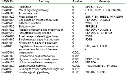
Table 2 Top 20 KEGG pathways enriched in advanced stage pancreatic carcinoma microarray data KEGG IDa Pathway P value Gene(s)a
hsa03010 Ribosome 0 RPS5, RPS26
hsa04010 MAPK signaling pathway 0 FOSB, TAOK1, EGFR, PRKAB2 hsa04360 Axon Guidance 0 -
hsa04510 Focal adhesion 0 EGF, PTEN, THBS1, VWF, EGFR hsa04514 Cell adhesion molecules (CAMs) 0 HLA-DOA, HLA-DQB1 hsa04520 Adherens junction 0 WASL, EGFR
hsa04530 Tight junction 0 PPP2R1B
hsa04612 Antigen processing and presentation 0 HLA-DOA, HLA-DQB 1 hsa04640 Hematopoietic cell lineage 0 HLA-DRB4, HLA-DRB5 hsa04660 T cell receptor signaling pathway 0 FOSB
hsa04662 B cell receptor signaling pathway 0 FOSB hsa04664 Fc epsilon RI signaling pathway 0 -
hsa04810 Regulation of actin cytoskeleton 0 EGF, WASL, EGFR
hsa00361 gamma-Hexachlorocyclohexane degradation 0.001 - hsa00460 Cyanoamino acid metabolism 0.001 -
hsa00561 Glycerophospholipid metabolism 0.001 PPAPDC1B hsa04120 Ubiquitin mediated proteolysis 0.001 UBE2D4
hsa04310 Wnt signaling pathway 0.001 THEM4(CTBP1), PPP2R1B hsa04670 Leukocyte transendothelial migration 0.001 -
hsa04910 Insulin signaling pathway 0.001 PRKAB2, SOCS3
aPathway identifiers as listed on the KEGG Pathway Finder at T1D Base (http://www.t1dbase.org). The genes
shown in the right-most column are representative genes that were used for KEGG pathway analysis.
compared to the pT2/pT3 carcinomas corresponding to 173 known genes (Supplementary Table 3). To determine the biologic significance of those genes or pathways most represented in this gene set, we performed Gene Set Enrichment Analysis (GSEA) [23]. GSEA revealed gene expression associated with advanced stage carcinomas was related to a variety of pathways including MAPK signaling, T and B cell receptor signaling, cell adhesion, cell adhesion and Wnt signaling (Table 2).
To verify our findings, we first focused upon four candidate upregulated genes (MAX Interacting Protein 1 (MXI1); Protein Kinase, AMP-Activated, Noncatalytic, Beta-2 (PRKAB2); Protein-Tyrosine Phosphatase, Receptor-Type Gamma (PTPRG) and Avian Musculo-aponeurotic Fibrosarcoma Oncogene Homolog (MAF)) and four candidate downregulated genes (Tribbles, Drosophila Homolog of 1 (TRIB1); Epidermal Growth Factor Receptor (EGFR); C-Terminal Modulator Protein (THEM4/CTMP); and Suppressor of Cytokine Signaling 3 (SOCS3)) in advanced stage carcinomas using semi-quantitative real-time PCR of cDNAs prepared from the same
carcinomas used for microarray analysis. All candidate upregulated genes were verified as having increased expression in advanced stage carcinomas compared to resectable carcinomas, ranging from 1.94 fold increased for MAF to 5.59 fold increased for MXI1 (data not shown). However, candidate down-regulated genes showed no difference in mRNA expression by quantitative PCR indicating these genes represent false positives in our statistical analysis of microarray data.
Figure 1 Verification of candidate upregulated genes in pancreatic cancers. Total mRNA from ten resected pancreatic cancers (stages pT1-pT3) and five advanced stage cancers with associated metastases (all pT4) was extracted and used to generate cDNAs for quantitative PCR of each gene. Ct values for each gene were normalized to that of beta-glucoronidase in the same sample and experiments were performed in triplicate. MXI1 is confirmed as having increased expression in pT4 carcinomas (p<0.05), whereas PRKAB2, PTPRG and MAF show no difference in expression.
found for MAF, PRKAB2 or PTPRG in either comparison.
To determine if increased MXI1 gene expression found by both microarrays and real-time PCR corresponds to an increase in protein expression as well, we performed immunolabeling for Mxi1 protein in an independent sampling of 13 pT2/pT3 paraffin embedded carcinomas and 10 pT4 paraffin-embedded carcinomas. Immunolabeling for Mxi1 was detected in the normal duct epithelium and in the neoplastic epithelium of all pancreatic cancers analyzed where it was localized to the nucleus although significant cytoplasmic labeling was also present (Figure 2). Labeling within normal ducts of the pancreas was confined to scattered cells
Figure 2 Mxi1 immunolabeling in pancreatic tissues. A. Normal pancreatic duct with scattered Mxi1 positive cells within the duct (indicated by arrows) with labeling present within the nucleus of positive cells. B. Mxi1 immunolabeling in a representative pT2 stage pancreatic cancer. The cancer glands are diffusely positive, although of less intensity than that of the representative pT4 stage pancreatic cancer shown in panel C.
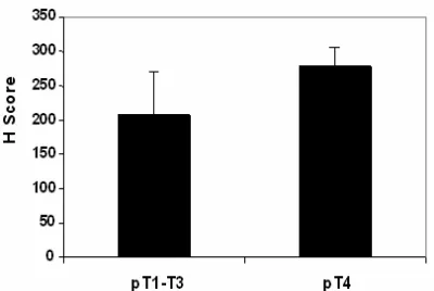
Figure 3 Summary of Mxi1 immunolabeling in pancreatic cancers. Shown are the mean histology (H) scores for the ten pT2/T3 and five pT4 pancreatic carcinomas immunolabeled for Mix1 protein. Mix1 labeling is consistently greater in the advanced stage carcinomas (p<0.003).
Discussion
Several studies have reported the gene expression profiles of infiltrating pancreatic cancers from surgical resection specimens [17, 35-42], but the expression profiles of advanced stage pancreatic cancers associated with locally destructive behavior and/or metastatic spread have not. This is largely due to the fact that advanced pancreatic cancer is not a surgical disease, thus beyond diagnosis tissue samples are often not available for research. We now report one of the first studies of advanced stage pancreatic cancer
using tissues obtained from rapid autopsy of patients with end stage pancreatic cancer.
Our comparisons among primary carcinomas and their matched metastases failed to show commonly deregulated genes. This is consistent with that shown in breast cancers [43, 44] although in the study by Ramaswamy et al [43] a 17 gene metastasis signature was found in the primary carcinomas that had matched metastases, whereas no such signature was found in the current study. While this may be a reflection of our sample sizes, another possibility is that our experimental findings support the common perception that all pancreatic cancers are metastatic, even at the time of surgical resection [45]. Similar profiles among matched primary and metastatic cancers have also been suggested as evidence for development of the metastatic phenotype during carcinogenesis time of tumor formation [43, 46] although the intriguing proposal that metastatic ability is programmed in normal tissues has also been put forth [47-49]. Thus, as consistent and robust changes in gene expression have been found in association with pancreatic carcinogenesis but not metastasis formation further study of the expression profiles of pancreatic cancer and correlation to outcomes is warranted to address this possibility.
[image:8.612.93.293.336.470.2]stage disease were found to be highly similar to that of resected cancers, our data nonetheless suggest that additional changes in gene expression may occur within the primary site that correlates with progression to advanced stage disease. One of the changes found and validated was increased expression of MXI1, a member of the MAD family of transcriptional repressors that negatively regulates c-Myc [50]. Amplification and overexpression of c-Myc has been well described in pancreatic cancer where it acts as a central regulator of gene transcription and predisposes cells to apoptosis under nutrient, growth factor or oxygen deprivation conditions [51, 52]. Thus, while c-Myc amplification and/or overexpression may promote pancreatic carcinogenesis, up-regulation of Mxi1 may occur during disease progression to refine c-Myc oncogenic signals and to protect cancer cells from c-Myc induced apoptosis [52]. To state that this refinement causes metastasis is unlikely. Rather, with continued growth of the neoplasm within an increasingly tenuous microenvironment, such as from small cancers confined to the pancreas to bulky unresectable cancers, subclones may constantly evolve with the neoplasm to maintain a positive net growth by modulation of oncogenic signaling. Indeed, the findings of Graeber et al support this possibility, as they have shown selection of clonal variants with diminished apoptotic potential in solid human tumors due to hypoxic environments [47].
Our findings from gene set enrichment analyses provide additional insight into the expression profiles associated with pancreatic cancer progression, such as the MAPK and WNT signaling pathways. Consistent with this finding, deregulation of the MAPK and Wnt signaling pathways have been implicated in epithelial-mesenchymal transition and in the metastasis of a variety of tumor types, including pancreatic cancer [53-58]. MAPK signaling is also a central component of the oxidative stress response, consistent with ongoing MAPK pathway deregulation with tumor growth at the primary site [59]. We also found evidence suggestive of immune deregulation in tumor progression. This finding is particularly intriguing considering the emerging role of immune-boosting therapies in the treatment of pancreatic cancer [60] and supports further investigations in this area particularly as the immune system relates to
tumor progression.
In summary, we provide examples of the expression profiles associated with pancreatic cancer progression and offer possibilities for further investigation into the mechanisms underlying the aggressive behavior of this disease.
Acknowledgements
This work was supported by NIH grant CA106610-01 to Christine A. Iacobuzio-Donahue), the Family of George Rubis, Sigma Beta Sorority, the Jeff Zgonina Foundation for Pancreatic Cancer Research and the Skip Viragh Foundation.
Please address all correspondences to Christine A. Iacobuzio-Donahue, MD, PhD, The Johns Hopkins Hospital, Division of Gastrointestinal/Liver Pathology, 1550 Orleans Street, CRBII Rm 343, Baltimore, MD 2123, USA. Tel: 410-955-3511; Fax: 410-614-0671; Email: ciacobu@jhmi.edu
References
[1] Jemal A, Siegel R, Ward E, Murray T, Xu J and Thun MJ. Cancer statistics, 2007. CA Cancer J Clin 2007;57:43-66.
2] Sohn TA, Yeo CJ, Cameron JL, Koniaris L, Kaushal S, Abrams RA, Sauter PK, Coleman J, Hruban RH and Lillemoe KD. Resected adenocarcinoma of the pancreas-616 patients: Results, outcomes, and prognostic indicators. J Gastrointest Surg 2000;4:567-579.
[3] Hansel DE, Kern SE and Hruban RH. Molecular pathogenesis of pancreatic cancer. Annu Rev Genomics Hum Genet 2003; 4:237-256. [4] Schneiderhan W, Diaz F, Fundel M, Zhou S,
Siech M, Hasel C, Moller P, Gschwend JE, Seufferlein T, Gress T, Adler G and Bachem MG. Pancreatic stellate cells are an important source of mmp-2 in human pancreatic cancer and accelerate tumor progression in a murine xenograft model and cam assay. J Cell Sci
2007;120:512-519.
[5] Ellenrieder V, Hendler SF, Ruhland C, Boeck W, Adler G and Gress TM. Tgf-beta-induced invasiveness of pancreatic cancer cells is mediated by matrix metalloproteinase-2 and the urokinase plasminogen activator system.
Int J Cancer 2001; 93:204-211.
61:4222-4228.
[7] Jungert K, Buck A, von Wichert G, Adler G, Konig A, Buchholz M, Gress TM and Ellenrieder V. Sp1 is required for transforming growth factor-beta-induced mesenchymal transition and migration in pancreatic cancer cells.
Cancer Res 2007; 67:1563-1570.
[8] Ijichi H, Chytil A, Gorska AE, Aakre ME, Fujitani Y, Fujitani S, Wright CV and Moses HL. Aggressive pancreatic ductal adenocarcinoma in mice caused by pancreas-specific blockade of transforming growth factor-beta signaling in cooperation with active kras expression. Genes Dev 2006; 20:3147-3160.
[9] Bardeesy N, Cheng KH, Berger JH, Chu GC, Pahler J, Olson P, Hezel AF, Horner J, Lauwers GY, Hanahan D and DePinho RA. Smad4 is dispensable for normal pancreas development yet critical in progression and tumor biology of pancreas cancer. Genes Dev 2006;20:3130-3146.
[10] Feldmann G, Dhara S, Fendrich V, Bedja D, Beaty R, Mullendore M, Karikari C, Alvarez H, Iacobuzio-Donahue C, Jimeno A, Gabrielson KL, Matsui W and Maitra A. Blockade of hedgehog signaling inhibits pancreatic cancer invasion and metastases: A new paradigm for combination therapy in solid cancers. Cancer Res 2007; 67:2187-2196.
[11] Yin T, Wang C, Liu T, Zhao G, Zha Y and Yang M. Expression of snail in pancreatic cancer promotes metastasis and chemoresistance. J Surg Res 2007;141:196-203.
[12] Tsutsumida H, Swanson BJ, Singh PK, Caffrey TC, Kitajima S, Goto M, Yonezawa S and Hollingsworth MA. Rna interference suppression of muc1 reduces the growth rate and metastatic phenotype of human pancreatic cancer cells. Clin Cancer Res 2006; 12:2976-2987.
[13] Kayed H, Kleeff J, Keleg S, Felix K, Giese T, Berger MR, Buchler MW and Friess H. Effects of bone sialoprotein on pancreatic cancer cell growth, invasion and metastasis. Cancer Lett
2007;245:171-183.
[14] Sawai H, Okada Y, Funahashi H, Matsuo Y, Takahashi H, Takeyama H and Manabe T. Integrin-linked kinase activity is associated with interleukin-1 alpha-induced progressive behavior of pancreatic cancer and poor patient survival. Oncogene 2006; 25:3237-3246. [15] Sawai H, Okada Y, Funahashi H, Matsuo Y,
Takahashi H, Takeyama H and Manabe T. Interleukin-1alpha enhances the aggressive behavior of pancreatic cancer cells by regulating the alpha6beta1-integrin and urokinase plasminogen activator receptor expression. BMC Cell Biol 2006;7:8.
[16] Chu CS, Xue B, Tu C, Feng ZH, Shi YH, Miao Y and Wen CJ. Nrage suppresses metastasis of melanoma and pancreatic cancer in vitro and in vivo. Cancer Lett 2007;250:268-275.
[17] Kim HN, Choi DW, Lee KT, Lee JK, Heo JS, Choi SH, Paik SW, Rhee JC and Lowe AW. Gene expression profiling in lymph node-positive and lymph node-negative pancreatic cancer.
Pancreas 2007;34:325-334.
[18] Embuscado EE, Laheru D, Ricci F, Yun KJ, de Boom Witzel S, Seigel A, Flickinger K, Hidalgo M, Bova GS and Iacobuzio-Donahue CA. Immortalizing the complexity of cancer metastasis: Genetic features of lethal metastatic pancreatic cancer obtained from rapid autopsy. Cancer Biol Ther 2005;4:548-554.
[19] Xin W, Yun KJ, Ricci F, Zahurak M, Qiu W, Su GH, Yeo CJ, Hruban RH, Kern SE and Iacobuzio-Donahue CA. Map2k4/mkk4 expression in pancreatic cancer: Genetic validation of immunohistochemistry and relationship to disease course. Clin Cancer Res 2004; 10:8516-8520.
[20] Sato N, Maitra A, Fukushima N, van Heek NT, Matsubayashi H, Iacobuzio-Donahue CA, Rosty C and Goggins M. Frequent hypomethylation of multiple genes overexpressed in pancreatic ductal adenocarcinoma. Cancer Res 2003; 63:4158-4166.
[21] Irizarry RA, Hobbs B, Collin F, Beazer-Barclay YD, Antonellis KJ, Scherf U and Speed TP. Exploration, normalization, and summaries of high density oligonucleotide array probe level data. Biostatistics 2003;4:249-264.
[22] Wettenhall JM and Smyth GK. Limmagui: A graphical user interface for linear modeling of microarray data. Bioinformatics 2004; 20:3705-3706.
[23] Subramanian A, Tamayo P, Mootha VK, Mukherjee S, Ebert BL, Gillette MA, Paulovich A, Pomeroy SL, Golub TR, Lander ES and Mesirov JP. Gene set enrichment analysis: A knowledge-based approach for interpreting genome-wide expression profiles. Proc Natl Acad Sci USA 2005;102:15545-15550. [24] Livak KJ and Schmittgen TD. Analysis of
relative gene expression data using real-time quantitative pcr and the 2(-delta delta c(t)) method. Methods 2001;25:402-408.
[25] Mudali SV, Fu B, Lakkur SS, Luo M, Embuscado EE and Iacobuzio-Donahue CA. Patterns of epha2 protein expression in primary and metastatic pancreatic carcinoma and correlation with genetic status. Clin Exp Metastasis 2006;23:357-365.
[26] Nichols LS, Ashfaq R and Iacobuzio-Donahue CA. Claudin 4 protein expression in primary and metastatic pancreatic cancer: Support for use as a therapeutic target. Am J Clin Pathol 2004; 121:226-230.
[28] Miyamoto Y, Maitra A, Ghosh B, Zechner U, Argani P, Iacobuzio-Donahue CA, Sriuranpong V, Iso T, Meszoely IM, Wolfe MS, Hruban RH, Ball DW, Schmid RM and Leach SD. Notch mediates tgf alpha-induced changes in epithelial differentiation during pancreatic tumorigenesis. Cancer Cell 2003;3:565-576. [29] Ripka S, Konig A, Buchholz M, Wagner M, Sipos
B, Kloppel G, Downward J, Gress T and Michl P. Wnt5a--target of cutl1 and potent modulator of tumor cell migration and invasion in pancreatic cancer. Carcinogenesis 2007;28:1178-1187. [30] Furukawa T, Sunamura M, Motoi F, Matsuno S
and Horii A. Potential tumor suppressive pathway involving dusp6/mkp-3 in pancreatic cancer. Am J Pathol 2003;162:1807-1815. [31] Yang AD, Camp ER, Fan F, Shen L, Gray MJ, Liu
W, Somcio R, Bauer TW, Wu Y, Hicklin DJ and Ellis LM. Vascular endothelial growth factor receptor-1 activation mediates epithelial to mesenchymal transition in human pancreatic carcinoma cells. Cancer Res 2006;66:46-51. [32] Tang RF, Wang SX, Peng L, Wang SX, Zhang M,
Li ZF, Zhang ZM, Xiao Y and Zhang FR. Expression of vascular endothelial growth factors a and c in human pancreatic cancer.
World J Gastroenterol 2006;12:280-286. [33] Michl P, Barth C, Buchholz M, Lerch MM, Rolke
M, Holzmann KH, Menke A, Fensterer H, Giehl K, Lohr M, Leder G, Iwamura T, Adler G and Gress TM. Claudin-4 expression decreases invasive-ness and metastatic potential of pancreatic cancer. Cancer Res 2003;63:6265-6271.
[34] Overall CM and Kleifeld O. Tumour microenvironment - opinion: Validating matrix metalloproteinases as drug targets and anti-targets for cancer therapy. Nat Rev Cancer
2006;6:227-239.
[35] Iacobuzio-Donahue CA, Ashfaq R, Maitra A, Adsay NV, Shen-Ong GL, Berg K, Hollingsworth MA, Cameron JL, Yeo CJ, Kern SE, Goggins M and Hruban RH. Highly expressed genes in pancreatic ductal adenocarcinomas: A comprehensive characterization and comparison of the transcription profiles obtained from three major technologies.
Cancer Res 2003;63:8614-8622.
[36] Logsdon CD, Simeone DM, Binkley C, Arumugam T, Greenson JK, Giordano TJ, Misek DE, Kuick R and Hanash S. Molecular profiling of pancreatic adenocarcinoma and chronic pancreatitis identifies multiple genes differentially regulated in pancreatic cancer.
Cancer Res 2003;63:2649-2657.
[37] Missiaglia E, Blaveri E, Terris B, Wang YH, Costello E, Neoptolemos JP, Crnogorac-Jurcevic T and Lemoine NR. Analysis of gene expression in cancer cell lines identifies candidate markers for pancreatic tumorigenesis and metastasis. Int J Cancer 2004;112:100-112. [38] Grutzmann R, Pilarsky C, Ammerpohl O, Luttges
J, Bohme A, Sipos B, Foerder M, Alldinger I,
Jahnke B, Schackert HK, Kalthoff H, Kremer B, Kloppel G and Saeger HD. Gene expression profiling of microdissected pancreatic ductal carcinomas using high-density DNA micro-arrays. Neoplasia 2004;6:611-622.
[39] Grutzmann R, Boriss H, Ammerpohl O, Luttges J, Kalthoff H, Schackert HK, Kloppel G, Saeger HD and Pilarsky C. Meta-analysis of microarray data on pancreatic cancer defines a set of commonly dysregulated genes. Oncogene
2005;24:5079-5088.
[40] Han H, Bearss DJ, Browne LW, Calaluce R, Nagle RB and Von Hoff DD. Identification of differentially expressed genes in pancreatic cancer cells using cdna microarray. Cancer Res
2002;62:2890-2896.
[41] Crnogorac-Jurcevic T, Efthimiou E, Nielsen T, Loader J, Terris B, Stamp G, Baron A, Scarpa A and Lemoine NR. Expression profiling of microdissected pancreatic adenocarcinomas.
Oncogene 2002;21:4587-4594.
[42] Iacobuzio-Donahue CA, Maitra A, Olsen M, Lowe AW, van Heek NT, Rosty C, Walter K, Sato N, Parker A, Ashfaq R, Jaffee E, Ryu B, Jones J, Eshleman JR, Yeo CJ, Cameron JL, Kern SE, Hruban RH, Brown PO and Goggins M. Exploration of global gene expression patterns in pancreatic adenocarcinoma using cdna microarrays. Am J Pathol 2003;162:1151-1162.
[43] Ramaswamy S, Ross KN, Lander ES and Golub TR. A molecular signature of metastasis in primary solid tumors. Nat Genet 2003;33:49-54.
[44] Weigelt B, Glas AM, Wessels LF, Witteveen AT, Peterse JL and van't Veer LJ. Gene expression profiles of primary breast tumors maintained in distant metastases. Proc Natl Acad Sci USA
2003;100:15901-15905.
[45] Ducreux M, Boige V and Malka D. Treatment of advanced pancreatic cancer. Semin Oncol
2007;34:S25-30.
[46] Bernards R and Weinberg RA. A progression puzzle. Nature 2002;418:823.
[47] Hunter KW. Host genetics and tumour metastasis. Br J Cancer 2004;90:752-755. [48] Malins DC, Gilman NK, Green VM, Wheeler TM,
Barker EA and Anderson KM. A cancer DNA phenotype in healthy prostates, conserved in tumors and adjacent normal cells, implies a relationship to carcinogenesis. Proc Natl Acad Sci USA 2005;102:19093-19096.
[49] Malins DC, Gilman NK, Green VM, Wheeler TM, Barker EA, Vinson MA, Sayeeduddin M, Hellstrom KE and Anderson KM. Metastatic cancer DNA phenotype identified in normal tissues surrounding metastasizing prostate carcinomas. Proc Natl Acad Sci USA 2004; 101:11428-11431.
[50] Hurlin PJ and Huang J. The max-interacting transcription factor network. Semin Cancer Biol
2006;16:265-274.
Osthus RC and Li F. The c-myc target gene network. Semin Cancer Biol 2006;16:253-264. [52] Corn PG, Ricci MS, Scata KA, Arsham AM,
Simon MC, Dicker DT and El-Deiry WS. Mxi1 is induced by hypoxia in a hif-1-dependent manner and protects cells from c-myc-induced apoptosis. Cancer Biol Ther 2005;4:1285-1294.
[53] Tan X, Tamori Y, Egami H, Ishikawa S, Kurizaki T, Takai E, Hirota M and Ogawa M. Analysis of invasion-metastasis mechanism in pancreatic cancer: Involvement of tight junction transmembrane protein occludin and mek/erk signal transduction pathway in cancer cell dissociation. Oncol Rep 2004;11:993-998. [54] Dissanayake SK, Wade M, Johnson CE,
O'Connell MP, Leotlela PD, French AD, Shah KV, Hewitt KJ, Rosenthal DT, Indig FE, Jiang Y, Nickoloff BJ, Taub DD, Trent JM, Moon RT, Bittner M and Weeraratna AT. The wnt5a/protein kinase c pathway mediates motility in melanoma cells via the inhibition of metastasis suppressors and initiation of an epithelial to mesenchymal transition. J Biol Chem 2007;282:17259-17271.
[55] Guo Y, Zi X, Koontz Z, Kim A, Xie J, Gorlick R, Holcombe RF and Hoang BH. Blocking wnt/lrp5 signaling by a soluble receptor modulates the epithelial to mesenchymal transition and
suppresses met and metalloproteinases in osteosarcoma saos-2 cells. J Orthop Res 2007; 25:964-971.
[56] Zhao CH, Bu XM and Zhang N. Hyper-methylation and aberrant expression of wnt antagonist secreted frizzled-related protein 1 in gastric cancer. World J Gastroenterol 2007; 13:2214-2217.
[57] Mendes O, Kim HT, Lungu G and Stoica G. Mmp2 role in breast cancer brain metastasis development and its regulation by TIMP2 and ERK1/2. Clin Exp Metastasis 2007;24:341-351.
[58] Hulit J, Suyama K, Chung S, Keren R, Agiostratidou G, Shan W, Dong X, Williams TM, Lisanti MP, Knudsen K and Hazan RB. N-cadherin signaling potentiates mammary tumor metastasis via enhanced extracellular signal-regulated kinase activation. Cancer Res 2007; 67:3106-3116.
[59] Wu JJ and Bennett AM. Essential role for mitogen-activated protein (map) kinase phosphatase-1 in stress-responsive map kinase and cell survival signaling. J Biol Chem
2005; 280:16461-16466.


![[N,N,N′,N′ Tetrakis(benzimidazol 2 ylmethyl)cyclohexane 1,2 diamine]nickel(II) dinitrate dihydrate](data:image/gif;base64,R0lGODlhAQABAIAAAP///wAAACH5BAEAAAAALAAAAAABAAEAAAICRAEAOw==)