Int J Clin Exp Pathol (2009) 2, 83-90
www.ijcep.com
/IJCEP803001
Original Article
Correlation of Macrophage Inflammatory Protein-2
Expression and Brain Edema in Rats after Intracerebral
Hemorrhage
He Wu1, #, Yuwei Cong1, #, Dandan Wang 2, #, Ruibo Zhao3 and JipingQi1
1Department of Pathology, First Clinical Hospital, Harbin Medical University, Harbin 150001, PR China;
2Department of Pathology,the Second Affiliated Hospital of Tsinghua University, Beijing 100049, PR China and
3Pathology Centralab, Harbin Medical University, Harbin 150086, PR China
Received 10 March 2008; Accepted in revision 22 May 2008; Available online 2 June 2008
Abstract: Brain edema is one of the most frequent and serious complications of intracerebral hemorrhage (ICH), but its underlying mechanism remains largely unknown. In order to understand whether inflammatory mediators released from the blood after cerebral hemorrhage plays a role in brain edema, we investigated the dynamic change of the inflammatory mediator macrophage inflammatory protein-2 (MIP-2) in rat brain after ICH. Our results indicate that the expression of MIP-2 increases 2 hours and peaks 2 days after ICH. The expression of MIP-2 correlates with NF-κB activation and brain water content. These results suggest that MIP-2 expression may play an important role in the development of brain edema after ICH in rats.
Key Words: MIP-2, NF-κB, brain edema, intracerebral hemorrhage
Introduction
Stroke is the third leading cause of death and the leading cause of long-term disability in the world. Intracerebral hemorrhage (ICH) is a common and one of the most devastating types of stroke. ICH accounts for 10-15% of all strokes, but it is responsible for approximately 50% of stroke-related deaths. It imparts some form of disability to 88% of its survivors [1, 2]. Secondary brain edema and brain damage may result in further deterioration in neurological functions after ICH. But the cause of secondary brain injury and edema formation is not completely understood. Previous reports showed that ICH was accompanied by inflammation. Whether the inflammation contributes to brain edema and neurological injury or plays a role in repairing the brain tissue is not fully known.
Macrophage inflammatory protein-2 (MIP-2) was first isolated from mouse macrophages in 1988
__________
# These authors contributed equally.
by Wolpe. MIP-2 is a chemoattractant [3] and an activating factor for neutrophils [4]. In neuropathological processes, MIP-2 is thought to play a pivotal role in the induction and perpetuation of inflammation in the central nervous system (CNS). The origin of MIP-2 in brain has not been fully defined. Recent studies have demonstrated that reactive glial cells also produced MIP-2 [5, 6]. In addition to astrocytes, microglial cells, endothelial cells, macrophages and some neuron cells may be potential sources of MIP-2 production in pathological states of the brain [7]. The release of MIP-2 into the tissue of the CNS may be caused by both in situ and infiltrated cells.
Materials and Methods
Animal Model of Intracerebral Hemorrhage
Ninety male Sprague-Dawley rats (200–250 g) were purchased from the Center for Experimental Animals, Harbin Medical University. NIH Guidelines for the Care and Use of Laboratory Animals were followed in all animal procedures.
We chose to inflict ICHby autologous blood to better mimic the sudden deposition ofblood in the brain parenchyma after the rupture of vessels inhumans [15]. Rats were anesthetized with chloral hydrate (350 mg/kg intraperitoneally). Animals were placed in a stereotaxic frame using modified ear-barsfitted with blunt rubber ends designed for rats. Proceduresfor induction of ICH by autologous blood were modified fromthe previous studies in rats [16] and mice [17]. A midline scalp incision was made,and a hole was drilled in the left skull (3mm lateral to midline, 0.2 mm anterior to bregma). Autologous blood (50 µL) from arteria caudilis was collected into a needleless sterile insulin syringe without any anti-coagulant. A 26-gauge needle was quickly attached to the syringe and stereotaxically introduced into the left caudate nucleus 5.5 mm below the surface of the hole inthe skull. The 50 µL blood was injected over 5min,and the needle was left in place for another 5 min to minimizebackflow. The syringe was then removed slowly. The burr holein the skull was sealed with bone wax, the scalp was sutured,and the animals were placed in a cage with ad libitum accessto food and water. Body temperature was maintained at 37°C with the use of a feedback-controlled heating pad. Blood pressure, rectal temperature and body weight were monitored periodically after ICH. Control animals were subjected to the same manipulations as ICH rats, in which no autologous blood but the same volume of saline was injected. Animals were sacrificed at 2h, 3h, 6h, 10h, 12h, 24h, 2d, or 5d after the injury for analyses. Control animals were killed at 2h after the injection.All experimental procedures were done in accordance with guidelines of the Chinese Council on Animal Care.
Histological Examination
Rats were overdosed with chloral hydrate at 2h, 3h, 6h, 10h, 12h, 24 h, 2d or 5d after ICHand were perfused through the heart with 300 mL
ice-cold 4% paraformaldehydein 0.1 mol/L PBS. Fixed brains were cut coronally approximately2 mm on either side of the needle entry site, which was identifiable on the brain surface. These brain slices were dehydratedand embedded in paraffin. Sections (4 µm) were cut and stained with hematoxylin and eosin. At the coronal level ofthe needle entry site, where the brain damage was maximal, a variety of histological and immunohistochemical stains were performed. Damaged brain areas were defined by the presence of blood, tissue rarefaction, or necrosis. These analyses were evaluated blindly by an experienced investigator to minimize observation bias.
Brain Water Content
Brain water content was determined by the dry-wet weight method. After decapitation, rat brain samples were immediately weighed on an electronic analytical balance (Changzhou instruments, Inc., China) to obtain the wet weight. Then brain samples were dried at 100°C in an Electric Blast Drying Oven (Chongqing Sida Apparatus, Inc, China) for 24 h to obtain the dry weight. The formula for water content calculation was as follows: wet weight - dry weight) / wet weight × 100%.
Immunohistochemistry
The procedures were processed according to the protocol recommended for MIP-2 and NF-κB immunohistochemistry kit. The rat brain sections werede-waxed and re-hydrated, rinsed with distilled water and PBS, quenchedwith 3% H2O2, blocked with 10% normal goat serum, and incubated with goat anti-MIP-2 polyclonal antibody (diluted at 1:150, Santa Cruz, USA), and rabbit anti-NF-κB polyclonal antibody (diluted at 1:100, Santa Cruz, USA) at 4°C overnight. Slides were then washedwith Triton PBS, incubated in biotinylated goat anti-rabbit IgG and rabbit anti-goat IgG(DAKO,USA) for 1 hour at room temperature, washed, incubated withstreptavidin peroxidase for 30 minutes at
room temperature, colored with
diaminobenzidine-H2O2 solution, washed, dehydrated in graded ethanol, immersed in xylene and coverslipped.Control sections were processed with omission of the primaryantibody.
Quantitative Real-time RT-PCR of MIP-2 mRNA
6h, 10h, 12h, 24 h, 2d or 5d after ICH were kept immediately in liquid nitrogen for the analysis of MIP2 mRNA by real-time RT-PCR. Two procedures were used for RNA extraction and reverse transcription (RT). Total RNA was isolated from rat brain using TRIzol reagent (Invitrogen, Carlsbad, CA, USA) according to the manufacturer's instructions. PCR reactions were performed in 96-well plates with an ABI PRISM 7300 Sequence Detection System (Applied Biosystems) using SYBR Green to monitor amplification. Reactions were done in 25 μL volumes containing 200 nM of each primer, 5 μL cDNA (corresponding to ~3 ng DNA), and 10 μL 2 × SYBR Green Master Mix Reagent (Applied Biosystems). Aliquots from the same cDNA sample were used with all primer sets in each experiment. Reactions were run using the manufacturer's recommended cycling parameters of 93°C for 2 min, 40 cycles of 93°C for 1 min and 55°C for 1 min. Each PCR reaction was completed in triplicate. Optical data obtained by real-time PCR was analyzed with the manufacturer's software. Primers used were 5’-cctgggaaggaagaacatgg-3’ (sense) and 5’-ggcacatcaggtacgatcca-3’ (antisense).
Western Blot Analysis for NF-κB
Animals were anesthetized before undergoing intracardiac perfusion with saline. The brains were sampled as described for brain water content. Western blot analysis was performed as previouslydescribed [19]. Briefly, 50 µg protein samples were separated by sodium dodecyl sulfate polyacrylamide gel electrophoresis and transferred to a Hybond-C pure nitrocellulose membrane. After blocking, membranes were probed with a 1:200 dilution of the primary antibody (rabbit polyclonal anti- NF-κB, Santa Cruz) and a 1:5000 dilution of the second antibody (peroxidase-conjugated goat anti-rabbit antibodies, Rockland). The antigen-antibody complexes were visualizedwith a chemiluminescence system (Amersham) and exposed to film (Kodak X-OMAT). The relative densities of bands were analyzedwith ODYSSEY infrared imaging system (LI-COR). GAPDH was used as an internal control. The experiments were repeated three times with similar results.
Cell Counting and Statistical Analysis
Two observers, blinded to the experimental surgery,evaluated the morphometric parameters in ten randomly chosen non-overlapping
high-power fields (hpf, original magnification×400, Olympus BX51, light microscope) from each section. All positive cells in the ten high-power fields were counted and the number of MIP-2 and NF-κB positive cells per high-power field was calculated. All data were reported as mean ± standard deviation (SD). The statistical significance of the data was analyzed by analysis of variance (ANOVA), post hoc tests and pearson correlation test with SPSS 13.0 for Windows (SPSS Inc). Values of P<0.05 were regarded as significant.
Results
Morphological Changes of Rat Brain after ICH
A spherical hematoma was observed in the caudate nucleus area at all time points after ICH. At 3h time point, a few scattered neutrophils were found at the periphery of the hematoma. Little morphological changes of neurons were observed. At 24h, brain edema around the hematoma was visible. The infiltrated inflammatory cells were mainly mononuclear with scattered neutrophils. At 48h, the brain edema around the hematoma was pronounced and the brain tissue was diffluent and necrotic. The hematoma was surrounded by a compact band of cells including viable neutrophils, some cell debris, a few macrophages and rare clusters of intact erythrocytes. At day 5 after ICH, the hematoma started to resolve with glia cell hyperplasia and abundant neovascularization. In contrast, no hematoma or significant changes were observed in the brain tissue of rats from the control group (Figures 1 A-C).
Immunocytochemical Analysis of MIP-2 Expression in Rat Brainafter ICH
Figure 1 Histopathology of cerebral cortex of rats from control (A), 3h after ICH (B) and 2d after ICH (C) (hematoxylin and eosin stain; scale bar = 100 µm). Mild morphological changes of neurons were observed 3h after ICH while significant changes were present 2d after ICH.
weakly expressed in normal rat brain. Compared to control rat brains, MIP-2 immunoreactivity showed a modest increase 2 hours after ICH, significant increase 3 hours and maximal increase 2 days after ICH (P< 0.01) (Figure 2D).
Quantitative RT-PCR Analyses of MIP-2 mRNA
Using real-time quantitative RT-PCR, we further investigated the expression of MIP-2 at mRNA level. MIP-2 mRNA continued to increase after
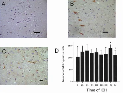
[image:4.612.97.518.355.629.2]Figure 3 NF-κB immunoreactivity in cerebral cortex of rats from control (A), 3h after ICH (B) and 2d after ICH (C). (immunohistochemical stain with anti NF-kB antibody; scale bar = 100 µm). NF-κB protein was weakly positive in the control, strongly cytoplasmic positive 3h after ICH, and cytoplasmic and nuclear positive 2d after ICH. Graphic presentation of the number of NF-kB positive cells per high power field after ICH (D). Vertical bars indicate the mean ± SD. # P<0.05 compared with control group rats; *P<0.05 compared with rats at a prior time point after ICH.
ICH and peaked at 3 hours (Figure 2E). The kinetics of MIP-2 mRNA expression is similar that of MIP-2 immunoreactivity.
NF-κB Immunocytochemistry in Rat Brainafter ICH
NF-κB protein in rat brain was expressed in cell bodies of neurons and astrocytes. NF-κB protein was also expressed in vascular endothelial cells and smooth muscle cells of the cerebral vasculature. NF-κB protein was weakly positive in the cytoplasm of the control group. It was translocated from cytoplasm to nucleus in the cells of the hemorrhage hemisphere (Figure 3 A-C). Similar to MIC-2, NF-κB immunoreactivity began to increase at 2 hours after ICH, dramatically increased at 3 hours and reached the maximum at 2 days after ICH (P<0.05 compared with control group) (Figure 3D).
[image:5.612.316.517.481.636.2]Figure 4 Western blot analysis of NF-κB in rat cerebral cortex. Expression of NF-κB protein increased 2 hours after ICH and reached the maximum at 1-2 days after ICH. GAPDH was used as an internal control. The experiments were repeated three times with similar results.
Western Blot Analysis of NF-κB in Rat Brain after ICH
Western blot analyses showed that NF-κB protein dramatically increased at 2 hours and reached the maximum at 1-2 days after ICH (Figure 4).
Brain Water Content
Our results indicated that cerebral edema was severe at the acute stage and relieved from 6 hours to 12 hours after ICH. However, after 24 hours, cerebral edema was dramatically severe (P<0.05 compared with control group) and reached the maximum at 2 days (Figure 5). There was a positive correlation between brain water content and MIP-2 as well as NF-κB expression. Cerebral edema and the expression
of MIP-2 was positively correlated at the 0.01 level (correlation coefficient is 0.548) on the whole. The expression of NF-κB was also positively correlated with cerebral edema at the 0.05 level (correlation coefficient is 0.314).
Discussion
A major side effect of inflammation is an increased permeability of the cerebral microvessels, resulting in vasogenic brain edema and a rise in intracranial pressure [10]. Cytokine increases the permeability of blood brain barrier, induces leukocytosis in cerebrospinal fluid and brain edema [11]. Experimentally, recruitment of neutrophils to the CNS is followed by a breeching of the blood-brain barrier. Administration of MIP-2 exacerbates damage of the blood-brain barrier [12] that may further contribute to inflammation by causing indiscriminate entry of leukocytes into the brain. The expression of MIP-2 mRNA correlates with injury to the blood-brain barrier as demonstrated by the appearance of serum proteins and leukocytes in cerebrospinal fluid and by the increase in brain water content [13].
To further study the role of MIP-2 in brain edema, we quantified mRNA and protein levels for MIP-2 in rat brain after ICH. We found that in addition to previously described inflammatory changes in the brain, stroke induced a complex, but organ specific, pattern of inflammatory factors in the brain as early as 2h after ICH. Pyramidal neurons in layers 3 and 5 of cortex showed more prominent MIP-2 immunoreactivity than those in the other cortical layers. The result of immunohistochemistry correlates with the mRNA levels by real-time quantitative RT-PCR.
Our results also demonstrate that the drastic inflammatory changes occurring in the damaged brain are dynamically reflected with the time course of ICH. The brain water content after ICH appears to follow the expression pattern of MIP-2. Based on the recent reports in the literature and our own findings, we speculate that MIP-2, as an important chemotactic cytokine, may increase blood brain barrier permeability and cause brain edema after ICH.
However, although MIP-2 is present in the brain parenchyma, neutrophils are unable to respond, and hence the subsequent activation-dependent step of leukocyte emigration cannot occur. The up-regulation of some chemokines has been attributed to constitutive activation of nuclear factor-kappaB (NF-κB) [14]. NF-κB is a transcription factor present in cytoplasm as a heterodimer composed of p50 and p65 subunits. After activation, NF-κB translocates from the cytoplasm to the nucleus of the cells, binds to specific DNA sequences, and initiates transcription. Several reports have indicated the
involvement of NF-κB in a large number of cellular processes such as inflammatory and immune response. Our study revealed that NF-κB protein in rat brain was mildly expressed in cytoplasm in control group, and translocated from cytoplasm to nucleus of the cells in the hemorrhage hemisphere after ICH. NF-κB binding sites are present in the promoter region of MIP-2 gene. When bound to MIP-2 gene, NF-κB activates transcription of MIP-2 [15]. Studies have indicated that expression of MIP-2 gene in rat lung epithelial cells is dependent on the transcription factor NF-κB [16]. NF-κB also plays a key role in secondary impairments of the tissues around the hematoma [17, 18] by inducing the expression of various genes related to cell injury and apoptosis. Here we demonstrated that both MIP-2 and NF-κB proteins began to increase 2 hours after ICH and reached the maximum 1-2 days after ICH. The expression of NF-κB follows the same pattern of MIP-2 during the entire course of ICH, which also correlated with the developmental course of brain edema. We speculate that NF-κB may induce MIP-2 expression, which subsequently increases the permeability of the blood-brain barrier and results in formation of brain edema after ICH. These findings may provide information for developing new therapeutic strategies and therapeutic targets for cerebral edema after ICH.
Acknowledgment
This work was supported by the National Natural Science Foundation of Heilongjiang Province (No. ZJY0705), and the postgraduate bring new ideas Foundation of Heilongjiang Province (No. YJSCX2007-0324HLJ). We also thank teachers in the Pathology Centralab of Harbin Medical University, Department of cerebral surgery laboratory and Clinical Analysis Centralab of the First Hospital of Harbin Medical University.
Please address all correspondences to Jiping Qi, Department of Pathology, First Clinical Hospital, Harbin Medical University, Harbin 150001, PR China.
Email: qi_jiping@hotmail.com
References
[1] Taylor CL, Selman WR and Ratcheson RA. Brain attack. The emergent management of hypertensive hemorrhage. Neurosurg Clin N Am
1997;8:237-244.
intracerebral hemorrhage. In Feldmann E (ed): Intracerebral Hemorrhage. Futura: Mount Kisco, NY; 1994. pp3-23.
[3] Diab A, Abdalla H, Li H, Shi FD, Zhu J, Hojberg B, Lindquist L, Wretlind B, Bakhiet M and Link H. Neutralization of macrophage inflammatory protein 2 (MIP-2) and MIP-1 α attenuate neutrophils recruitment in the central nervous system during experimental bacterial meningitis.
Infect Immun 1999;67:2590-2601.
[4] Tekamp-Olson P, Gallegos C, Bauer D, McClain J, Sherry B, Fabre M, van Deventer S and Cerami A Cloning and characterization of cDNA for murine macrophage inflammatory protein and its human homologues. J Exp Med 1990;172: 911-919. [5] Yates SL, Burgess LH, Kocsis-Angle J, Antal JM,
Dority MD, Embury PB, Piotrkowski AM and Brunden KR. Amyloid beta and amylin fibrils induce increases in proinflammatory cytokine and chemokine production by THP-1 cells and murine microglia. J Neurochem 2000;74:1017- 1025.
[6] Giri RK, Selvaraj SK and Kalra VK. Amyloid peptide-induced cytokine and chemokine expression in THP-1 monocytes is blocked by small inhibitory RNA duplexes for early growth response-1 messenger RNA. J Immunol 2003; 170:5281-5294.
[7] Tomita M, Holman BJ, Santoro CP and Santoro TJ. Astrocyte production of the chemokine macrophage inflammatory protein-2 is inhibited by the spice principle curcumin at the level of gene transcription. J Neuroinflammation 2005; 2:8.
[8] Zhang X, Li H, Hu S, Zhang L, Liu C, Zhu C, Liu R and Li C. Brain edema after intracerebral hemorrhage in rats: the roles of inflammation.
Neurol India 2006;54:402-407
[9] Butcher K and Laidlaw J. Current intracerebral haemorrhage management. J Clin Neurosci
2003;10:158-167.
[10] Pfister HW, Koedel U and Paul R. Acute meningitis. Curr Infect Dis Rep 1999;1:153- 159.
[11] Saukkonen K, Sande S, Cioffe C, Wolpe S, Sherry B, Cerami A and Tuomanen E.The role of cytokines in the generation of inflammation and tissue damage in experimental gram-positive meningitis. J Exp Med 1990;1:171:439-448. [12] Bell MD, Taub DD and Perry VH. Overriding the
brain's intrinsic resistance to leukocyte recruitment with intraparenchymal injections of recombinant chemokines. Neuroscience 1996; 74:283-292.
[13] Galdiero M, D'Amico M, Gorga F, Di Filippo C, D’Isanto M, Vitiello M, Longanella A and Tortora A. Haemophilus influenzae porin contributes to signaling of the inflammatory cascade in rat brain. Infect Immun 2001;69:221-227.
[14] Dhawan P and Richmond A. Role of CXCL1 in tumorigenesis of melanoma. J Leukoc Biol
2002;72:9-18.
[15] Widmer U, Mangoue KR, Cerami A and Sherry B. Genomic cloning of promoter analysis of macrophage inflammatory protein MIP-2, MIP-1, and member of the chemokine super family of proinflammatory cytokines. J Immunol 1993; 150:4996-5012.
[16] Driscoll KE. TNFalpha and MIP-2: role in particle-induced inflammation and regulation by oxidative stress. Toxicol Lett 2000;112-113: 177-183.
[17] Karibe H, Niizuma H, Ohyama H, Shirane R and Yoshimoto T. Hepatitis C virus (HCV) infection as a risk factor for spontaneous intracerebral hemorrhage: Hospital based case-control study. J
Clin Neurosci 2001;8:423-425.
[18] Hang CH, Shi JX, Li JS, Wu W and Yin HX. Concomitant upregulation of nuclear factor-kB activity, proinflammatory cytokines and ICAM-1 in the injured brain after cortical contusion trauma in a rat model. Neurol India 2005;53:312-317. [19] Xi G, Keep RF, Hua Y, Xiang J and Hoff JT.