Introduction
Reactive oxygen species (ROS), which cause cellular damage via oxidation, have been impli-cated in the pathogenesis of diabetes mellitus [1, 2]. Persistent hyperglycemia in diabetes in-duces ROS production by glucose autoxidation [3, 4], activation of protein kinase C (PKC), and increased flux through the hexosamine pathway [1]. An important source of ROS production is nicotinamide adenine dinucleotide phosphate (NADPH) oxidase, which has been implicated in the pathogenesis of diabetic complications, in-cluding neuropathy [5, 6].
Hyperbaric oxygen (HBO) is applied in treatment of various diseases including diabetic patients with non-healing foot ulcers [7]; however, this
treatment also increases the formation of ROS, which are known to result in cellular damage through the oxidation of lipid, protein, and DNA [8]. The oxidative effects of HBO have been in-vestigated in animals and humans [9-11]; how-ever, the side effects of HBO treatment in dia-betic patients and animals have been little in-vestigated. In a previous study, we showed that a clinically recommended HBO treatment signifi-cantly decreased both mRNA expression and activities of antioxidant enzymes in the strepto-zotocin (STZ)-induced diabetic rat, suggesting that HBO causes oxidative stress [12].
STZ-induced β-cell death in the pancreas is as-sociated with oxidative stress caused by the production of excess intracellular ROS [13, 14]; furthermore, STZ may damage pancreatic tissue
www.ijcep.com/IJCEP1101009
Original Article
Enhancement of reactive oxygen species and induction of
apoptosis in streptozotocin-induced diabetic rats under
hyperbaric oxygen exposure
Tokio Matsunami, Yukita Sato, Yuki Hasegawa, Satomi Ariga, Haruka Kashimura, Takuya Sato, Masayoshi Yukawa
Laboratory of Biomedical Science, Department of Veterinary Medicine, College of Bioresource Sciences, Nihon University, Fujisawa 252-0880, Japan.
Received January 31, 2011; accepted February 17, 2011; Epub February 20, 2011; published March 31, 2011
Abstract: An important source of reactive oxygen species (ROS) production is nicotinamide adenine dinucleotide phosphate (NADPH) oxidase, which on activation induces superoxide production viaoxidation in the mitochondria, inflammation and stress; such ROS are implicated in the pathogenesis of diabetic complications, including neuropa-thy. Hyperbaric oxygen (HBO) treatments are applied various diseases including diabetic patients with unhealing foot ulcers, however, and also increases the formation of ROS. In a previous study, we showed that a clinically recom-mended HBO treatment significantly enhanced oxidative stress of pancreatic tissue in the diabetic rats. However, no study has been undertaken with regard to the effects of HBO on the activity and gene expression of the NADPH oxi-dase complex and on apoptosis in the pancreas of diabetic animals. The purpose of this study was to investigate the effect of HBO exposure on gene expression of the NADPH complex, and pancreatic expression of genes related to apoptosis via the mitochondria, using the NADPH oxidase inhibitor apocynin. The mRNA expression of genes related to NADPH oxidase complex and apoptosis increased significantly (P < 0.05) in the pancreas of diabetic rats under HBO exposure. Similarly, activities of NADPH oxidase and caspase-3 changed in parallel with mRNA levels. These results suggest that oxidative stress caused by HBO exposure in diabetic animals induces further ROS production and apoptosis, potentially through the up-regulation of NADPH oxidase complex. Thus, this study can contribute to devel-opment of a better understanding of the molecular mechanisms of apoptosis via the mitochondria in diabetes, under HBO exposure.
via imposition of oxidative as well as nitrosative stresses, which in turn can induce apoptosis in pancreatic cells [15]. In addition, the oxidative stress caused by HBO exposure induces apop-tosis via the mitochondrial pathway [16]. How-ever, to our knowledge, no study has been un-dertaken on the effects of HBO on the activity and gene expression of the NADPH oxidase complex in the pancreas of diabetic animals. In the present study, to evaluate the effects of HBO in the pancreas of STZ-induced diabetic rats, we examined the activity and gene expres-sion of the NADPH complex; we also examined pancreatic expression of genes related to apop-tosis via the mitochondria, and evaluated the effect of apocynin, an NADPH oxidase inhibitor [17-19].
Materials and methods
Animals and experimental design
Male Wistar rats (weight range, 250–270 g) were purchased from Japan SLC Inc (Shizuoka, Japan) at 7 weeks of age. They were housed at 23 - 25 °C with light from 7:00 AM to 7:00 PM and free access to water at all times. All rats were fed a commercial diet during the experi-ment. All study procedures were implemented in accordance with the Institutional Guidelines for Animal Experiments at the College of Biore-source Sciences, Nihon University under the permission of the Committee for Experimental Animals in our College. Rats were allowed to acclimatize for 1 week prior to treatment, and at the start of the experiment they were randomly divided into four groups, 6 rats per group. The groups were as follows: non-diabetic induction and non-HBO group (Control), non-diabetic in-duction and HBO group (HBO), diabetic induc-tion and non-HBO group (DM), and diabetic in-duction and HBO group (DM + HBO). In this study, diabetes developed on the third day after diabetic induction, and rats in the DM + HBO group were then exposed to HBO once daily for 7 days. Rats in the HBO group were also ex-posed to HBO once daily for 7 days. Similarly, rats in the four groups were injected with apo-cynin daily for 7 days, and these groups were designated “treated with apocynin”. The mean body weight of animals in all groups was meas-ured at the start and end of the study period.
Diabetes induction and apocynin treatment
In four groups (DM and DM + HBO groups
treated or untreated with apocynin), diabetes was induced by a single intraperitoneal (i.p.) injection of streptozotocin (STZ: 40 mg/kg body weight) dissolved in 0.05 M citrate buffer (pH 4.5), as described previously [12]. Control and HBO groups treated or untreated with apocynin were treated with an equal volume and concen-tration of STZ injection vehicle, citrate buffer. Diabetes was confirmed in the STZ-treated rats by measuring the fasting plasma glucose con-centration 48 h post‑injection. After an over-night fast, blood was obtained from the tail vein, centrifuged at 3,000 × g for 5 min, and the plasma collected. Fasting plasma glucose and insulin concentrations were measured every day using commercially available enzyme-linked colorimetric diagnostic kits (DRI-CHEM4000, FUJIFLIM, Tokyo, Japan) and Rat Insulin ELISA KIT; AKRIN-130 (Shibayagi, Gunma, Japan), re-spectively. Diabetes was considered to have been induced when the blood glucose level reached at least 14 mmol/l [20]. Two days af-ter the induction of diabetes, rats injected with citrate buffer were orally administered saline (Control and HBO groups) or 15 mg/kg per day apocynin [21]; rats injected with STZ were orally administered saline (DM and DM + HBO groups) or 15 mg/kg per day apocynin. The treatment period was 1 week.
HBO exposure
For all experiments, HBO was applied at a clini-cally used pressure of 2.4 ATA for 90 min, once daily for 7 days, in a hyperbaric chamber for small animals (Nakamura Tekkosho, Tokyo, Ja-pan). The ventilation rate was 4–5 L/min. Each exposure was started at the same hour in the morning (10 AM) to exclude any confounding issues associated with changes in biological rhythm.
Tissue preparation and blood sampling
stored at -80°C until analysis. Additionally, pan-creatic samples were collected in RNAlater solu-tion (Qiagen, Hilden, Germany) for molecular biologic studies, and stored at -80°C until analy-sis.
Biochemical analysis
Fasting plasma glucose was measured using commercially available enzyme-linked colorimet-ric diagnostic kits (DRI-CHEM4000, Fujifilm, Tokyo, Japan), and the plasma free fatty acid (FFA) concentration was measured by an en-zyme method using a JCA-BM2250 (JEOL Ltd., Tokyo, Japan). Fasting plasma insulin concen-trations were determined using a rat insulin ELISA kit (AKRIN-010H, Shibayagi, Gunma, Ja-pan).
Analysis of antioxidant defense enzymes, lipid peroxidation, and NADP+/NADPH concentra-tions
Pancreatic homogenates were prepared as a 1:10 (w:v) dilution in 10 mM potassium phos-phate buffer, pH 7.4, using an Ultra-Turrax® ho-mogenizer (IKA® Japan, Nara, Japan). Samples were centrifuged at 3000 rpm for 10 min at 4° C, and the supernatants were collected and immediately assayed for enzyme activities. For total glutathione (GSH), ~50 μg of liver was ho-mogenized in 5% trichloroacetic acid at a ratio of 1:10 (w:v) and centrifuged for 5 min at 8000 rpm and 4°C. Total GSH was measured in the tissues as previously described [22]. Total su-peroxide dismutase (SOD), catalase, and total glutathione peroxidase (Gpx) activities were measured according to Sun et al. [23], Aebi [24], and Paglia and Valentine [25], respec-tively. Lipid peroxidation levels were measured by the thiobarbituric acid (TBA) reaction using the method of Ohkawa et al. [26]. Quantitation of NADP+/NADPH concentrations in pancreatic samples was undertaken using the Enzy-Chrom™ NADP+/NADPH assay kit (BioAssay Systems, CA, U.S.A). This kit is based on a glu-cose dehydrogenase cycling reaction, in which a tetrazolium dye (MTT) is reduced by NADPH in the presence of phenazine methosulfate. The intensity of the reduced product color is propor-tionate to the NADP+/NADPH concentration in the sample as described previously [27].
Terminal deoxynucleotidyl transferase-mediated dUTP nick end labeling (TUNEL) assay
An in situ cell death detection kit (Roche) was
used to visualize apoptotic pancreatic cells. Af-ter being dewaxed in xylene and absolute etha-nol, slides were rehydrated with decreasing con-centrations of ethanol and rinsed in PBS buffer. Rehydrated sections were incubated at room temperature for 30 min with proteinase K (20 μg/ml in 10 mM Tris–HCl, pH 8). After rinsing with PBS, sections were permeabilized with 0.1% Triton X-100 in PBS for 2 min at 4 °C. Posi-tive control sections were treated for 10 min at 37°C with DNase. Slides were rinsed with PBS and incubated for 1 h at 37 °C in the TUNEL reaction mixture (terminal deoxynucleotidyl transferase enzyme (TdT) with nucleotide-fluorescein-conjugated mixture in reaction buffer). In negative controls, TdT enzyme was omitted from the reaction mixture. The slides were rinsed in 0.01 M PBS buffer, pH 7.4, and mounted in fluorescent mounting medium (Dako, Carpinteria, CA, USA).
Apoptotic nuclei analysis
The counts of apoptotic nuclei in each group of rats were compared using images obtained with the Image J software version 1.8 systems (Wayne Rasband National Institutes of Health, USA). One hundred islets of Langerhans were chosen at random from each group of individual rats. The percentage of apoptotic nuclei was calculated for each group.
RNA extraction and Quantitative Real- time PCR
of 15 s at 95°C and 1 min at 59°C, using Power SYBR® Green PCR Master Mix (Applied Biosystems) with each primer at a concentra-tion of 400 nM/L. After PCR, a melting curve analysis was performed to demonstrate the specificity of the PCR product, which was dis-played as a single peak (data not shown). All samples were analyzed in triplicate. The rela-tive expression ratio (R) of a target gene was expressed for the sample versus the control in comparison to 18S rRNA [28]. R was calcu-lated based on the following equation [29]: R = 2-ΔΔCt , where Ct represents the cycle at which the fluorescence signal was significantly from background and ΔΔCt was (Ct, target – Ct, 18s rRNA) treatment – (Ct,target – Ct,18s rRNA) control.
Determination of caspase-3 activity
Caspase-3 activity was determined using the Caspase- 3/CPP32 Fluorometric Assay Kit (Biovision, Inc., Mountain View, Calif., USA). For each assay, 50 μg of pancreatic tissue was used. Samples were read in a microplate spec-trofluorometer (Gemini EM, Molecular Devices, CA, and USA) with a 400-nm excitation and a 505-nm emission filter. Fold increase in cas-pase-3 activity was determined by comparison with fluorescence of 7-amino-4-trifluoromethyl coumarin in controls and with data reported from other groups [30].
Data analysis
Results are expressed as mean ± standard
er-ror of the mean (SEM). Statistical significance was determined by unpaired t-test with a P value of < 0.05.
Results
The baseline body weight of rats at the begin-ning of the study was similar in all groups. At the end of the treatment, there was no difference in body weight between control and HBO rats treated or untreated with apocynin (mean group weights ranged from 253 to 282 g). However diabetic rats presented with weight loss, and the body weight of DM + HBO rats (238 ± 8 g) in particular, decreased significantly, when com-pared with the rats in the other 3 groups (control, 275 ± 7 g; HBO, 260 ± 7 g; DM, 251 ± 5 g). In addition, there was no difference in body weight between DM and DM + HBO rats treated or untreated with apocynin (data not shown).
Biochemical findings
Hyperglycemia occurred within 3 days after STZ administration. Fasting plasma glucose concen-trations in the DM + HBO group were signifi-cantly higher than those in the DM group (Table 2). Moreover, the fasting plasma insulin concen-trations of the DM + HBO group were signifi-cantly decreased compared with the DM group (Table 2). The fasting plasma glucose and insu-lin concentrations in the DM and DM + HBO groups, and the fasting plasma glucose concen-trations in the DM and DM + HBO groups treated with apocynin were decreased com-Table 1. Primers sequences used for real-time PCR reactions
Gene 5' - 3'
gp91phox Forward CGGAATCTCCTCTCCTTCCT
Reverse GCATTCACACACCACTCCAC
p22phox Forward TGTTGCAGGAGTGCTCATCTGTCT
Reverse AGGACAGCCCGGACGTAGTAATTT
p47phox Forward AGGTTGGGTCCCTGCATCCTATTT
Reverse TGGTTACATACGGTTCACCTGCGT
Bcl-2 Forward GGGAGCGTCAACAGGGAGA
Reverse CAGCCAGGAGAAATCAAACAGA
Bax Forward CCAAGAAGCTGAGCGAGTGT
Reverse GCAAAGTAGAAGAGGGCAACC
Caspase-3 Forward CAGAGCTGGACTGCGGTATTGA
Reverse AGCATGGCGCAAAGTGACTG
18s rRNA Forward GTAACCCGTTGAACCCCATT
[image:4.612.81.533.96.295.2]pared with the corresponding groups untreated with apocynin (Table 2); in addition, the insulin concentrations in the DM and DM + HBO groups treated with apocynin were increased (Table 2). The fasting FFA concentrations in the DM and DM + HBO groups were significantly higher than in the control and HBO groups (Table 2). Simi-larly, no significant differences were observed between the DM and DM + HBO groups treated with or without apocynin.
Antioxidant defense activities
TBARS levels in the pancreatic tissues exam-ined are presented in Figure 1. TBARS levels in the HBO, DM, and DM + HBO groups treated with or without apocynin were significantly in-creased compared with those of the control group. In addition, TBARS levels were higher in the HBO and DM + HBO groups than in the DM
group (Figure 1). In addition, TBARS levels in the HBO, DM, and DM + HBO groups treated with apocynin were decreased compared with those groups untreated with apocynin (Figure 1). The GSH, total SOD and CAT activities in the pancre-atic tissues examined in the HBO, DM, and DM + HBO groups treated with or without apocynin were significantly decreased compared with those of the control group, whereas the Gpx activities in the DM + HBO groups treated with or without apocynin were increased compared with those of the control group (Table 3). More-over, the GSH, total SOD and CAT activities in the DM + HBO group treated with apocynin were increased compared with those of the DM + HBO group untreated with apocynin (Table 3).
NADP+ and NADPH concentrations
NADP (including NADP+ and NADPH) is likely to
Group Apocynin
(-/+) Plasma glucose(mmol/l)
Plasma insulin (pmol/l)
Control (-) 5.5 ± 0.2 126 ± 9
(+) 5.3 ± 0.6 131 ± 6
HBO (-) 5.7 ± 0.4 122 ± 7
(+) 5.4 ± 1 121 ± 7
DM (-) 23.8 ± 2.5 57 ± 11
(+) 22.2 ± 3.1 † 84 ± 7 †
DM + HBO (-) 26.5 ± 4 * 26 ± 5 *
(+) 23.9 ± 2.5 † 49 ± 6 †
Values are expressed as mean ± SEM (n = 6).
Non-diabetic rats in the non-HBO (control), non-diabetic rats in the HBO (HBO), diabetic rats in the non-HBO
(DM), and diabetic rats in the HBO (DM + HBO) groups.
* Represents significance at P < 0.05 compared with the DM group untreated with apocynin.
[image:5.612.80.532.105.225.2]† Represents significance at P < 0.05 compared with the same group treated with or without apocynin.
Table 2. Plasma glucose and insulin concentrations in experimental groups treated with or without apocynin.
[image:5.612.85.335.309.446.2]belong to the class of fundamental common mediators of diverse biological processes, in-cluding energy metabolism, mitochondrial func-tions, prevention/generation of oxidative stress, and cell death. In particular, NADP modulates multiple key factors in cell death, such as apop-tosis-inducing factor [27] (Vilcheze et al., 2005). In the present study, we determined the levels of NADP+ and NADPH in pancreas of all groups, then calculated the NADP+/NADPH ratio. The NADP+/NADPH ratio was significantly higher in the HBO, DM, and DM + HBO groups treated
with or without apocynin than in the control group (Figure 2). The NADP+/ NADPH ratios in the HBO and DM + HBO groups treated with apocynin were decreased compared with the corre-sponding groups untreated with apocynin (Figure 2).
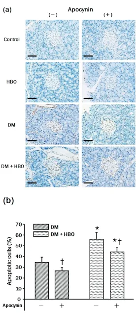
Immunodetection of apoptotic nuclei in pancreatic tissue
In the present study, apoptosis of islets of Langerhans in pancreas was de-tected by the TUNEL method and the nuclei were counted and indicated as a percentage of total nuclei (see Materi-als and Methods). As shown in Figure 3a and 3b, STZ treatment induced a higher percentage of apoptosis of islets of Langerhans in the DM and DM + HBO groups treated without apocynin (DM; 33.5 ± 6.5%, DM + HBO; 55.3 ± 9.2%). The percentage of apoptosis in the DM and DM + HBO groups treated with apocynin decreased compared with the corresponding groups untreated with apocynin (Figure 3b).
Expression of genes of NADPH oxidase complex (gp91phox, p22phox, p47phox)
[image:6.612.81.532.97.228.2]Gene expression of NADPH complex compo-nents studied is summarized in Figure 4. We measured the expression of NADPH oxidase complex, gp91phox, p22phox, and p47phox. In the Table 3. Antioxidant enzyme activities in experimental groups treated with or without apocynin.
Group Apocynin GSH Cu-Zn SOD Catalase Gpx
(-/+)
Control (-) 76 ± 8 13.8 ± 2 282 ± 18 0.6 ± 0.15
(+) 72 ± 5 13.9 ±3 290 ± 16 0.5 ± 0.07
HBO (-) 55 ± 7 * 6.3 ± 0.9 * 166 ± 9 * 0.7 ± 0.1
(+) 58 ± 6 * 11.5 ± 2.5 † 205 ± 22 *† 0.6 ± 0.14
DM (-) 67 ± 4 * 8.2 ± 1.2 * 233 ± 26 * 0.6 ± 0.14
(+) 70 ± 3 * 10.7 ± 2.9 266 ± 37 0.5 ± 0.1
DM + HBO (-) 49 ± 5 * 5.8 ± 0.9 * 94 ± 17 * 0.9 ± 0.12 *
(+) 60 ± 3 *† 7.4 ± 0.7 *† 134 ± 36 *† 0.8 ± 0.1 *
Values are expressed as mean ± SEM (n = 6). Non-diabetic rats in the non-HBO (control), non-diabetic rats in the HBO (HBO), diabetic rats in the non-HBO (DM), and diabetic rats in the HBO (DM + HBO) groups.Levels of glu-tathione (GSH) were measured and expressed as nmol/mg protein. The activities of superoxide dismutase (SOD), catalase, and glutathione peroxidase (Gpx) are expressed as U/mg of protein. * Represents significance at P < 0.05 compared with the control group untreated with apocynin. † Represents significance at P < 0.05 compared with the same group treated with or without apocynin.
Figure 2. NADPH oxidase activity (NADP+/NADPH ratios) in the
[image:6.612.80.336.334.447.2]DM and DM + HBO groups untreated with apocynin, gp91phox (also called Nox2) and p22phox were significantly up-regulated (gp91phox; 2.6-, and 2.4-fold, p22phox ; 2.5-, and 2.7-fold, re-spectively) compared with the control group (Figure 4a). In addition, there was no difference in the expression of gp91phox and p22phox between DM and DM + HBO rats treated with or without apocynin (data not shown). The expression of p47phox in the HBO, DM, and DM + HBO groups un-treated with apocynin was signifi-cantly up-regulated (3.3-, 2.0-, and 5.2-fold, respectively) compared with the control group (Figure 4a). More-over, the expression of p47phox in the HBO and DM + HBO groups treated with apocynin was down-regulated compared with the corresponding groups untreated with apocynin (Figure 4a).
Expression of genes of apoptosis (Bcl-2, Bax, and Caspase-3)
[image:7.612.80.356.74.707.2]As shown in Figure 4b, the expres-sion of Bcl-2 in the HBO, DM, and
DM + HBO groups untreated with apocynin was significantly down-regulated (0.8-, 0.43-, and 0.3-fold, respectively) compared with the control group. In addition, the expression of Bcl-2 in the HBO and DM + HBO groups treated with apo-cynin was up-regulated compared with the cor-responding groups untreated with apocynin (Figure 4b). Expression of Bax in the HBO, DM, and DM + HBO groups untreated with apocynin was significantly down-regulated (0.8-, 0.43-, and 0.3-fold, respectively) compared with the control group. Moreover, the expression of Bax in the HBO, DM, and DM + HBO groups treated with apocynin was down-regulated compared with the corresponding groups untreated with apocynin (Figure 4b). The expression of caspase -3 in the DM and DM + HBO groups untreated with apocynin was up-regulated (8.8-, and 7.8-fold, respectively) compared with the control group (Figure 4c). No difference in the mRNA expression of caspase-3 was found between DM and DM + HBO rats treated with or without apocynin (data not shown).
Caspase-3 activity in pancreatic tissue
Caspase-3 activity was significantly higher in the DM and DM + HBO groups untreated with apo-cynin compared with the control group (Figure
5). No difference in caspase-3 activity was found between DM and DM + HBO rats treated with or without apocynin (data not shown).
Discussion
In the present study, we showed that develop-ment of hyperglycemia and hypoinsulinemia was facilitated in diabetic rats receiving HBO treatment, and that activity of the NADPH com-plex was also significantly increased, suggesting the production of ROS and apoptosis-related caspase-3 activity. Moreover, we have also shown, for the first time, the dynamic panel of mRNA expression of genes related to ROS and apoptotic factors in diabetic rat pancreas ex-posed to HBO.
[image:8.612.83.530.86.284.2]Hyperglycemia subsequent to diabetes causes oxidative stress, mainly leading to enhanced production of mitochondrial ROS [1]. STZ has been proposed to act as a diabetogenic agent due to its ability to destroy pancreatic β-islet cells, possibly via the formation of excess free radicals [15, 31]. Furthermore, STZ-induced β -cell death is associated with oxidative stress caused by the production of excess intracellular ROS [13, 14]. Moreover, the oxidative stress caused by HBO exposure induces apoptosis via
Figure 4. Real-time PCR analysis of NADPH oxidase complex (a), apoptosis (b), and caspase-3 gene expression in the pancreas of diabetic rats in the HBO (control), diabetic rats in the HBO (HBO), diabetic rats in the non-HBO (DM), and diabetic rats in the non-HBO (DM + non-HBO) groups. Values are expressed as mean ± SEM (n = 6). * Repre-sents significance at P < 0.05 compared with the control group untreated with apocynin. † Represents significance at
P < 0.05 compared with the same group treated with or without apocynin. gp91phox, gp91phox protein; p22phox, p22phox
the mitochondria [16]. Thus, our demonstration of changes in activity and mRNA expression related to ROS and apoptosis may correlate with oxidative stress induced and enhanced by both diabetes induction and HBO exposure.
An important source of ROS production is NADPH oxidase [5, 6]. Increased activation of NADPH oxidase-dependent superoxide produc-tion has a role in hypertension, hypercholes-terolemia, diabetes and increased superoxide bioavailability [32-34]. However, to our knowl-edge, no study has been undertaken of the ef-fects of HBO on the activity and gene expression of the NADPH oxidase complex of pancreatic tissue in animals with or without diabetes. In the present study, we showed that the activity of NADPH oxidase in the HBO, DM, and DM + HBO groups was significantly increased, while such activity was significantly decreased in the HBO and DM + HBO groups treated with the NADPH oxidase inhibitor apocynin, compared to the corresponding activity in the absence of apo-cynin. These results suggest that even clinically used HBO may induce NADPH oxidase activity in pancreatic tissue of diabetic rats.
Activation of NADPH oxidase induces superox-ide production via processes such as oxidation in the mitochondria, inflammation, and stress [35]. NADPH oxidase is a multi-component en-zyme complex consisting of the subunits gp91phox and p22phox, as well as the cytosolic factors p47phox and p67phox, and the small
GTP-binding protein Rac-1 [19]. Recent re-ports have provided evidence for in-creased mRNA and/or protein expres-sion of these subunits in pancreatic is-lets of diabetic animals and human pa-tients [36, 37]. Apocynin, an NADPH oxidase inhibitor, prevents the translo-cation of p47phox to gp91phox, and im-pedes p47phox subunit assembly within the membrane complex in human and animal endothelial cells, thereby inhibit-ing the activity of NADPH oxidase and production of superoxide [38, 39]. In the present study, there was no difference in the expression of gp91phox and p22phox between HBO and DM + HBO groups treated with or without apocynin, whereas the expression of p47phox was significantly down-regulated. In addition, the activity of NADPH oxidase in HBO and DM + HBO groups treated with apo-cynin was significantly decreased compared with the corresponding groups without apo-cynin. These results suggest that apocynin may have inhibited the production of ROS by specifi-cally preventing the translocation of p47phox to gp91phox [39]. Furthermore, the expression of p47phox in the HBO group untreated with apocynin was significantly up-regulated compared to that in the DM group, suggesting that HBO exposure may have specifically promoted p47phox mRNA expression. Further analysis would be necessary of the detailed mechanism of translocation of p47phox to gp91phox under HBO exposure.
In the present study, activities of the antioxidant enzymes GSH, Cu-Zn SOD, catalase, and Gpx in the DM + HBO group untreated with apocynin were decreased compared with the other groups. On the other hand, in the DM + HBO group treated with apocynin, GSH, Cu-Zn SOD, and catalase activities were increased compared with activities in the same group untreated with apocynin. These results suggest that an increase in the activity of the antioxidant enzyme is attributable to apocynin, which decreases the expression of p47phox and activity of the NADPH oxidase.
The mechanisms by which hyperglycemia exerts its deleterious effects on β-cells include genera-tion of ROS; the latter cause cellular apoptosis via oxidation and have been implicated in the pathogenesis of diabetes mellitus [1].
[image:9.612.81.335.85.200.2]over, the oxidative stress caused by HBO expo-sure is linked to the mitochondrial pathway of apoptosis [16]. However, there is no study with regard to the effects of HBO on the activity and expression of genes related to apoptosis via mitochondria in the pancreas of diabetic ani-mals. In the present study, we demonstrated down-regulation of the expression of the anti-apoptotic factor Bcl-2, in contrast to up-regulation of mRNA levels of the apoptotic fac-tor, Bax in the DM + HBO group untreated with apocynin. In addition, we found that the decrease of apoptotic nuclei in islets of Langerhans in the DM + HBO group treated with apocynin was more than that in the same group untreated with apocynin. These results suggested that exposure of pancreas to apoptosis in diabetic animals is attributable to HBO. The present study, however, showed that there was no difference in caspase-3 activity and mRNA expression between the DM + HBO group treated with or without apocynin. This suggests that the production of ROS caused by HBO exposure may involve enhanced apoptosis in diabetic rat pancreas via some other route, such as the main death receptor Fas [16]. However, Weber et al reported that the death receptor Fas is down-regulated by HBO, suggesting that such down-regulation may be a protective action of the cell in response to stress [16]. Recently, the apoptotic signal of death receptors has been shown to be transduced to caspase-8 via FADD [40]; inhibition of caspase-8 by HBO blocks the signal to caspase-3 and inhibits execution of apoptosis [16]. Thus, these reports suggest that HBO has some other route by which it induces apoptosis of pancreatic tissue. Further studies are necessary to clarify the mechanism of apoptosis due to HBO exposure in diabetic animals.
An important pathogenetic mechanism of pan-creatic β-cell damage during experimental STZ-induced diabetic animals is increased expres-sion of pro-inflammatory cytokines and in-creased ROS production in pancreatic islets [13, 14, 41]. Recently, Manna et al reported that TNFα stimulated by ROS production, which was induced in pancreatic tissue of STZ-intoxicated animals, up-regulated the expression of phos-pho-extracellular signal-regulated kinase (ERK) 1/2 and phospho-p38; furthermore, activation of phosphorylated kinases was suggested to be a critical component in the oxidative stress-induced apoptotic process [15]. In the present study, we showed that the percentage of
apop-tosis of islets of Langerhans in the DM and DM + HBO groups treated with apocynin decreased compared with the corresponding groups untreated with apocynin; however, there was no difference in caspase-3 activity and mRNA expression between DM and DM + HBO rats treated with or without apocynin. Thus, apocynin may inhibit apoptosis via pancreatic ERK1/2 and p38 in STZ-induced diabetic animals due to HBO exposure; however, no further information is available to date on this. The present results have the potential to be applied to further studies of the mechanisms of apoptosis via ERK1/2 and p38.
In conclusion, this study demonstrates various gene expression dynamics of the NADPH oxi-dase complex and apoptosis in the pancreatic tissue of STZ-induced diabetic rats exposed to HBO. This study further suggests that oxidative stress caused by HBO exposure in diabetic ani-mals induces further ROS production and apop-tosis, potentially through the up-regulated ex-pression of genes related to NADPH oxidase complex, and down-regulated expression of anti-apoptotic factors in the mitochondria. Thus, the present study advances understanding of the molecular mechanisms of apoptosis in the dia-betic pancreas under HBO exposure; however, the role of other molecules involved in the apop-totic signal cascade such as Fas, ERK1/2 and p38 remains unclear. Further studies are also needed to address the detailed mechanisms operating in the apoptotic pathway under HBO exposure and diabetes induction. These obser-vations suggest that identification of a protec-tive mechanism against ROS production caused by HBO exposure may be beneficial in humans with essential diabetes mellitus. Furthermore, our study model could serve as a useful model for toxicological evaluation of the side effects of HBO treatment.
Acknowledgements
This study was partially supported by the Aca-demic Frontier Project “Surveillance and control for zoonoses” and Strategic Research Base De-velopment Program "International research on epidemiology of zoonoses and training for young researchers" from Ministry of Education, Cul-ture, Sports, Science and Technology, Japan.
and Fax: +81-466-84-3445, E-mail address: sato.yukita@nihon-u.ac.jp
References
[1] Brownlee M. Biochemistry and molecular cell biology of diabetic complications. Nature 2001; 414:813-820.
[2] Kowluru RA and Chan PS. Oxidative stress and diabetic retinopathy. Exp Diabetes Res 2007; 2007:43603.
[3] Hunt JV, Smith CC and Wolff SP. Autoxidative glycosylation and possible involvement of per-oxides and free radicals in LDL modification by glucose. Diabetes 1990;39:1420-1424. [4] Wolff SP, Jiang ZY and Hunt JV. Protein glycation
and oxidative stress in diabetes mellitus and ageing. Free Radic Biol Med 1991;10:339-352. [5] Opara EC. Oxidative stress, micronutrients,
diabetes mellitus and its complications. J R Soc Health 2002; 122:28-34.
[6] Asaba K, Tojo A, Onozato ML, Goto A, Quinn MT, Fujita T and Wilcox CS. Effects of NADPH oxi-dase inhibitor in diabetic nephropathy. Kidney Int 2005;67:1890-1898.
[7] Feldmeier JJ and Undersea and Hyperbaric Medical Society: Hyperbaric oxygen 2003: indi-cations and results: the Hyperbaric Oxygen Therapy Committee report. Edited by Feldmeier JJ. North Carolina, Durham, 2003, pp. 141 [8] Speit G, Dennog C, Radermacher P and
Roth-fuss A. Genotoxicity of hyperbaric oxygen. Mutat Res 2002; 512:111-119.
[9] Eken A, Aydin A, Sayal A, Ustündağ A, Duydu Y and Dündar K. The effects of hyperbaric oxygen treatment on oxidative stress and SCE frequen-cies in humans. Clin Biochem 2005; 38:1133-1137.
[10] Oter S, Korkmaz A, Topal T, Ozcan O, Sadir S, Ozler M, Ogur R and Bilgic H. Correlation be-tween hyperbaric oxygen exposure pressures and oxidative parameters in rat lung, brain, and erythrocytes. Clin Biochem 2005; 38:706-711. [11] Korkmaz A, Oter S, Sadir S, Topal T, Uysal B,
Ozler M, Ay H and Akin A. Exposure time related oxidative action of hyperbaric oxygen in rat brain. Neurochem Res 2008;33:160-166. [12] Matsunami T, Sato Y, Sato T, Ariga S,
Shimo-mura T and Yukawa M. Oxidative stress and gene expression of antioxidant enzymes in the streptozotocin-induced diabetic rats under hy-perbaric oxygen exposure. Int J Clin Exp Pathol 2010; 3:177-188.
[13] Peschke E, Ebelt H, Brömme HJ and Peschke D. 'Classical' and 'new' diabetogens--comparison of their effects on isolated rat pancreatic islets in vitro. Cell Mol Life Sci 2000; 57:158-164. [14] González E, Jawerbaum A, Sinner D, Pustovrh C,
White V, Capobianco E, Xaus C, Peralta C and Roselló-Catafau J. Streptozotocin-pancreatic damage in the rat: modulatory effect of 15-deoxy delta12,14-prostaglandin j(2) on
nitrider-gic and prostanoid pathway. Nitric Oxide 2002;6:214-220.
[15] Manna P, Sinha Ma and Sil PC. Protective role of arjunolic acid in response to streptozotocin-induced type-I diabetes via the mitochondrial dependent and independent pathways. Toxicol-ogy 2009;257:53-63.
[16] Weber SU, Koch A, Kankeleit J, Schewe JC, Siekmann U, Stüber F, Hoeft A and Schröder S. Hyperbaric oxygen induces apoptosis via a mi-tochondrial mechanism. Apoptosis 2009; 14:97 -107.
[17] Stolk J, Rossie W and Dijkman JH. Apocynin improves the efficacy of secretory leukocyte protease inhibitor in experimental emphysema. Am J Respir Crit Care Med 1994; 150:1628-1631.
[18] Gill PS and Wilcox CS. NADPH oxidases in the kidney. Antioxid Redox Signal 2006;8:1597-1607.
[19] Thallas-Bonke V, Thorpe SR, Coughlan MT, Fu-kami K, Yap FY, Sourris KC, Penfold SA, Bach LA, Cooper ME and Forbes JM. Inhibition of NADPH oxidase prevents advanced glycation end product-mediated damage in diabetic nephropathy through a protein kinase C-alpha-dependent pathway. Diabetes 2008;57:46-469. [20] Matkovics B, Kotorman M, Varga IS, Hai DQ and Varga C. Oxidative stress in experimental diabe-tes induced by streptozotocin. Acta Physiol Hung 1997; 85:29-38.
[21] Cotter MA and Cameron NE. Effect of the NAD (P) H oxidase inhibitor, apocynin, on peripheral nerve perfusion and function in diabetic rats. Life Sci 2003; 73:1813-1824.
[22] Meister A. Glutathione deficiency produced by inhibition of its synthesis, and its reversal; ap-plications in research and therapy. Pharmacol Ther 1991; 51:155-194.
[23] Sun Y, Oberley LW and Li Y. A simple method for clinical assay of superoxide dismutase. Clin Chem 1988; 34:497-500.
[24] Aebi H. Catalase in vitro. Methods Enzymol 1984; 105:121-126.
[25] Paglia DE and Valentine WN. Studies on the quantitative and qualitative characterization of erythrocyte glutathione peroxidase. J Lab Clin Med 1967; 70:158-169.
[26] Ohkawa H, Ohishi N and Yagi K. Assay for lipid peroxides in animal tissues by thiobarbituric acid reaction. Anal Biochem 1979; 95:351-358.
[27] Vilchèze C, Weisbrod TR, Chen B, Kremer L, Hazbón MH, Wang F, Alland D, Sacchettini JC and Jacobs WR Jr. Altered NADH/NAD+ ratio mediates coresistance to isoniazid and ethiona-mide in mycobacteria. Antimicrob Agents Chemother 2005;49:708-720.
2000;46:69-81.
[29] Pfaffl MW. A new mathematical model for rela-tive quantification in real-time RT-PCR. Nucleic Acids Res 2001; 29:e45.
[30] Jafari Anarkooli I, Sankian M, Ahmadpour S, Varasteh AR and Haghir H. Evaluation of Bcl-2 family gene expression and Caspase-3 activity in hippocampus STZ-induced diabetic rats. Exp Diabetes Res 2008; 2008:638467.
[31] Szkudelski T. The mechanism of alloxan and streptozotocin action in β cells of the rat pan-creas. Physiol Res 2001; 50:537-546.
[32] Cai H and Harrison DG. Endothelial dysfunction in cardiovascular diseases: the role of oxidant stress. Circ Res 2000; 87:840-844.
[33] Hink U, Li H, Mollnau H, Oelze M, Matheis E, Hartmann M, Skatchkov M, Thaiss F, Stahl RA, Warnholtz A, Meinertz T, Griendling K, Harrison DG, Forstermann U and Munzel T. Mechanisms underlying endothelial dysfunction in diabetes mellitus. Circ Res 2001; 88:E14-22.
[34] Li JM and Shah AM. Endothelial cell superoxide generation: regulation and relevance for cardio-vascular pathophysiology. Am J Physiol Regul Integr Comp Physiol 2004; 287:R1014-1030. [35] Hancock JT, Desikan R and Neill SJ. Role of
reactive oxygen species in cell signalling path-ways. Biochem Soc Trans 2001; 29:345-350. [36] Lupi R, Del Guerra S, Bugliani M, Boggi U,
Mo-sca F, Torri S, Del Prato S and Marchetti P. The direct effects of the angiotensin-converting enzyme inhibitors, zofenoprilat and enalaprilat, on isolated human pancreatic islets. Eur J En-docrinol 2006; 154:355-361.
[37] Uchizono Y, Takeya R, Iwase M, Sasaki N, Oku M, Imoto H, Iida M and Sumimoto H. Expression of isoforms of NADPH oxidase components in rat pancreatic islets. Life Sci 2006; 80:133-139.
[38] Johnson DK, Schillinger KJ, Kwait DM, Hughes CV, McNamara EJ, Ishmael F, O'Donnell RW, Chang MM, Hogg MG, Dordick JS, Santhanam L, Ziegler LM and Holland JA. Inhibition of NADPH oxidase activation in endothelial cells by ortho-methoxy-substituted catechols. Endo-thelium 2002; 9:191-203.
[39] Stefanska J and Pawliczak R. Apocynin: molecu-lar aptitudes. Mediators Inflamm 2008; 2008:106507.
[40] Krammer PH. CD95's deadly mission in the immune system. Nature 2002;407:789-795. [41] Haluzík M and Nedvídková J. The role of nitric