Introduction
Approximately 40,000 new cases of pancreatic ductal adenocarcinoma were diagnosed every year [1]. The median overall survival is only 18 months even for the 15-20% patients with re-sectable tumor at initial presentation [2, 3]. Although gemcitabine-based chemotherapy and chemoradiation therapy are used widely as an adjuvant treatment for resectable pancreatic adenocarcinoma [4–6], many patients show poor response. Once local recurrence or metas-tasis occurs, the options for these patients are often limited to palliative or supportive care [2, 3].
Efforts to address gemcitabine resistance mechanism of pancreatic adenocarcinoma have been focusing on the role of key players in-volved in the transport and metabolism of
gem-citabine [7]. Among these proteins such as de-oxycytidine kinase, human equilibrative nucleo-side transporter 1 (hENT1), and ribonucleotide reductase M1 (RRM1), ribonucleotide reductase small subunit M2 (RRM2), the catalytic subunit of ribonucleotide reductase [8], is also associ-ated with tumor progression and resistance to gemcitabine [9]. Preclinical studies in pancre-atic cancer cell lines demonstrated the relation-ship between gemcitabine resistance and RRM2 overexpression [10]. The transfection of siRNA to RRM2 decreased pancreatic cancer cell invasiveness as well as gemcitabine resis-tance [11], suggesting that RRM2 is a potential therapeutic target in treating pancreatic adeno-carcinoma.
From the results of these in vitro studies, RRM2 seemed to have features that are independent of RRM1, which has been demonstrated as a
Original Article
Ribonucleotide reductase M2 does not predict survival in
patients with resectable pancreatic adenocarcinoma
Hao Xie1, Jingmei Lin3, Dafydd G Thomas4, Wei Jiang1, Xiuli Liu1
1Anatomic Pathology, Cleveland Clinic, Cleveland, OH; 2Cleveland Clinic Lerner College of Medicine of Case Western
Reserve University, Cleveland, OH; 3Department of Pathology and Laboratory Medicine, Indiana University School of
Medicine, Indianapolis, IN; 4Departments of Pathology, University of Michigan Health System, Ann Arbor, MI, USA.
Received March 28, 2012; accepted April 13, 2012; Epub April 16, 2012; Published May 30, 2012
Abstract: Background: Ribonucleotide reductase M2 (RRM2) was associated with pancreatic tumor progression and resistance to gemcitabine. This study aimed to determine if RRM2 protein expression was prognostic in patients with resectable pancreatic adenocarcinoma and predictive of adjuvant gemcitabine benefit. Methods: 117 patients under-went tumor resection for pancreatic adenocarcinoma from 10/1999 to 12/2007. We constructed tissue microarrays from paraffin-embedded tumors and determined RRM2 protein expression using immunohistochemistry and grouped as negative or positive. We estimated overall survival (OS) and progression-free survival (PFS) using the Kaplan-Meier method and examined the prognostic and predictive value of RRM2 expression using Cox proportional hazards model. Results: RRM2 expression showed no prognostic value in the entire group regarding OS (median OS 30.9 months in RRM2-positive versus 13.7 months in RRM2-negative, P = 0.26) and PFS (median OS 20.6 months in RRM2-positive versus 11.8 months in RRM2-negative, P = 0.46). RRM2 expression did not predict adjuvant gemcit-abine benefit in the subgroup of 44 patients who received gemcitgemcit-abine therapy (median OS 31.2 versus 15.2 months, P = 0.62; median PFS 11.3 versus 14.0 months, P = 0.35). Cox proportional hazards regression showed no prognostic effect of RRM2 expression on OS and PFS in the subgroup of 44 patients. However, the number of positive lymph nodes and perineural invasion were prognostic factors for OS (HR 1.2, P = 0.005) and for PFS (HR 5.5, P = 0.007), respectively. Conclusion: RRM2 protein expression in pancreatic adenocarcinoma is neither prognostic nor predictive of adjuvant gemcitabine benefit in patients with resectable pancreatic adenocarcinoma.
predictive factor for adjuvant gemcitabine bene-fit to overall survival in pancreatic adenocarci-noma [12]. However, the clinical evidence pre-sented so far were contradictory. Giovannetti and coworkers [13] studied 102 patients with pancreatic adenocarcinoma and 67 of them treated with adjuvant gemcitabine. The results showed neither prognostic nor predictive value of RRM2 mRNA level for survival. However, a recent study by Fujita and coworkers [14] on 40 patients treated with adjuvant gemcitabine showed that low mRNA expression of RRM2 was predictive of treatment benefit of gemcitabine in patients with resected pancreatic adenocarci-noma. Furthermore, RRM2 expression was also prognostic for survival in a univariate analysis of the entire cohort of 70 patients.
To elucidate the role of RRM2, we evaluated 117 patients with resectable pancreatic adeno-carcinoma. We aimed to determine whether RRM2 protein expression level assessed by im-munohistochemistry is prognostic in patients with resectable pancreatic adenocarcinoma or is predictive of adjuvant gemcitabine benefit.
Materials and methods
We retrospectively reviewed the medical records of 117 patients who underwent surgical resec-tion for pancreatic adenocarcinoma at the Cleveland Clinic from October 1999 to Decem-ber 2007. We included the patients with the diagnosis of pancreatic ductal adenocarcinoma and pathologic stage of T(1-3)N(0,1,x)M0. We excluded the patients with unresectable or me-tastatic disease, R2 resection, ampullary carci-noma, and indolent pancreatic tumors such as mucinous cystadenoma, mucinous cystadeno-carcinoma, and islet cell tumor. This study was approved by the institutional review board at the Cleveland Clinic.
All 117 pancreatic adenocarcinoma tissue sam-ples were either formalin-fixed (103 patients, 88.0%) or Hollande’s-fixed (14 patients, 12.0%), and paraffin-embedded. We centrally reviewed the tumor-containing hematoxylin and eosin stained slides. Tissue microarrays of tumors (duplicate, 2 mm core from the area with the highest density of tumor cells) were constructed and used for the determination of RRM2 pro-tein. Immunohistochemistry staining was per-formed on the DAKO Autostainer (DAKO, Carpin-teria, CA) using DAKO LSAB+ and
diaminoben-zadine (DAB) as the chromogen. De-paraffinized sections at five-micron thickness were labeled with RRM2 antibody (Goat polyclonal antibody, SC-10846, 1:1000, Santa Cruz Biotechnology, Santa Cruz, CA). Microwave citric acid epitope retrieval was employed. Appropriate negative (no primary antibody) were stained in parallel with each set of tumors studied. RRM2 immuno-reactivity was assessed for the percentage of nuclear immunoreactivity in tumor cells. Results were grouped into the following categories: no nuclear staining, negative; with nuclear staining, positive.
Statistical analysis was primarily descriptive in nature in this retrospective study. Thus, we did neither sample size calculation nor power esti-mation. Categorical data were summarized as frequency counts and percentages. Continuous measures were summarized as means, stan-dard deviations (s.d.), medians, and ranges. We compared continuous data with student t-test and categorical data with Fisher’s exact test. OS was measured from the time of tumor resection to death. PFS was measured from the time of tumor resection to disease progression or death. Time-to-event data was plotted using the Kaplan-Meier method with surviving patients censored at the date of last follow-up. The dif-ferent groups of patients were evaluated using the log-rank tests. We used Cox proportional hazards model for multivariable analyses of potential prognostic indicators in a stepwise fashion. The criterion for entry variables was P < 0.1. The retention criterion was P < 0.05. Haz-ard ratio (HR), 95% confidence interval (CI) for the HR, and corresponding P-value were used to present Cox proportional hazards regression results. All the statistical analyses were per-formed using SAS 9.2 (SAS Institute, Cary, NC).
Results
Prognostic value of RRM2 in the entire cohort of patients with resectable pancreatic adenocarci-noma
(82.1%), distal pancreatectomy in 19 patients (16.2%), subtotal pancreatectomy in 1 patient (0.9%), and uncinate excision in 1 patient (0.9%). 36 patients (30.8%) had a positive surgi-cal margin. The mean tumor size was 3.4 cm
(s.d. 1.4 cm). As to pathologic stage, the tumor of 11 patients (9.4%) was T1, of 50 patients (42.7%) was T2, of 56 patients (47.9%) was T3, of 47 patients (40.1%) was N0, and of 69 (59.0%) patients was N1. The tumors were well differentiated in 5 patients (4.3%), moderately differentiated in 67 patients (57.3%), and poorly differentiated in 45 patients (38.5%). The mean number of positive lymph nodes was 2.2 (s.d. 3.3). Lymphovascular invasion was present in the tumor of 56 patients (47.9%). Perineural invasion was present in the tumor of 75 pa-tients (64.1%). 24 papa-tients (20.5%) did not un-dergo any form of adjuvant treatment. 59 pa-tients (50.4%) underwent adjuvant chemoradia-tion therapy. 16 patients (13.7%) had adjuvant chemotherapy only. The treatment history of 15 patients (12.8%) was unknown. 44 patients (37.6%) received adjuvant gemcitabine treat-ment or gemcitabine-based regimens. The me-dian follow-up time was 13.4 months (range: 0.2-105.6). At the time of the last follow-up, 19 patients (16.2%) were still alive, whereas 98 (83.8%) patients were dead.
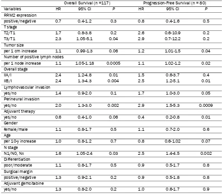
For OS analysis, 97 patients were grouped into RRM2 negative expression group, 20 patients into RRM2 positive expression group. For PFS analysis, 67 patients were grouped into RRM2 negative expression group, 13 patients into RRM2 positive expression group. These were carried on to univariate analysis of prognostic factors for survival. The variables included RRM2 protein expression, T stage, tumor size, number of positive lymph nodes, overall stage, lymphovascular invasion, perineural invasion, adjuvant therapy, gender, age, N stage, tumor differentiation, status of surgical margins, and adjuvant gemcitabine. As shown in Table 2, number of positive lymph nodes (HR 1.1 per 1 more, P = 0.0005), overall stage IIA/I (HR 2.4, P
= 0.01), IIB/I (HR 2.4, P = 0.004), perineural invasion (HR 2.0, P = 0.002), N stage (HR 1.6, P
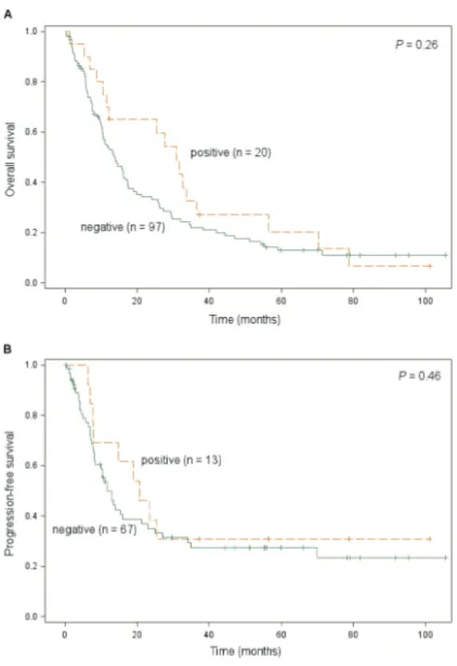
[image:3.612.78.292.99.632.2]= 0.03) were associated with decreased OS. Similarly, number of positive lymph nodes (HR 1.1 per one more, P = 0.02), tumor size (HR 1.2, per 1 cm increase, P = 0.04), perineural invasion (HR 2.9, P = 0.0009), and N stage (HR 2.5, P = 0.002) were associated with decreased PFS. RRM2 expression and adjuvant gemcit-abine were not prognostic factors for either OS or PFS. As depicted in Figure 1, patients with negative RRM2 expression in tumors did not have significantly longer OS than patients with positive RRM2 expression in tumors (median OS Table 1. Patient Demographics, Clinical
Char-acteristics, and Histopathologic Features (n = 117)
Variables Number of Patients (%) Gender
male/female 68 (58.1)/ 49 (41.9) Age
median [range] 65 [35-93] Surgical Procedure
Whipple 96 (82.1)
Distal pancreatec-tomy
19 (16.2)
Subtotal
pancreatec-tomy 1 (0.9)
Uncinate excision 1 (0.9) Tumor size, cm
mean ± s.d. 3.4 ± 1.4 median [range] 3.2 [0.1-8] Pathologic stage
T1/T2/T3 11 (9.4)/ 50 (42.7)/ 56 (47.9)
N0/N1/Nx 47 (40.1)/ 69 (59.0)/ 1 (0.9)
M0 117 (100)
Overall stage
IA/IB/IIA/IIB 5 (4.3)/ 17(14.7)/ 24 (20.7)/ 71 (60.7) Differentiation
well/moderate/poor 5 (4.3)/ 67 (57.3)/ 45 (38.5)
Number of positive lymph nodes mean ± s.d. 2.2 ± 3.3 median [range] 1 [0-21] Lymphovascular invasion
Yes/No 56 (47.9)/ 61 (52.1) Perineural invasion
Yes/No 75 (64.1)/ 42 (35.9) Surgical margins
negative/positive 81 (69.2)/ 36 (30.8) Adjuvant therapy
Chemoradiation 59 (50.4) Chemotherapy only 16 (13.7)
None 24 (20.5)
Unknown 15 (12.8)
Adjuvant gemcitabine
yes/no/unknown 44 (37.6)/ 55 (47.0)/ 18 (15.4)
Status at follow-up
13.7 months versus 30.9 months, P = 0.26). The same conclusion stayed true for PFS (median PFS 11.8 months versus 20.6 months,
P = 0.46).
Multivariable analyses identified prognostic fac-tors for survival based on the pre-defined entry and retention criteria (Table 3). The presence of perineural invasion was associated with both decreased OS (HR 1.9, P = 0.009) and de-creased PFS (HR 2.9, P = 0.0009). In addition, number of positive lymph nodes (HR 1.1 per 1 more, P = 0.0003) was associated with de-creased OS. The presence of adjuvant therapy was associated with increased OS (HR 0.6, P = 0.02) and increased PFS (HR 0.4, P = 0.01). RRM2 expression was not associated with
ei-ther OS or PFS.
Prognostic value of RRM2 in the subgroup of resectable pancreatic adenocarcinoma patients treated with adjuvant gemcitabine
Similar analyses for a subpopulation of 44 pa-tients who received adjuvant gemcitabine treat-ment were also performed. Univariate analysis revealed that number of positive lymph nodes (HR 1.2 per 1 more, P = 0.005) was associated with decreased OS. Only perineural invasion (HR 5.52, P = 0.007) was associated with de-creased PFS. RRM2 expression did not predict the treatment benefit of adjuvant gemcitabine for either OS or PFS (Table 4). As depicted in Figure 2, patients with negative RRM2 expres-Table 2. Univariate Analysis for Overall Survival and Progression-Free Survival in the Entire Cohort
Overall Survival (n =117) Progression-Free Survival (n = 80)
Variables HR 95% CI P HR 95% CI P
RRM2 expression
positive/negative 0.7 0.4-1.2 0.3 0.8 0.4-1.6 0.5
T stage
T2/T1 1.7 0.8-3.8 0.2 2.6 0.6-10.9 0.2
T3/T1 2.3 1.05-5.1 0.04 2.9 0.7-12.2 0.2
Tumor size
per 1 cm increase 1.1 0.99-1.3 0.06 1.2 1.01-1.5 0.04 Number of positive lymph nodes
per 1 node increase 1.1 1.05-1.18 0.0005 1.1 1.02-1.2 0.02 Overall stage
IIA/I 2.4 1.2-4.8 0.01 1.5 0.6-3.7 0.4
IIB/I 2.4 1.3-4.3 0.004 2.5 1.2-5.1 0.01
Lymphovascular invasion
yes/no 1.4 0.9-2.0 0.1 1.7 1.0-3.0 0.05
Perineural invasion
yes/no 2.0 1.3-3.0 0.002 2.9 1.5-5.3 0.0009
Adjuvant therapy
yes/no 0.6 0.4-1.0 0.06 0.4 0.2-0.8 0.01
Gender
female/male 1.1 0.8-1.7 0.5 1.1 0.7-2.0 0.6
Age
per 10-y increase 1.0 0.8-1.2 0.7 0.8 0.6-1.02 0.07 N stage
N1/N0, Nx 1.6 1.05-2.4 0.03 2.5 1.4-4.5 0.002
Differentiation
poor/moderate 1.1 0.8-1.7 0.5 0.9 0.5-1.7 0.8
Surgical margin
positive/negative 1.3 0.9-2.1 0.2 0.9 0.5-1.8 0.8
Adjuvant gemcitabine
[image:4.612.78.532.94.492.2]sion in tumors did not have significantly longer OS than patients with positive RRM2 expression (median OS 15.2 months versus 31.2 months,
P = 0.62). The same conclusion stayed true for PFS (median PFS 14.0 months versus 11.3 months, P = 0.35).
The results from multivariable analyses shown in Table 5 were consistent with the aforemen-tioned findings. Number of positive lymph nodes and perineural invasion were predictors of shorter OS (HR 1.2 per 1 more, P = 0.005) and shorter PFS (HR 5.5, P = 0.007), respectively. RRM2 expression was not associated with ei-ther OS or PFS.
Discussion
In this retrospective study, we reviewed the
medical records of patients from a single terti-ary medical center. It turned out that the me-dian age, gender, treatment, and the survival of these patients were not statistically different from those in the previous large studies of pa-tients with resectable pancreatic adenocarci-noma [13, 14]. The results from univariate analyses demonstrate common histopathologi-cal prognostic factors for resectable pancreatic adenocarcinoma [15, 16], including tumor size, stage, number of positive lymph nodes, the presence of lymphovascular invasion or per-ineural invasion, and the absence of adjuvant therapy.
[image:5.612.80.291.83.390.2]In addition to the traditional morphological markers, molecular markers associated with the prognosis have also been extensively studied; especially those involved in the transport and Figure 1. Overall survival and progression-free
[image:5.612.324.532.84.381.2]sur-vival by RRM2 expression in the entire cohort (A) Overall survival by RRM2 expression (RRM2-negative: median OS 13.7 months; RRM2-positive: median OS 30.9 months). (B) Progression-free survival by RRM2 expression (RRM2-negative: median PFS 11.8 months; RRM2-positive: median PFS 20.6 months).
metabolism of gemcitabine [7], the mainstay chemotherapeutic agent of pancreatic adeno-carcinoma [5]. RRM1, the ribonucleotide reduc-tase large subunit for substrate and allosteric regulator bindings [8] has been established as the prognostic and predictive factor of survival in patients with various types of cancers includ-ing pancreatic adenocarcinoma treated with gemcitabine [12]. RRM2, the ribonucleotide
reductase small subunit, was believed to be biochemically unrelated to the enzyme overall activity and substrate specificity as a protein radical provider [8]. However, in preclinical stud-ies, it was associated with tumorigenesis, inva-siveness and gemcitabine resistance [10, 11].
Currently, contradictory evidence of prognostic value and predictive value of RRM2 exists. Gio-Table 3. Multivariable Analysis for Overall Survival and Progression-free Survival in the Entire Cohort
OS PFS
Variables HR 95% CI P HR 95% CI P
Number of positive lymph nodes
per 1 node increase 1.1 1.06-1.2 0.0003 NA NA NA
Perineural invasion
yes/no 1.9 1.2-3.2 0.009 2.9 1.5-5.4 0.0009
Adjuvant therapy
yes/no 0.6 0.3-0.9 0.02 0.4 0.2-0.8 0.01
[image:6.612.79.530.97.202.2]NA: not applicable
Table 4. Univariate Analysis for Overall Survival and Progression-Free Survival in Patients Treated with Adjuvant Gemcitabine
Overall Survival (n =44) Progression-Free Survival (n = 33)
Variables HR 95% CI P HR 95% CI P
RRM2 expression
positive/negative 0.8 0.4-1.8 0.6 1.5 0.6-3.8 0.4
T stage
T2/T1 2.7 0.8-9.5 0.1 1.1 0.2-5.0 0.9
T3/T1 3.6 1.04-12.3 0.04 1.1 0.2-4.8 0.9
Tumor size
per 1 cm increase 1.3 0.9-1.7 0.1 0.9 0.6-1.4 0.6
Number of positive lymph nodes
per 1 node increase 1.2 1.1-1.4 0.005 1.1 0.9-1.3 0.3 Overall stage
IIA/I 1.7 0.6-4.8 0.3 0.6 0.2-2.3 0.5
IIB/I 2.0 0.8-5.1 0.1 1.6 0.6-4.5 0.4
Lymphovascular invasion
yes/no 0.8 0.4-1.5 0.5 1.4 0.6-3.4 0.4
Perineural invasion
yes/no 1.6 0.8-3.2 0.2 5.5 1.6-18.9 0.007
Gender
female/male 1.1 0.6-2.1 0.7 0.7 0.3-1.8 0.5
Age
per 10-y increase 0.96 0.7-1.4 0.8 0.6 0.4-1.01 0.05 N stage
N1/N0, Nx 1.5 0.8-2.8 0.2 2.1 0.9-4.8 0.1
Differentiation
poor/moderate 1.7 0.8-3.5 0.2 1.5 0.5-4.3 0.4
Surgical margin
[image:6.612.79.533.254.591.2]vannetti and coworkers [13] stratified 67 pa-tients with pancreatic adenocarcinoma treated with gemcitabine into two or three equally di-vided groups based on RRM2 mRNA level. No significant correlations between RRM2 expres-sion and survival were found. However, this con-clusion was challenged by a Japanese study of 31 patients. Itoi and coworkers [17] reported that RRM2 mRNA level of fine needle aspiration biopsy specimen was a key predictive marker of survival and response in gemcitabine-treated patients. This was further supported by the Fu-jita study also from Japan [14], where 70 pa-tients with early stage pancreatic adenocarci-noma were included and 40 patients were treated with gemcitabine. Recursive partitioning analysis was used to stratify patients based on the RRM2 mRNA level in their tumors. De-creased RRM2 mRNA level was associated with increased OS and PFS in both the entire cohort and the subpopulation who received gemcit-abine.
The disease stage of the patients in our study was similar to those in the Fujita study [14]. However, RRM2 protein expression determined by immunohistochemistry appeared to be nei-ther prognostic nor predictive in both the entire cohort of 117 patients with resectable pancre-atic adenocarcinoma and 44 patients subse-quently treated with adjuvant gemcitabine after tumor resection. Our results were more consis-tent with the conclusions of the Giovannetti study [13].
[image:7.612.79.531.108.179.2]Several possible explanations were proposed for the apparent disagreements. First, different quantification methods were used to examine RRM2 expression. All the previous studies used QRT-PCR for RRM2 mRNA quantification with only one reference gene either β-actin or glyceraldehyde-3-phosphate dehydrogenase [13, 14, 17]. However, further analyses with Norm-Finder (v19) and geNorm (v3.5) packages
demonstrated that β-actin was not as stable as the two reference genes (RPL13A and HMBS) we used in pancreatic cancer (unpublished data). In addition, compared to immunohisto-chemistry, RNA-based quantification methods often suffer from the availability reduction of target mRNA to the cDNA probe due to cross-linking proteins and RNA caused by the fixation process. Thereby we chose to measure RRM2 protein expression with pancreatic-specific tis-sue array and immunohistochemistry using an anti-RRM2 antibody, which is known to work reliably on both Hollande’s and formalin-fixed paraffin-embedded specimens. Second, these studies involved patients with different stages of pancreatic adenocarcinoma. A significant proportion of patients in the Giovannetti [13] and the Itoi studies [17] contained unresectable disease. Most of the patients studied in the Fu-jita study [14] had stage II disease although patients with stage III and IV disease were also included. As demonstrated in clinical practice, patients with resectable or unresectable pan-creatic adenocarcinoma have drastically distinct response to therapy as well as prognosis [2]. Thereby, it is reasonable to suspect different RRM2 protein expression and its roles in the different stages of pancreatic adenocarcinoma. In this study, we exclusively evaluated patients with stage I and II disease that were all re-sectable to eliminate this possible issue that was probably encountered in previously re-ported studies. Third, different methods were used to choose the cut-off value for RRM2 stratification [13, 14]. The cut-off values from methods such as RPA and dichotomization based on median are dependent on the patient population included in a specific study. The di-chotomization method based on the presence or absence of chemoluminescence in our study was independent to patient population and can be readily translated into clinical practice. Last, genotypic variations between Japanese popula-tion [14, 17] and Caucasian patients in the Gio-Table 5. Multivariable Analysis for Overall Survival and Progression-free Survival in Patients Treated with Adjuvant Gemcitabine
OS PFS
Variables HR 95% CI P HR 95% CI P
Number of positive lymph nodes
per 1 node increase 1.2 1.1-1.4 0.005 NA NA NA
Perineural invasion
yes/no NA NA NA 5.5 1.6-18.9 0.007
vannetti [13] and our studies may also need to be considered.
Our study has several limitations. First, as a retrospective study, complete demographic and histopathological data, therapeutic details, and even outcome data were not available for all patients. This information was collected in a time span of 8 years. Thus it is unlikely to com-pletely eliminate all potential selection bias and confounders. Second, interobserver variability is an intrinsic problem associated with the inter-pretation of the signal intensity in immunostain-ing. In the current study, central review process and dichotomization simply based on the pres-ence or abspres-ence of RRM2 protein expression greatly reduced the interobserver discrepancy to a lesser extent.
This study demonstrated that RRM2 protein expression level determined by immunohisto-chemistry on paraffin-embedded pancreatic adenocarcinoma tissue is not prognostic of sur-vival in the entire cohort of patients with re-sectable pancreatic adenocarcinoma. In addi-tion, RRM2 protein expression level does not predict the treatment benefit of adjuvant gem-citabine in patients with resectable pancreatic adenocarcinoma.
Address correspondence to: Dr. Xiuli Liu, Department of Anatomic Pathology, Cleveland Clinic, 9500 Euclid Avenue/L25, Cleveland, Ohio 44195 Tel: 216-445-8745; Fax: 216-445-6967; E-mail: liux3@ccf.org
References
[1] Jemal A, Siegel R, Ward E, Hao Y, Xu J, Thun MJ. Cancer statistics, 2009. CA Cancer J Clin 2009; 59: 225-249.
[2] Hidalgo M. Pancreatic cancer. N Engl J Med 2010; 362: 1605-1617.
[3] Vincent A, Herman J, Schulick R, Hruban RH, Goggins M. Pancreatic cancer. Lancet 2011; 378: 607-620.
[4] Neoptolemos JP, Stocken DD, Friess H, Bassi C, Dunn J a, Hickey H, Beger H, Fernandez-Cruz L, Dervenis C, Lacaine F, Falconi M, Pederzoli P, Pap A, Spooner D, Kerr DJ, Büchler MW. A ran-domized trial of chemoradiotherapy and che-motherapy after resection of pancreatic cancer. N Engl J Med 2004; 350: 1200-1210.
[5] Oettle H, Post S, Neuhaus P, Gellert K, Langrehr J, Ridwelski K, Schramm H, Fahlke J, Zuelke C, Burkart C, Gutberlet K, Kettner E, Schmalen-berg H, Weigang-Koehler K, Bechstein W-O, Niedergethmann M, Schmidt-Wolf I, Roll L, Do-erken B, Riess H. Adjuvant chemotherapy with
gemcitabine vs observation in patients under-going curative-intent resection of pancreatic cancer: a randomized controlled trial. JAMA 2007; 297: 267-277.
[6] Regine WF, Winter KA, Abrams RA, Safran H, Hoffman JP, Konski A, Benson AB, Macdonald JS, Kudrimoti MR, Fromm ML, Haddock MG, Schaefer P, Willett CG, Rich TA. Fluorouracil vs gemcitabine chemotherapy before and after fluorouracil-based chemoradiation following resection of pancreatic adenocarcinoma: a randomized controlled trial. JAMA 2008; 299: 1019-1026.
[7] Okazaki T, Javle M, Tanaka M, Abbruzzese JL, Li D. Single nucleotide polymorphisms of gemcit-abine metabolic genes and pancreatic cancer survival and drug toxicity. Clin Cancer Res 2010; 16: 320-329.
[8] Reichard P. From RNA to DNA, why so many ribonucleotide reductases? Science 1993; 260: 1773-1777.
[9] Jordheim LP, Sève P, Trédan O, Dumontet C. The ribonucleotide reductase large subunit (RRM1) as a predictive factor in patients with cancer. Lancet Oncol 2011; 12: 693-702. [10] Zhou B, Mo X, Liu X, Qiu W, Yen Y. Human
ribo-nucleotide reductase M2 subunit gene amplifi-cation and transcriptional regulation in a homo-geneous staining chromosome region responsi-ble for the mechanism of drug resistance. Cyto-genet Cell Genet 2001; 95: 34-42.
[11] Duxbury MS, Ito H, Zinner MJ, Ashley SW, Whang EE. RNA interference targeting the M2 subunit of ribonucleotide reductase enhances pancreatic adenocarcinoma chemosensitivity to gemcitabine. Oncogene 2004; 23: 1539-1548. [12] Akita H, Zheng Z, Takeda Y, Kim C, Kittaka N,
Kobayashi S, Marubashi S, Takemasa I, Na-gano H, Dono K, Nakamori S, Monden M, Mori M, Doki Y, Bepler G. Significance of RRM1 and ERCC1 expression in resectable pancreatic adenocarcinoma. Oncogene 2009; 28: 2903-2909.
[13] Giovannetti E, Del Tacca M, Mey V, Funel N, Nannizzi S, Ricci S, Orlandini C, Boggi U, Cam-pani D, Del Chiaro M, Iannopollo M, Bevilacqua G, Mosca F, Danesi R. Transcription analysis of human equilibrative nucleoside transporter-1 predicts survival in pancreas cancer patients treated with gemcitabine. Cancer Res 2006; 66: 3928-3935.
[14] Fujita H, Ohuchida K, Mizumoto K, Itaba S, Ito T, Nakata K, Yu J, Kayashima T, Souzaki R, Tajiri T, Manabe T, Ohtsuka T, Tanaka M. Gene expression levels as predictive markers of out-come in pancreatic cancer after gemcitabine-based adjuvant chemotherapy. Neoplasia 2010; 12: 807-817.
outcomes, and prognostic indicators. J Gastro-intest Surg 2000; 4: 567-579.
[16] Brennan MF, Kattan MW, Klimstra D, Conlon K. Prognostic nomogram for patients undergoing resection for adenocarcinoma of the pancreas. Ann Surg 2004; 240: 293-298.


![PE Structure [Deok9] pdf](data:image/gif;base64,R0lGODlhAQABAIAAAP///wAAACH5BAEAAAAALAAAAAABAAEAAAICRAEAOw==)