Original Article
Complement membrane attack complex is related with
immune-mediated necrotizing myopathy
Lu Cong, Chuan-Qiang Pu, Qiang Shi, Qian Wang, Xiang-Hui Lu
Department of Neurology, The Chinese PLA General Hospital, Beijing, China
Received May 8, 2014; Accepted June 25, 2014; Epub June 15, 2014; Published July 1, 2014
Abstract: This study is to investigate the expression of complement membrane attack complex (C5b-9) in the skel-etal muscle of patients with necrotizing myopathy (NM), and to investigate the relationship between C5b-9 and NM. Thirteen patients with NM and control patients with polymyositis and muscular dystrophy were enrolled in this study. Examinations including creatine kinase (CK) and L-lactate dehydrogenase (LDH) in the serum, electromyogram and muscle pathological examination were performed. C5b-9 expression in the skeletal muscle was determined by im-munohistochemistry and analyzed by Image Plus Pro 6.0. C5b-9 expression was particularly prominent in necrotic
muscle fibers, and also positive in blood vessels. C5b-9 diffusely expressed in vascular endothelial cells and smooth
muscle layer. But the intensity was not related with the elevated level of serum CK. So, C5b-9 is strongly expressed
in the necrotic muscle fiber and blood vessels, and may contribute to the pathogenesis of NM.
Keywords: Necrotizing myopathy, complement membrane attack complex, C5b-9, muscle fiber, endothelial cell
Introduction
Necrotizing myopathy (NM) is defined as the necrosis of skeletal muscle fiber caused by immunological mediation, malignant tumor, virus infection, statins [1] or connective tissue diseases. It usually consists of immune-medi-ated and secondary NM. The pathology fea-tures include diffused or focal muscle fiber necrosis, inflammatory cells infiltration, signifi -cant thinning of vascular and remnant wall. Most of the patients presented with progres-sive limb inability, muscular atrophy, and high creatine kinase (CK) in the serum. The severity of the disease was various. Respiratory muscle could be involved, which could lead to death. The pathogenesis of NM has not yet been clari-fied. NM is believed to be autoimmune with upragulated MHC-1, which could be triggered by unidentified endogenous or exogenous myo -toxic factors [2, 3]. NM is often misdiagnosed as toxic myopathy, metabolic myopathy, or other inflammatory myopathies such as poly -myositis, though there is no T cell infiltrates or MHC-I expression as seen in polymyositis [4]. Muscular dystrophy, a general term for a group of inherited disorders which are characterized
by progressive degeneration of skeletal mus-cles, also should be paid attention in the differ-ential diagnosis of NM.
C5b-9, also called membrane attack complex (MAC), is of great importance in many inflamma -tory reactions. It is the common terminal effect production of complement system activated by some potentially destructive complement con-stituents, including anaphylotoxins and opso-nins in classical, alternative and lectin ways [5]. C5b-9 has been associated with kidney dis-ease, nervous system disdis-ease, blood disdis-ease, atherosclerosis and pregnancy hypertension disease. C5b-9 is obviously expressed in merular endothelial and epithelial cells in glo-merulonephritis, and it plays a crucial role in the process of cell necrotizing. Hence, it was hypothesized that C5b-9 was involved in the pathogenesis of NM. The role of C5b-9 in NM has been rarely reported. Our previous study demonstrated the phenomenon that C5b-9 deposited in vascular wall of NM [6].
Table 1. Clinical characteristics of patients with necrotizing myopathy, polymyositis, and muscular dystrophy
Case NumberCase Gen-der set (years)Age of on- Duration of dis-order (months) CK (U/L) LDH (U/L) GPT (U/L) GOT (U/L) Necrotizing myopathy 1 M 14 2 3897.6 829.1 156.5 62.5
2 M 72 12 129.4 292.8 17.7 24.4
3 F 31 12 1746.2 339.8 114.3 117.2
4 F 64 6 4486.1 991.3 138.4 136.9
5 F 52 3 4678.3 1353.1 406.2 206.9
6 M 53 2 2508.5 540.2 290.1 183.3
7 F 32 15 12619.9 2240.3 98.6 181.7
8 M 29 2 8462.8 1254 338 255
9 F 63 5 7297.3 1064 202.5 141.3
10 M 56 4 132 492 209 179
11 M 57 24 6252 964.9 194.5 227.8
12 M 53 4 4770.3 999.8 537.9 314.9
13 M 59 36 3840 618 185 130
Polymyositis 1 F 34 72 8502 897.7 89.7 69.5
2 F 13 96 939.9 598.6 57.1 142.6
3 F 30 24 3412 450 66 81
4 M 7 84 2140 295 50 76
5 F 15 36 251 247 27 26
6 M 42 60 291 268 82 49
Muscular dystrophy 1 F 44 6 5997 1399 286.6 353.8
2 M 56 7 9452.8 1649 187.3 218.3
3 F 46 24 451.5 444.8 58.6 53.1
4 F 19 3 4218.1 944 82 140.5
5 F 22 1 1932.6 530.5 84.9 86
in this study we focus on the expression of C5b-9 in muscular tissues of patients with NM, polymyositis, and muscular dystrophy, in order to explore the difference of C5b-9 expression among the three diseases and demonstrate the potential role of C5b-9 in the mechanism of NM.
Methods
Patients and clinical data collection
Open muscle biopsy was performed after par-ticipants or their guardians provided their writ-ten informed consent, and this study has been reviewed and approved by the Ethic Committee of PLA General Hospital. Totally 32 patients, including 13 NM, 6 polymyositis and 5 muscu-lar dystrophy, who admitted at neurology department of PLA general hospital from January 2010 to October 2011, were enrolled in this study. Diagnosis of NM, polymyositis, and muscular dystrophy was based on the
com-bination of clinical data, laboratory examina-tions and the diagnosis criteria by open skeletal muscle biopsies. General data including age, sex, time of onset, and duration was collected.
Laboratory and pathological examination
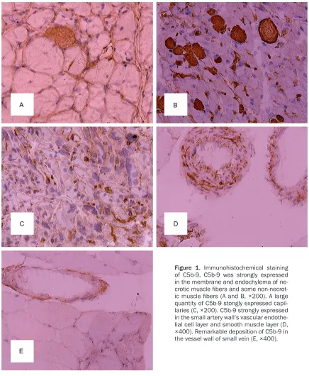
Figure 1. Immunohistochemical staining of C5b-9, C5b-9 was strongly expressed in the membrane and endochylema of
ne-crotic muscle fibers and some non-necrot
-ic muscle fibers (A and B, ×200). A large
quantity of C5b-9 stongly expressed
capil-laries (C, ×200). C5b-9 strongly expressed
in the small artery wall’s vascular endothe-lial cell layer and smooth muscle layer (D,
×400). Remarkable deposition of C5b-9 in
the vessel wall of small vein (E, ×400).
Immunohistochemical staining of C5b-9
C5b-9 protein in the all of the muscle samples was detected by immunohistochemical stain-ing. Rabbit human C5b-9 polyclonal anti-body (Abcam, America, 1:100) was used as pri-mary antibody. Sections of the muscles were dried at room temperature for 20 minutes and incubated with primary antibody for 1 hour. After three times of wash, the sections were
Figure 2. Immunohistochemical staining of C5b-9, the number of positive muscle cells (yellow granulation in cell
mem-brane or endochylema) in the inflamma -tory myopathies group except necrotizing
myopathy (B, ×200) and muscular dystro
-phy group (C, ×200) were less observed
than that in necrotizing myopathy
pa-tients (A, ×200). In necrotizing myopathy
patients, the numbers of C5b-9 positive
cells were significantly higher.
membrane and endochylema, or yellow color of vascular intima were considered as positive reactions [7]. Average optical density (integrat-ed optical density/area) of positive reactions was analyzed with Image-Pro Plus 6.0 software.
Data analysis
SPSS 13.0 statistical software (SPSS Inc., Chicago IL, USA) was used for statistical analy-sis. Measurement data were expressed as mean ± SD, and count data were expressed as frequency counts and percentages. After the Bartlett homogeneity of variance, comparison of measurement data was performed by T-test or nonparametric test. All tests were two-tailed and P < 0.05 was considered to be statistically significant.
Results
Clinical characteristics of NM patients
Age, sex, time of onset, duration, and level of biochemical enzymes (CK, LDH, GPT, GOT) in the serum of patients with NM, polymyositis,
and muscular dystrophy were displayed in Table 1. The 13 NM patients included 8 male (61.5%) and 5 female (38.5%), with the ratio of male to female of 1.6:1. There was no signifi -cant difference on onset age between male and female (t = 1.94, P = 0.053 > 0.05). The 13 NM patients showed symptom of weakness of limbs (84.6%, 11/13), bulbar muscle involve-ment (23.1%, 3/13), cervical muscle weakness (61.5%, 8/13), and respiratory muscle involve-ment (23.1%, 3/13). The major symptoms were displayed as weakness of four limbs. Weakness of distal limbs was more obvious than that of proximal limbs. Complain of muscle pain or ten-derness and muscle atrophy was recorded in 53.8% (7/13) and 46.2% (6/13) of the patients. The level of CK in the serum ranged from 45 IU/L to 12619.9 IU/L (average: 4678.5 ± 3449.05 IU/L).
C5b-9 expression in the muscle from patients with NM
NM may be associated with immune-media-tion. The main clinical features of NM are acute or subacute onset and progressive limb weak-ness. The main pathological characteristics are a large number of necrotic muscle fibers with -out significant inflammatory cell infiltration [8]. Serum creatine kinase (CK) is elevated to dif-ferent degrees. In recent years, many studies have focused on the relationship between ele-vated serum sign recognition particle antibody (SRP) and NM [9]. To date, the pathogenesis of NM has not been fully elucidated, which may be resulted from a variety of reasons, such as immune-mediation, tumor, infections [10, 11], and application of statin [12]. Both the antigen-antibody reaction and positive expression of cytokines (such as TNF, IL-2) were considered as the evidences to support the opinion that immune abnormalities played a crucial role in the pathogenesis of NM. NM caused by unex-plained reasons is more sensitive to immuno-suppressant, so it is also called the steroid reactive NM or immune-mediated NM. Maybe the clinical and pathological features are differ-ent in secondary and non-secondary NM. how-ever, the pathological change of muscle fiber necrosis is alike. Therefore, it is speculated that there may be a common pathway that involved in muscle fiber necrosis, but until now, the precise mechanism has not yet been reported.
C5b-9, as well as the membrane attack com-plex, plays an important role in a variety of physiological and pathological process. Soane’s investigation released that sub C5b-9 can increase survival ability of oligodendrocytes by up-regulating Bcl-2 protein and inhibiting cas-pase-3 activation in the demyelination process mediated by inflammation and immune system [13, 14]. Sub C5b-9 shows a positive effect in the antiapoptotic process and promotes the formation of myelin sheath. After sedimenta-tion in the vascular endothelial cells, C5b-9 can induce intracellular exocytosis secretion of vWF, which can mediate the combination of platelet and exposed GBM, as well as play a hemostatic function after depositing in the damaged vessel wall. C5b-9 also participated in the antiviral mechanism (enveloped virus as influenza virus, reverse transcribing virus as HIV) by combining with phosphatide to entirety disintegrate lipid double membrane. This effect is a very important mechanism in lysing envel-Figure 3. Immunohistochemical staining of C5b-9 of
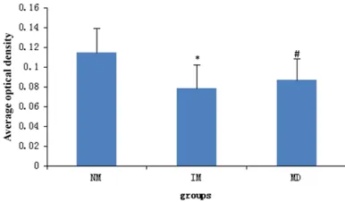
muscle tissues. NM: necrotizing myopathy group (n
= 13), IM: inflammatory myopathies group, excluding
necrotizing myopathy (n = 6), MD: muscular dystro-phy group (n = 5). Muscular cells containing yellow granulation (arrow showing) in the cell membrane and endochylema, or yellow color of vascular intima were considered as C5b-9 positive reactions. The C5b-9 positive areas increased in the NM group in comparison to IM group and MD group. Data are ex-pressed as means ± SD. P = 0.038 < 0.05 in com-parison to IM group, P = 0.013 < 0.05 in comparison to MD group.
staining intensity of C5b-9 ranged from positive to strongly positive even in the broken particles of muscle fibers. A few non-necrotic fibers also showed weakly positive expression of C5b-9. Immunoreactivity of C5b-9 could be found in blood vessels of muscle tissue of all the NM patients. C5b-9 expressed in vascular endothe-lial cells and muscular layer of small arteries, veins and capillaries, with different intensity. No expression of C5b-9 was observed in adven-titia of the vascular wall and perivascular tissue (Figure 1C-E).
Difference of C5b-9 expression in NM, poly-myositis and muscular dystrophy
Positive staining (brown granules) was found in cell membrane and endochylema of muscle tis-sues from the NM group (Figure 2A), but rela-tively less in other two groups (Figure 2B and 2C). The average intensity of C5b-9 in positive cells was significantly higher in NM patients compared with those in the polymyositis group (P = 0.013, Figure 3, and the muscular dystro-phy group ( P = 0.038, Figure 3). These results suggest that C5b-9 was overexpressed in NM patients.
Discussion
[image:5.612.92.285.72.185.2]oped virus. C5b-9 may also cause membrane irreversible damage in some specific pathologi -cal process. It increases permeability by insert-ing into the membrane, leadinsert-ing to internal flow of water, ions, and small molecules while mac-romolecules and proteins are incapable of escaping the cytoplasm. The cellosmotic pres-sure of internal and external become unbal-anced, activating intracellular signal transduc-tion pathways to promote cell synthesis and the release of certain inflammatory mediators and cytokines, leading to the cells secondary injury even necrosis, which plays a role in the patho-genesis and progression of many diseases. As we all know, the damage to the cell membrane caused by C5b-9 is an important factor which acts as a trigger for a variety of kidney diseases such as glomerulonephritis. Cybulsky’s study shows that C5b-9 can form transmembrane ion channels when inserting Glomerular epithelial cell membrane (GEC) [15]. After that, the chan-nel causes Ca2+- inhalation and induces cPLA2-activation. Free arachidonic acid (AA) can be released via deacylation after the combination of cPLA2 and intracellular membranes. Prostaglandin (PG) and thromboxane A2 (TXA2), which can be transformed by AA, play a variety of pathophysiological functions in the occur-rence and development of membranous nephropathy, but there was no report about the influence of C5b-9 on the occurrence of NM.
Our study showed that C5b-9 positively expressed in necrotic muscle fibers, atrophic muscle fibers, small arteries, veins and capillar -ies of muscle tissue of patients with NM. This is consistent with other studies. In 1991, Emslie-Smith firstly proposed the the concept of NM [16]. Immunohistochemical detection of mus-cle specimens found muscular fibers degenera -tion, necrosis, and deposition of membrane attack complex. In 2002, Miller reported 7 cases of NM with decreased muscle tissue cap-illaries and C5b-9 deposition in which [17]. In 2004, Bleecker et al. further confirmed C5b-9 deposition in some endomysial capillaries and arteriolar wall around muscle bundles [18]. Previous studies have demonstrated the phe-nomenon of membrane attack complex deposi-tion in vascular wall of NM, but there is still much controversy and discussion regarding the role of C5b-9 plays in the mechanism of NM. It was speculated that muscle fiber necrosis may be related to antibody-dependent
complement-mediated immune reactions caused by the dis-solution of the sarcolemma [19, 20].
Our findings suggest that C5b-9 may play an important role in the pathogenesis of NM. C5b-9 may function as follows: (1) C5b-9 accu-mulates in the vascular endothelium. Then the permeability of wall is increased and some immune substances ooze out of the wall, lead-ing to stenosis or occlusion of the blood vessel. Finally it results in ischaemia, even the isch-emic necrosis of the muscle fibers of the peri -vascular region. (2) C5b-9 directly goes around or seeps into the muscle fibers, causing destruction of muscle fibers.
This study offers a new vision for the treatment of NM, that is, the purpose of therapy can be achieved by disturbing and preventing the destructive effects of C5b-9, which contributes to muscle fibers and blood vessels. Our obser -vations have led us to make a conclusion that there was no significant correlation between C5b-9 expression intensity and the level of serum CK. This result may not be reliable due to the limited number of cases and the inaccu-rate quantitation of C5b-9. Further study is needed to explore the relationship between C5b-9 and clinical index by expanding the num-ber of cases and using a quantitative method. In conclusion, C5b-9 is strongly expressed in the necrotic muscle fiber and blood vessels, and may contribute to the pathogenesis of nec-rotizing myopathy.
Disclosure of conflict of interest
None.
Address correspondence to: Dr. Chuan-Qiang Pu, Department of Neurology, Chinese PLA General Hospital, No.28, Fuxing Road, Beijing 100853, China. Tel: 861066939251; Fax: 861066939251; E-mail: [email protected]
References
[1] Grable-Esposito P, Katzberg HD, Greenberg SA, Srinivasan J, Katz J, Amato AA. Immune-medi-ated necrotizing myopathy associImmune-medi-ated with statins. Muscle Nerve 2010; 41: 185-90. [2] Dalakas MC. Toxic and drug-induced
[3] Needham M, Fabian V, Knezevic W, Panegyres P, Zilko P, Mastaglia FL. Progressive myopathy with up-regulation of MHC-I associated with statin therapy. Neuromuscul Disord 2007; 17: 194-200.
[4] Hengstman GJ, Vree Egberts WT, Seelig HP, Lundberg IE, Moutsopoulos HM, Doria A, Mos-ca M, Vencovsky J, van Venrooij WJ, van Enge-len BG. Clinical characteristics of patients with myositis and autoantibodies to different frag-ments of the Mi-2 beta antigen. Ann Rheum Dis 2006; 65: 242-245.
[5] Webster S, Lue LF, Brachova L, Tenner AJ, Mc-Geer PL, Terai K, Walker DG, Bradt B, Cooper NR, Rogers J. Molecular and Cellular Charac-terization of the Membrane Attack Complex, C5b-9, in Alzheimer’s Disease. Neurobiol Aging 1997; 18: 415-421.
[6] Cong L, Pu C, Mao Y, Lu X, Wang Q. Role of C5b-9 expression in skeletal muscle blood vessels in necrotizing myopathy. Nan Fang Yi Ke Da Xue Xue Bao 2012; 32: 714-7.
[7] Feng Y, Ni L, Wang Q. Administration of
cathep-sin B inhibitor CA-074Me reduces inflamma -tion and apoptosis in polymyositis. J Dermatol Sci 2013; 72: 158-67.
[8] Dalakas MC. Pathophysiology of inflammatory
and autoimmune myopathies. Presse Med 2011; 40: e237-47.
[9] Mammen AL. Autoimmune myopathies: auto-antibodies, phenotypes and pathogenesis. Nat Rev Neurol 2011; 7: 343-54.
[10] Yao PP, Qian L, Xia Y, Xu F, Yang ZN, Xie RH, Li X, Liang WF, Huang XX, Zhu ZY, Zhu HP. Entero-virus 71-induced neurological disorders in young gerbils, Meriones unguiculatus: devel-opment and application of a neurological dis-ease model. PLoS One 2012; 7: e51996. [11] Crum-Cianflone NF. Bacterial, fungal, parasitic,
and viral myositis. Clin Microbiol Rev 2008; 21: 473-94.
[12] Hamann PD, Cooper RG, McHugh NJ, Chinoy H. Statin-induced necrotizing myositis - a discrete autoimmune entity within the “statin-induced myopathy spectrum”. Autoimmun Rev 2013; 12: 1177-81.
[13] Soane L, Rus H, Niculescu F, Shin ML. Inhibi-tion of oligodendrocyte apoptosis by sublytic C5b-9 is associated with enhanced synthesis of Bcl-2 and Mediated by Inhibition of Cas-pase-3 Activation. J Immunol 1999; 163: 6132-6138.
[14] Soane L, Cho HJ, Niculescu F, Rus H, Shin ML. C5b-9 terminal complement complex protects oligodendrocytes from death by regulating bad through phosphatidylinositol 3-kinase/akt pathway. J Immunol 2001; 167: 2305-2311. [15] Cybulsky AV, Takano T, Papillon J, McTavish AJ.
Complement-induced phospholipase A2 acti-vation in experimental membranous nephrop-athy. Kidney Int 2000; 57: 1052-1062. [16] Emslie-Smith AM, Engel AG. Necrotizing
myop-athy with pipestem capillaries, microvascular deposition of the complement membrane
at-tack complex (MAC), and minimal cellular infil -tration. Neurology 1991; 41: 936-9.
[17] Miller T, Al-Lozi MT, Lopate G, Pestronk A. My-opathy with antibodies to the signal recogni-tion particle: clinical and pathological features. J Neurol Neurosurg Psychiatry 2002; 73: 420-8.
[18] Bleecker JD, Vervaet V, Van den Bergh P. Nec-rotizing myopathy with microvascular deposi-tion of the complement membrane attack complex. Clin Neuropathol 2004; 23: 76-9. [19] Hoogendijk JE, Amato AA, Lecky BR, Choy EH,
Lundberg IE, Rose MR, Vencovsky J, de Visser M, Hughes RA. 119th ENMC international workshop: tria design in adult idiopathic
in-flammatory myopathies,with the exception of
inclusion body myositis, 10-13 October 2003, Naarden, The Netherlands. Neuromuscul Dis-ord 2004; 14: 337-45.
[20] Authier FJ, Kondo H, Ghnassia RT, Revuz J, Gh-erardi RK. Necrotizing myopathy with pipestem
capillaries and minimal cellular infiltration: a