RESEARCH ARTICLE
Modelling colour constancy in fish: implications for vision and
signalling in water
Lucas Wilkins1, N. Justin Marshall2, Sönke Johnsen3and D. Osorio1,*
ABSTRACT
Colour vision and colour signals are important to aquatic animals, but light scattering and absorption by water distorts spectral stimuli. To investigate the performance of colour vision in water, and to suggest how photoreceptor spectral sensitivities and body colours might evolve for visual communication, we model the effects of changes in viewing distance and depth on the appearance of fish colours for three teleosts: a barracuda,Sphyraena helleri, which is dichromatic and two damselfishes,Chromis veraterandChromis hanui, which are trichromatic. We assume that photoreceptors light-adapt to the background, thereby implementing the von Kries transformation, which can largely account for observed colour constancy in humans and other animals, including fish. This transformation does not, however, compensate for light scattering over variable viewing distances, which in less than a metre seriously impairs dichromatic colour vision, and makes judgement of colour saturation unreliable for trichromats. The von Kries transformation does substantially offset colour shifts caused by changing depth, so that from depths of 0 to 30 m modelled colour changes (i.e. failures of colour constancy) are sometimes negligible. However, the magnitudes and directions of remaining changes are complex, depending upon the specific spectral sensitivities of the receptors and the reflectance spectra. This predicts that when judgement of colour is important, the spectra of signalling colours and photoreceptor spectral sensitivities should be evolutionarily linked, with the colours dependent on photoreceptor spectral sensitivities, and vice versa.
KEY WORDS: Colour, Vision, Fish, Colour constancy, Communication, Evolution
INTRODUCTION
Fish are known for their bright colours, but how do these colours evolve and how can they work as signals? It is thought that land animals detect form and motion mostly by luminance, while colour serves object recognition. This is because the pattern of light and shade make it difficult to judge the overall reflectance (grey level) of a surface, whereas the spectral composition of reflected light is a relatively stable cue to material properties (e.g. pigmentation; Rubin and Richards, 1982; Livingstone and Hubel, 1988; Gegenfurtner and Kiper, 2003; Osorio and Vorobyev, 2005; Baddeley and Attewell, 2009). Nonetheless, terrestrial illumination spectra do vary, so that judgement of a reflectance spectrum–known as‘object colour’or‘absolute colour’ –requires colour constancy: that is the
ability to discount the effects of illumination on colour appearance. Colour vision can therefore be understood as a means to recover reflectance spectra from photoreceptor signals (Barlow, 1982; Buchsbaum and Gottschalk, 1983; Maloney, 1986; Osorio and Vorobyev, 2005).
At short ranges (<0.1 m) in shallow water, colour vision can operate much as it does on land, but natural waters scatter and absorb light far more than air, which makes colour constancy difficult (Figs 1 and 2; Jerlov, 1976; Mobley, 1994; Osorio et al., 1997; Johnsen, 2012; Cronin et al., 2014). Vorobyev (2001) and others (Marshall and Vorobyev, 2003) modelled colour constancy based on the von Kries transformation (see below), for the red and brown
fish Scarus spinus and magenta and yellow fish Pseudochromis
paccagnellae, and concluded that it failed to compensate for
changes in the colour with varying distance. Consequently, aquatic animals have been thought to be less concerned with the representation of reflectance spectra (or object colour) than with the detection of visual contrast–either within the coloration pattern itself, or against the background. Notably, the chromatic offset hypothesis proposes that aquatic animals evolve multiple cone classes to enhance the visual contrast of objects seen in open water (McFarland and Munz, 1975; Lythgoe, 1979; Sabbah and Hawryshyn, 2013). Supporting this account, Marshall and others (2006) examined the colours used by several fish species as communication signals by comparing visual systems and their performance over depth in various marine light environments. The study did not consider colour constancy, but its conclusion that a fish’s pattern could be a more reliable signal than its colour (Marshall et al., 2006), is consistent with evidence that cichlid cone sensitivities are well adapted for detecting patterns (Sabbah and Hawryshyn, 2013).
From the foregoing arguments it follows that where colour is used for communication over distances of greater than roughly 0.1 m (depending on turbidity) or at varying depths, it is the patterns rather than the colours themselves that are the primary signals (Marshall et al., 2006); a conclusion that contrasts with the emphasis on object colour as the primary signal for land animals (Hill and Montgomerie, 1994; Osorio and Vorobyev, 2008). Nonetheless, object colour is thought to be important to fish communication (Houde, 1997; Seehausen et al., 2008; Elmer et al., 2009; Maan and Sefc, 2013), so one can ask under what conditions it might be used: are some colours expected to offer more reliable signals with variable depth and/or viewing distance than others? Will the best set of receptors be general for all spectra in a given visual environment? Or will it depend on the specific reflectance spectra?
Colour constancy in water
Perceptual constancies allow an observer to perceive the cause of a stimulus (e.g. an object), despite variation in the stimulus received by the sense organs. Human colour constancy involves both low-level (e.g. retinal) and high-low-level (e.g. cortical) mechanisms
Received 15 February 2016; Accepted 27 March 2016 1
School of Life Sciences, University of Sussex, Brighton BN1 9QG, UK. 2
Queensland Brain Institute, University of Queensland, Brisbane, Queensland 4072, Australia.3Biology Department, Duke University, Durham, NC 27708, USA.
*Author for correspondence ([email protected])
D.O., 0000-0002-5856-527X
Journal
of
Experimental
(Brainard and Freeman, 1997; Smithson, 2005; Foster, 2011), but it is logical to start with physiologically and mathematically the simplest colour constancy mechanism, namely the von Kries transformation, whereby each photoreceptor’s response is normalised to the average for that receptor class across the image (Eqns 3,4; Worthey and Brill, 1986; Smithson, 2005; Foster, 2011). The von Kries transformation can, at least formally, be attributed to light adaptation, which takes place in photoreceptors and other early stages of visual processing (Vanleeuwen et al., 2007; Sabbah et al., 2013), and given the universality of light adaptation, it is not surprising that all animals tested, including insects, terrestrial vertebrates and fish, have colour constancy (Dörr and Neumeyer, 1997, 2000; Chittka et al., 2014). It is, however, difficult to identify the specific mechanism; for example, Neumeyer and co-workers (2002) found that goldfish colour constancy is consistent with a von Kries transformation, but there is evidence that colour constancy in guppies improves with experience (Intskirveli et al., 2002), which is indicative of higher-level processes. Also, it is has been suggested that the spectral opponent responses of horizontal cells in teleost
retinas have a role in colour constancy (Kamermans et al., 1998). As horizontal cells receive multiple, and often colour opponent, receptor inputs, their involvement implies a role for interactions between different spectral receptors, which is inconsistent with a von Kries mechanism (Vanleeuwen et al., 2007).
The model
Here, we evaluate the potential and limitations of colour vision and colour signalling in water by modelling of the propagation of light in coral reef water to a depth of 30 m. We estimate the responses of fish photoreceptors viewing a set of 25 fish reflectance spectra over a range of depths and distances (Figs 1–3).
To implement the von Kries transformation the model receptor responses are normalised, either to the horizontal space light–i.e. the background radiance in open water with a horizontal line of sight
–or to an achromatic background. These two idealised backgrounds are fundamentally different because the spectral composition of light from a reflecting surface changes with viewing distance, whereas the light from open water is fixed.
We consider three coral reef teleost fish (Fig. 2): a barracuda,
Sphyraena helleriJenkins 1901, which like many open-water fish is
dichromatic (see the Materials and Methods), and two damselfishes,
Chromis veraterJordan and Metz 1912 andChromis hanuiRandall
and Swerdloff 1973. Both damselfishes are trichromatic, but they have markedly different photoreceptor spectral sensitivities, with that ofC. hanuibeing more widely separated and extending into the UV. We do not model tetrachromatic fish vision (Neumeyer, 1992), but we expect this to be qualitatively similar to that for trichromats (Kelber and Osorio, 2010).
Our aim is not to predict any particular optimal system for colour communication, which would require details of the fish’s vision, colours, behaviour and visual environment, but rather to understand the adaptive landscape on which fish colours and colour vision co-evolve (Seehausen et al., 2008; Miyagi et al., 2012). Specifically, we aim to: (1) compare trichromacy and dichromacy; (2) examine the effects of varying photoreceptor spectral tuning in trichromats; (3) model how the reflectance spectrum affects colour constancy; and (4) determine whether performance is sensitive to an open-water or a reflective surface background.
MATERIALS AND METHODS Illumination and viewing conditions
Light scatter and absorption mean that, in water, the illumination spectrum falling on a surface is dependent on its orientation (Figs 1 and 2; Johnsen, 2012). We assume here that the surface being viewed is Lambertian (matte) and oriented perpendicular to a horizontal line of sight. The background is either open water
300 400 500 600
0 2 4
⫻1017
A
B
S M L
L S
300 400 500 600
30 m 20 m 10 m
0 m
Wavelength (nm)
Normalised sensitivity
Light flux (photons m
–2
s
–1
[image:2.612.54.297.56.251.2]nm)
Fig. 2. Illumination spectra and photoreceptor spectral sensitivities.(A) Modelled illumination spectra in coral reef water at depths of 0, 10, 20 and 30 m (see the Materials and Methods). Note that the light flux at 10 m exceeds that at the surface in the 450-500 nm range, this is due to scattered light, and is dependent on the orientation of the stimulus relative to the surface. (B) Spectral sensitivities of the fish photoreceptors used in our models: the barracudaSphyraena helleri, a dichromat (top panel) and the trichromatsChromis hanui
(bottom panel, solid lines) andChromis verater
(bottom panel, dotted lines). S, M, L: short, medium and long wavelength photoreceptors, respectively.
3 2
1
4
[image:2.612.51.389.599.732.2]5
Fig. 1. The visual scene.The object fish ( purple) is illuminated directly from above (1) via both single and multiple scattering events in the water (2). The observer fish (green) is at the same depth. Light reaching the observer from the direction of the stimulus is a combination of light scattered by the water (3) and light reflected from the stimulus (4). Light reflected by the stimulus is lost though scattering and absorption (5). We model the object viewed against a background, which is either horizontal space light, that is the light seen in open water, or a surface reflecting equally at all wavelengths (not illustrated). Note that light reaching the eye from the achromatic background changes with the viewing distance of the object, whereas the open-water background is fixed.
Journal
of
Experimental
(‘space-light’; Johnsen, 2012) or a matte spectrally neutral surface (i.e. with equal reflectance across the spectrum) at the same distance as the object. The key difference is that light from a reflecting surface varies with distance, whereas space-light is constant. In fact, the reflectance spectra of natural backgrounds, such as sand or coral rubble, are probably not achromatic, but tend to increase linearly with wavelength (giving a brownish colour), but any difference would have minimal impact on our conclusions (Osorio et al., 1997).
Aquatic illumination, absorption and scattering
Clear tropical coastal waters, such as those of coral reefs, have maximum transmission at about 500 nm (Fig. 2A; Jerlov, 1976). We model spectrally selective scatter by suspended particles following Mobley (1994) and Johnsen (2012). The main optical processes, schematised in Fig. 1, can be formalised by a differential equation (Eqn 1), which equates the change in horizontal radiance viewing distance with: (1) a positive contribution, denotedS, that describes the amount of light, of wavelength λ, entering the ray, which is predominantly via scattering; and (2) a negative contribution that describes its attenuation (absorption and out-of-ray scattering), proportional to the radiance, which is denoted by a constantα. The horizontal viewing condition makes it possible to treat the medium as uniform along the viewing axis, soSandαdo not depend on viewing distance (although they do change with depth). Thus:
d
dxLðxÞ ¼SaLðxÞ; ð1Þ
wherexis the distance from the subject andL(x) is the radiance. Constants α and S were calculated using Hydrolight (Sequoia Scientific) for a Case I bio-optical model, assuming a chlorophyll concentration of 0.5 mg m−3.
Eqn 1 can be rewritten in terms of the radiance at the objectL0 (viewing distance of zero) and a‘space-light’termLb–equal toS/α
– which is the radiance of open water (viewing distance in the infinite limit):
LðxÞ ¼ L0eaxþLbð1eaxÞ; ð2Þ
whereL(x) is the radiance at distancexfrom the object,αis again the attenuation coefficient, which equals the sum of the absorption coefficient and the scattering coefficient. In this form, it is evident that the horizontal radiance is a mixture of the reflected radiance and the space-light, weighted by an exponentially decreasing function of distance.
Photoreceptor responses
We model receptor responses of three teleosts,S. helleri,C. verater
and C. hanui (Fig. 2), which live in or around corals reefs. The
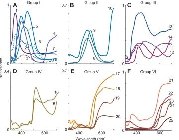
fishes’photoreceptor sensitivities are derived from photopigment absorbances and the transmission of their ocular media (Losey et al., 2003). The 25 reflectance spectra are from freshly captured coral reef fish in Hawaii, which were measured with illumination normal to the surface, and the detector at 45 deg (Fig. 3; Marshall et al., 2003a).
For modelling receptor responses with light adaptation, photoreceptor quantum catchesqifor each receptor are defined as:
qi¼
ð
L
LðlÞriðlÞdl; ð3Þ
where ri is the rate at which photons activate the photopigment (assuming all photopigment molecules are available for transduction), andΛ represents the wavelength range over which the integral is performed, in this case 300 to 700 nm.
The responses are transformed to a von Kries adapted value,vi, by
division by the quantum catch from the adapting background radiancebi:
vi¼qi=bi: ð4Þ
18 17 1
6
400 600
0 0.7
0.7
9
8
10 1
12 11 13
0 1
23
25 24 21
22
400 600
0 0.4
15 16
0 0
1
2
14
3
7 5
4
400 600
0
19
20
Reflectance
Wavelength (nm)
Group I Group II Group III
Group IV Group V
F
Group VIC
A
B
[image:3.612.50.410.57.339.2]E
D
Fig. 3. Fish reflectance spectra.Twenty-five reflectance spectra from coral reef fish (Losey et al., 2003) used for the models. The line colours are given by the CIE loci of the spectra, and so approximate their appearance to a human. (A–F) To identify natural categories of spectra (as opposed to classifications based on visual responses) they are placed into six groups (I–VI in A–F, respectively) by normalising them to their respective maxima, square-root transformation (to reduce effects of overall reflectance) and then classifying them with the MatLab (v.2012a)k-means clustering algorithm, using the‘correlation’parameter. This classification is a convenient way to group the colours according to their reflectance spectra, as opposed to photoreceptor excitations and it is interesting to note how they cluster and shift in the fish colour spaces (Figs 4, 5, 7 and 8).
Journal
of
Experimental
The transformed values are converted into normalised chromaticity coordinates, ni, by division by total photoreceptor
quantum catch:
ni¼vi=
X
i
vi: ð5Þ
These two steps normalise the response relative to the background radiance.
We then assume that receptor responses are compared by opponent mechanisms to give chromatic signals (Kelber et al., 2003). Normalisation of these signals (discounting overall intensity) allows us to represent the dichromat’s chromatic signal using the formula:
X¼ ðLSÞ=ðLþSÞ ð6Þ
and to project the trichromatic space in a two-dimensional chromaticity diagram (Maxwell’s triangle). The projection gives two chromaticity values by a linear transform, namely:
X¼
ffiffiffi
2
p
2 ðn1n3Þ ¼
ffiffiffi
2
p
2 ðLSÞ; ð7Þ
Y¼
ffiffiffi
2 3
r
½n2 ðn1þn3Þ=2 ¼
ffiffiffi
2 3
r
½M ðLþSÞ=2; ð8Þ
withnibeing ordered by the wavelength of peak sensitivity (λmax) from short to long.L,MandSrefer to the responses of the long, medium and short wavelength sensitive photoreceptor responses, respectively (Fig. 2), either before or after normalisation to the background (Eqn 4). Note that although scattered light in clear water generally looks blue to divers and objects become bluer with increasing distance, it is implicit in our model that object colours would move to the achromatic point with increasing distance.
Modelling discrimination thresholds
A failure of colour constancy can be behaviourally significant only if the shift exceeds the colour discrimination threshold, or one just-noticeable difference (JND; here 1 JND will be detected 75% of the time from two alternatives). We consider only chromatic signals (i.e. changes in hue and saturation) and assume that colour thresholds are
independent of light intensity (i.e. Weber’s law holds; Kelber et al., 2003), with receptor noise equivalent to a contrast of 0.05 in each cone type (Figs 4,6,7; eqns 3,4 in Vorobyev and Osorio, 1998). This estimate of the JND is similar to a recent estimate for a bird (Olsson et al., 2015), although in reality, the effects of the ambient illumination–which changes with depth–on receptor photon catch are likely to affect the discrimination thresholds (Marshall and Vorobyev, 2003).
Notes on terminology
The terms hue, saturation and brightness refer to aspects of human colour perception (Wyszecki and Stiles, 1982), which cannot at present be defined for any animal (Kelber and Osorio, 2010). Here, we use geometric definitions that parallel the human terms. We decompose the space into a brightness axis, and an
n−1 dimensional chromaticity space. The location in a chromaticity space is given by dividing the receptor catch coordinates by the sum of receptor values (nominally brightness). Saturation is the distance from the centre of the chomaticity space and hue is the remaining dimension(s). It follows that a dichromat does not distinguish hue, a trichromat has one dimension of hue and a tetrachromat has two. Note also that then-chromacy (di-, tri- etc.) is defined not by the number of spectrally distinct cone photoreceptors in the eye but by the number of primaries needed to match any colour. Here, in the absence of direct behavioural evidence, we assume thatS. helleri
is a dichromat and theChromis species are trichromats.
RESULTS
We model photoreceptor responses of three fish to fish reflectance spectra (Fig. 3) in coral reef water. The models predict how varying the viewing distance, depth and background (Fig. 1) will affect receptor responses and chromatic signals after photoreceptor adaptation to the background (Eqns 3 and 4). Modelled colours are plotted in chromaticity diagrams, which represent the colour based on photoreceptor quantum catches (Eqns 3-5, 7,8), in terms of the chromatic aspects of colour (i.e. hue and saturation for humans; Wyszecki and Stiles, 1982), independent of intensity (or brightness). A dichromat has a single chromatic dimension, so
VI III I
IV
V II
21 22 11 13 14 1 2 3
7 5 6
15 16
19 18 20 9 10
A
B
–0.2 0 0.2 0.4 –0.2 0 0.2 0.4
JNDs
JNDs
(L–S)/(L+S)
4
8
12
17
23 24 25
21 22 11 13 14 1 2 3
7 5 6
15 16
19 18 20 9 10 4
8
12
17
23 24 25
Distance Depth Fig. 4. Colour shifts for the dichromat
S. hellerias a function of depth and
distance.Plots show modelled shifts of fish spectra in Fig. 3. (A) With distance at 2 m depth against an achromatic background, tickmarks show the chromatic signal at 0, 1 and 2 m, the first metre is coloured as in Fig. 3. (B) With depth from 0 m to 30 m at a distance of 0.3 m (c.f. Figs 5 and 7), tickmarks indicate 10 m intervals, coloured from red to blue with increasing depth. It is assumed that photoreceptors are adapted to the achromatic background (Fig. 1). The origin corresponds to the achromatic point. Numbers along the left and right margins identify the spectra, arranged in the six groups identified in Fig. 3, and the scale along the upper and lower margins indicates the just noticeable differences (JNDs) for the chromatic signal, assuming a Weber fraction of 0.05 in both cone types.
Journal
of
Experimental
[image:4.612.51.411.506.733.2]colours are represented on a line (Fig. 4), whereas trichromats have two dimensions, which are represented by a plane (Figs 5–7).
Variation in distance
We modelled the effects of varying viewing distance from 0 to 100 m against open water and a spectrally neutral reflector at the
same distance as the object. Visibility falls rapidly, so 100 m is in effect infinity (Figs 4 and 5; Loew and Lythgoe, 1975; Cronin et al., 2014). Light scatter and absorption (Fig. 1) cause colours to become less saturated with increasing distance, shifting them towards the achromatic point (Figs 4 and 5), which is by definition the background colour. For the trichromaticChromisspecies, spectrally selective absorption has a slight effect, causing hue shifts, which are seen as‘hooks’on the plots in the chromaticity diagram (Fig. 5), evident at ranges exceeding 3 m.
An open-water background does not change with viewing distance, so that the photoreceptor adaptation state is fixed, and von Kries colour constancy can have no effect. By comparison, a reflecting background in the same plane as the object changes with distance in a similar manner to the object, which does allow the von Kries transform to take effect. However, the transform corrects for multiplicative effects (effects of illumination or absorption in most real-world cases), which do not apply to scattering and, in fact, the modelled colour changes for the open-water and solid backgrounds are qualitatively similar, with colours moving toward the achromatic point (Fig. 4A and Fig. 5). Thus, the model implies that receptor adaptation to the background will not affect colour changes caused by varying viewing distance, because scatter dominates light absorption by water (Figs 1 and 2).
Variation in depth
We modelled receptor responses of the three fish species for depths of 0–30 m (Fig. 2), with a viewing distance of 0.3 m. Here, photoreceptor adaptation substantially offsets the effects of changing depth on the relative rates of photon absorption by the different spectral receptors (Fig. 4B, Figs 6 and 7). Nonetheless, residual changes (Fig. 4B, Figs 7 and 8) may exceed the colour discrimination threshold, and so might cause failures of colour constancy.
For the dichromatS. helleri, which has one chromatic dimension, all the residual changes are towards the achromatic point with increasing depth, but they vary in magnitude for different spectra (Fig. 4B), ranging from <1 to >3 JNDs. The larger shifts are for spectra that reflect strongly at long wavelengths, which lie to the right of the neutral point.
For the trichromatic Chromis species, residual shifts vary substantially in their magnitudes and their directions in colour
–0.4 0 0.4
–0.4 0 0.4 0.8
420 nm
460 nm
500 nm
540 nm
S L
M
–0.4 0 0.4
440 nm 480 nm
520 nm
560 nm
S L
M
400 nm
A
B
x
[image:5.612.55.293.56.298.2]y
Fig. 6. Shifts of trichromatic colour loci due to photoreceptor adaptation at depths from 0 m to 30 m (lines) and colour discrimination thresholds (ellipses).The lines shows the correction imposed by receptor adaptation for a depth range of 0-30 m (red: shallow, blue: deep) forC. verater(A) andC. hanui(B) adapted to horizontal space light. The magnitudes of the transformation vary across the colour space. Differences between the two species are related to the differences in receptor sensitivities (Fig. 2). Corrections are dependent on the chromatic locus; they are smallest for spectra that excite single photoreceptors and largest for those that excite both long (L) and medium (M) wavelength receptors.C. hanuihas larger corrections thanC. veraterprobably because of the greater spectral separation of the photoreceptors. Ellipses show approximate colour discrimination thresholds estimated assuming that receptor noise limits performance with a Weber fraction of 0.05 in all three cones (Vorobyev and Osorio, 1998). The boundary lines plot the monochromatic loci for depths 0 m and 30 m. Shifts produced by adaptation to an achromatic background are similar. S, short wavelength photoreceptor.
–0.2 0 0.2
0 0.2
1
9
12 8
19
16
2
18 20
13 14
23
17 3
7
21
22 5
4
500 nm –0.1
0 0.1
–0.1 0.1
1
9 8
19
16 2
18 20
11
23 17
3 7
21 22 5
4 440 nm
A
B
x
[image:5.612.151.467.519.652.2]y
Fig. 5. Colour shifts for trichromats with varying viewing distance.The fish spectra in Fig. 3 viewed at varying distance at 2 m depth from 0 to 100 m (effectively infinity) with von Kries normalisation of modelled receptor responses to an achromatic background. Tickmarks show the chromatic signal at 1 m intervals, with the first metre coloured as in Fig. 3. Plots are receptor-based chromaticity diagrams for (A)C. veraterand (B)C. hanui, which represent the colours on a plane of equal brightness (Fig. 6; Eqns 3–5, 7 and 8). With increasing distance, the predominant shift is a decrease in saturation, with colours moving towards the achromatic point (i.e. the origin) as a result of scattered light. Minor hue shifts are apparent in the curvature of the lines, and the variable‘hooks’seen at long ranges. The effects are very similar for an open-water background and at greater depths.
Journal
of
Experimental
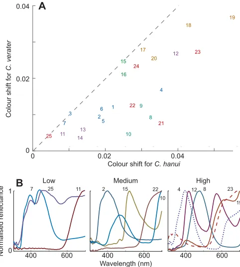
space (Figs 7 and 8). ForC. hanui, the shifts range from 0.005 to 0.05, with a mean of 0.025 units, in thex–ycolour space, whereas
forC. verater, shifts are smaller, ranging from 0.005 to 0.04, with a
mean of 0.015 units. These values can be compared with the JND, which ranges from 0.01 to 0.02 units, depending on location and direction in the colour space (Figs 6 and 7). The difference between the two species is probably due mainly to the spectral sensitivities of
C. veraterphotoreceptors being more closely spaced than those of
C. hanui, but the particular spectral locations of the receptors is also
relevant (Fig. 2; Worthey and Brill, 1986; Osorio et al., 1997) and it is evident that the direction and magnitude of shifts depend upon the specific set of photoreceptors, the spectra and the viewing conditions (illumination spectrum and adapting background). Also, there are examples of metamerism, where different spectra have the same colours, for instance, spectra 15 and 16 are almost identical forC. hanui, but not forC. verater(Fig. 3D and Fig. 7).
DISCUSSION
To examine how absorption and scattering of light might affect colour vision and communication in water (Fig. 1), we modelled chromatic signals for three species of fish viewing fish reflectance spectra. There are four scenarios: either the distance from the viewer to the object varies at a fixed depth (Fig. 4A and Fig. 5) or the depth varies at a fixed distance (Fig. 4B, Figs 6–8) and the background is either open water or a grey surface at the same location as the object. We assume that colour constancy is provided by normalisation of receptor responses to the background (Smithson, 2005; Foster, 2011; Neumeyer et al., 2002). Fish may have additional retinal (Kamermans et al., 1998, Vanleeuwen et al., 2007) and higher-level
mechanisms (Intskirveli et al., 2002; Smithson, 2005; Foster, 2011) but is it logical to start with von Kries constancy.
Variation in viewing distance
As the distance to the object changes, scatter and absorption remove light and light is scattered into the path. Scatter is fairly spectrally neutral, but the absorption is spectrally selective, removing long and short wavelengths and leaving blue light (Fig. 1), which is then available to be scattered into the path. This moves the spectrum towards that of the open water so that the visibility of the fish declines to near zero over a few metres (Fig. 4A and Fig. 5). Furthermore, because an open-water background has a fixed spectrum (Figs 1, 4 and 5) colour constancy based on adaptation to the background is useless. When the background is a surface at the same distance as the object, von Kries constancy could theoretically have an effect, but in fact, because of the effects of scattered light, the modelled changes of colour are almost identical for open-water and reflective backgrounds, with spectral loci moving toward the achromatic point as distance increases (Fig. 4A and Fig. 5).
The model implies that the failure of colour constancy with varying distance could not be corrected unless the viewer takes account of both the distance to the object and the turbidity of the water, which is probably difficult (but see Schechner et al., 2003). These observations lead to two conclusions: first, that for trichromats an object’s hue will be more constant than its saturation, and second that the range over which a colour can be detected will increase with increasing saturation (relative to the background). These considerations could account for the intense
4
–0.1 0.1
0
440 nm
–0.2 0 0.2
–0.1 0 0.1 0.2
440 nm
420 nm 420 nm
–0.2 0 0.2
1
12
8
19 2
20
11
10
13 14
23 25 24 3
7
21 22 5
6
9
15 16
18
1
1 1
12
8
19 2
20
11 10
13 14
23 25 24 3
7
21 22 5
6
9
15 16
18 17
12 8
2 20
11 10
13 14
23 25
24 3 7
21
22 5
6
9
15 16
18 17
4
12 8
19 2
20
11 10
13 14
23 25
24 3
7
21 22 5
6
9
15 16
18 17
4
A
B
C
D
Open-water background Achromatic background
19
17 4
x
[image:6.612.71.550.56.345.2]y
Fig. 7. Colour shifts for trichromats after correction by the von Kries transformation.Shifts of loci in the colour spaces ofC. verater(A,C) andC. hanui
(B,D) to the fish spectra (Fig. 3) after receptor adaptation to background radiance from 0 m (red) to 30 m depth (blue), with tickmarks at 10 m intervals, for horizontal space light (open water) and achromatic backgrounds. The axes and plotting conventions are as for Fig. 4B, Figs 5 and 6. The residual changes vary substantially in magnitude and direction. Grey ellipses are the 1 JND values as plotted in Fig. 6. Numbers identify the spectra as given in Fig. 3 and are coloured schematically according to their group (I–VI) in that figure.
Journal
of
Experimental
colours of some aquatic animals. Furthermore, at least for dichromats like S. helleri (Losey et al., 2003), which cannot distinguish hue from saturation (see Materials and Methods), this conclusion is consistent with the view that colour vision is concerned more with pattern recognition than object colour (Munz and McFarland, 1973; Marshall et al., 2006; Sabbah and Hawryshyn, 2013).
Variation in depth
With varying depth but at a fixed viewing distance, spectrally selective light attenuation by water alters colours. In the absence of receptor adaptation – or some equivalent colour constancy mechanism–all colours shift towards the illumination locus with increasing depth (Fig. 6). However, as on land (Smithson, 2005; Foster, 2011), the von Kries transformation would be effective, so that a fish viewing an object from a fixed distance can achieve useful colour constancy over a range of depths. Sometimes, over tens of metres, the residual shifts–which correspond to failures of colour constancy – are negligible, falling below the discrimination threshold (Fig. 4B, Figs 7 and 8). As expected from theory (Worthey and Brill, 1986; Osorio et al., 1997), the more widely separated receptors ofC. hanuisuffer shifts that are, on average, 40% larger than C. verater. In theory, higher-level mechanisms might compensate for such failures, but the fact that the residual shifts vary in magnitude and direction (Figs 7 and 8) would complicate any such compensation.
Assuming that accurate judgement of colour over depth is relevant, what are the consequences of the evolution and co-evolution of fish photoreceptor spectral sensitivities and reflectance spectra? It is notable that colour changes for different spectra vary both in their magnitudes and in their directions in the trichromatic colour spaces (Figs 3, 7 and 8). Many spectra shift towards the short wavelength (bottom left) corner of the colour triangle, but blue spectra (e.g. spectra 8, 9 and 10) shift towards the long wavelength corner (bottom right). Similarly, the magnitudes of shifts in the trichromat colour spaces are not easily predictable, either from the location of the colours in their chromaticity diagrams (Figs 7 and 8) or from their grouping identified by the k-means clustering algorithm (Figs 3 and 8): the largest shifts tend to be for spectra with high reflectance at longer wavelengths, such as those in group V, and the smallest shifts being for those such as group III with high reflectance at short wavelengths, but there is much variation between related spectra, especially for the reddish colours in group I. Moreover, shifts can be different for spectra that have similar colour loci: for example, for S. helleri spectra 6 and 11 (Fig. 4B), and forC. hanui, spectra 11 and 12 (Fig. 7B); the latter difference probably arises because spectrum 12 is double peaked (Fig. 3). The unpredictability of these colour changes implies that it would be difficult to apply a simple rule to compensate for them and that the stability of the colour of a given spectrum is contingent upon the local visual environment and the colour vision of the receiver.
Colour and communication in water
Communication depends on a receiver being able to discriminate different possible states of the signaller. Much work implies that object colour is important for fish communication, as it is on land (see Introduction; Osorio and Vorobyev, 2008), but the widespread occurrence of dichromacy in coral reef fish, coupled with recognition of the problem of colour constancy (Marshall et al., 2003b; Marshall and Vorobyev, 2003) suggests that this view may be simplistic. Instead, it is argued that receptor sensitivities evolve to benefit contrast with the background, as proposed by the offset hypothesis (Loew and Lythgoe, 1975; Sabbah and Hawryshyn, 2013) and likewise, that the displays of reef fish are adapted to produce conspicuous body patterns (Marshall et al., 2003b).
Despite the problems faced by colour vision in water, we find that, at least for trichromatic fish (and by implication for tetrachromatic species), colour constancy can effectively limit colour shifts associated spectral absorption of light at varying depths, but not light scattering with varying distance. It follows that if the level of pigmentation, which typically affects saturation, is an informative component of a colour signal (Milinski and Bakker, 1990; Hill and Montgomerie, 1994), decisions about object colour, for instance in mate choice, even in clear water should be made at fixed ranges of less than 1 m. Similarly, it follows that for colour variation in saturation, but not so much in hue, the opposite sex always looks better when nearer. Therefore, fish gain from coming closer– although signals are really only fairly compared if they originate from the same distance.
Trichromats can separate hue from saturation, and hue is affected little by veiling light (Fig. 5). Taking account of both scattering and absorption, this implies that the best colours for signalling in water should be saturated, with minimal hue shift following receptor adaptation to the background. Hue changes would then be robust and potentially informative. In general, von Kries colour constancy favours small photoreceptor separations (Figs 2, 7 and 8; Osorio et al., 1997) but beyond this, both the magnitudes and the directions of changes are variable, being dependent upon interactions between
0 0
Colour shift for C. hanui
0.02 0.04
0.02 0.04
4
1
12
8
19
2
20
11 13 10 14
23
25
24
3
7 21
22
5
6 9
15
16
18
17
Colour shift for
C. verater
400 600
8
1 15 12
0
400 600
400 600
19
7 11 2
10
25 22 4 23
Low Medium High
Normalised reflectance
B A
[image:7.612.54.295.58.326.2]Wavelength (nm)
Fig. 8. Colour shifts due to colour constancy failure differ between the
twoChromisspecies and between related spectra.(A) Modelled colour
shifts experienced between 0 and 30 m. Shifts are maximum displacements over the depth range 0–30 m for an open-water background (Fig. 7). The mean shifts forC. veraterandC. hanuiare 0.0152 and 0.0256, respectively; the correlation coefficient between shifts is 0.78. A JND is approximately 0.01– 0.02 units (Fig. 7). Numbers identify the spectra as given in Fig. 3, and are coloured schematically according to their group (I–VI). (B) Examples of spectra giving low (<0.015), medium (0.015–0.03) and high (>0.03) shifts forC. hanui. One of each of the spectra from classes I–VI (Fig. 3) that fall into the relevant range is illustrated. The spectra are normalised, and numbered as in Fig. 3.
Journal
of
Experimental
the photoreceptor spectral sensitivities, reflectance spectra and the visual environment. For example, in clear coastal seawater for
C. verater, with its closely spaced photoreceptor spectral
sensitivities, many of the bluish spectra would be satisfactory, (Figs 2, 3, 7 and 8). By comparison, the larger spectral separation of
C. hanuiphotoreceptors increases chromatic signals (Figs 5 and 7),
but this advantage may be negated by the failure of colour constancy (Figs 7 and 8; and also by reduced quantum catch). Saturated colours, such as 4, 15 and 18 (Fig 3A,D,E), where constancy failures with depth cause shifts in saturation would potentially be useful, because their hue can offer a reliable signal, whereas colour 23 (Fig. 3F), which has substantial hue shift would be less good.
Our prediction that there will be co-evolutionary interactions between the spectral sensitivities of photoreceptors used for colour vision by aquatic animals and the signalling colours directed at them (Osorio and Vorobyev, 2008; Cheney et al., 2009; Cheney and Marshall, 2009; Hofmann et al., 2009) contrasts with the sensitivity hypotheses, which proposes that fish photoreceptor spectral sensitivities tend to match the ambient illumination spectrum (Lythgoe, 1979; Bowmaker et al., 1994). It may therefore be worthwhile to take account of how colour constancy might affect the evolution and co-evolution of fish colours and of photoreceptor spectral sensitivities (Seehausen et al., 2008; Miyagi et al., 2012; Maan and Sefc, 2013).
Acknowledgements
We thank Tim Caro and Devi Stuart-Fox for comments on the manuscript.
Competing interests
The authors declare no competing or financial interests.
Author contributions
D.O., L.W. and N.J.M. conceived the study; L.W. and S.J. did the modelling. All authors contributed to writing the paper.
Funding
This work was funded by a Biotechnology and Biological Sciences Research Council PhD scholarship to L.W.; an Australian National University Visiting Fellowship and the Wissenschaftskolleg zu Berlin to D.O. N.J.M. was supported by the Australian Research Council.
References
Baddeley, R. and Attewell, D.(2009). The relationship between language and the environment: information theory shows why we have only three lightness terms.
Psychol. Sci.20, 1100-1107.
Barlow, H. B.(1982). What causes trichromacy? A theoretical analysis using comb-filtered spectra.Vision Res.22, 635-643.
Bowmaker, J. K., Govardovskii, V. I., Shukolyukov, S. A., Zueva, L. V., Hunt, D. M., Sideleva, V. G. and Smirnova, O. G.(1994). Visual pigments and the photic environment: the cottoid fish of Lake Baikal.Vision Res.34, 591-605.
Brainard, D. H. and Freeman, W. T.(1997). Bayesian color constancy.J. Opt. Soc. Am. A.14, 1393-1411.
Buchsbaum, G. and Gottschalk, A.(1983). Trichromacy, opponent colours coding and optimum colour information transmission in the retina.Proc. R. Soc. B Biol. Sci.220, 89-113.
Cheney, K. L. and Marshall, N. J.(2009). Mimicry in coral reef fish: how accurate is this deception in terms of color and luminance?Behav. Ecol.20, 459-468.
Cheney, K. L., Grutter, A. S., Blomberg, S. P. and Marshall, N. J.(2009). Blue and yellow signal cleaning behavior in coral reef fishes.Curr. Biol.19, 1283-1287.
Chittka, L., Faruq, S., Skorupski, P. and Werner, A.(2014). Colour constancy in insects.J. Comp. Physiol. A.200, 435-448.
Cronin, T. W., Johnsen, S., Marshall, N. J. and Warrant, E. J.(2014).Visual Ecology. Princeton, NJ: Princeton University Press.
Dörr, S. and Neumeyer, C.(1997). Simultaneous color contrast in goldfish–a quantitative study.Vision Res.37, 1581-1593.
Dörr, S. and Neumeyer, C.(2000). Color constancy in goldfish: the limits.J. Comp. Physiol. A Sens. Neural Behav. Physiol.186, 885-896.
Elmer, K. R., Lehtonen, T. K. and Meyer, A.(2009). Color assortative mating contributes to sympatric divergence of neotropical cichlid fish. Evolution 63, 2750-2757.
Foster, D. H.(2011). Color constancy.Vision Res.51, 674-700.
Gegenfurtner, K. R. and Kiper, D. C.(2003). Color vision.Ann. Rev. Neurosci.26, 181-206.
Hill, G. E. and Montgomerie, R. (1994). Plumage colour signals nutritional condition in the house finch.Proc. R. Soc. B Biol. Sci.258, 47-52.
Hofmann, C. M., O’Quin, K. E., Marshall, N. J., Cronin, T. W., Seehausen, O. and Carleton, K. L.(2009). The eyes have it: regulatory and structural changes both underlie cichlid visual pigment diversity.PLoS Biol.7, e1000266.
Houde, A. E.(1997).Sex, Color, and Mate Choice in Guppies. Princeton, NJ: Princeton University Press.
Intskirveli, I. E., Roinishvili, M. O. and Kezeli, A. R.(2002). Experience-dependent color constancy in guppies (Poecilia reticulata).Neural Plasticity9, 205-216.
Jerlov, N. G.(1976).Marine Optics. Amsterdam: Elsevier.
Johnsen, S.(2012).The Optics of Life: A Biologist’s Guide to Light in Nature. Princeton, NJ: Princeton University Press.
Kamermans, M., Kraaij, D. A. and Spekreijse, H.(1998). The cone/horizontal cell network: a possible site for color constancy.Visual Neurosci.15, 787-797.
Kelber, A. and Osorio, D.(2010). From spectral information to animal colour vision: experiments and concepts.Proc. R. Soc. B Biol. Sci.277, 1617-1625.
Kelber, A., Vorobyev, M. and Osorio, D.(2003). Animal colour vision: behavioural tests and physiological concepts.Biol. Rev. Camb. Philos. Soc.78, 81-118.
Livingstone, M. and Hubel, D.(1988). Segregation of form, color, movement, and depth: anatomy, physiology, and perception.Science240, 740-749.
Loew, E. R. and Lythgoe, J. N.(1975). The ecology of cone pigments in teleost fishes.Vision Res.18, 715-722.
Losey, G. S., McFarland, W. N., Loew, E. R., Zamzow, J. P., Nelson, P. A. and Marshal, N. J.(2003). Visual biology of Hawaiian coral reef fishes. I. Ocular transmission and visual pigments.Copeia2003, 433-454.
Lythgoe, J. N.(1979).The Ecology of Vision.Oxford: Oxford University Press.
Maan, M. E. and Sefc, K. M.(2013). Colour variation in cichlid fish: developmental mechanisms, selective pressures and evolutionary consequences.Semin. Cell Dev. Biol.24, 516-528.
Maloney, L. T.(1986). Evaluation of linear models of surface spectral reflectance with small numbers of parameters.J. Opt. Soc. Am. A3, 1673-1683.
Marshall, N. J. and Vorobyev, M.(2003). The design of color signals and color vision in fishes. InSensory Processing in the Aquatic Environment(ed. N. J. Marshall and S. P. Collin), pp. 194-222. New York: Springer.
Marshall, N. J., Jennings, K., McFarland, W. N., Loew, E. R. and Losey, G. S.
(2003a). Visual biology of Hawaiian coral reef fishes. II. Colors of Hawaiian coral reef fish.Copeia2003, 455-466.
Marshall, N. J., Jennings, K., McFarland, W. N., Loew, E. R. and Losey, G. S.
(2003b). Visual biology of Hawaiian coral reef fishes. III. Environmental light and an integrated approach to the ecology of reef fish vision.Copeia2003, 467-480.
Marshall, N. J., Vorobyev, M. and Siebeck, U. E.(2006). What does a reef fish see when it sees a reef fish? Eating‘Nemo’. InCommunication in Fishes (ed. F. Ladich, S. P. Collin, A. P. Moller and B. G. Kapoor), pp. 393-422. Endfield, NH: Science Publishers Inc.
McFarland, W. N. and Munz, F. W. (1975). The evolution of photopic visual pigments in fishes.Vision Res.15, 1071-1080.
Milinski, M. and Bakker, T. C. M.(1990). Female sticklebacks use male coloration in mate choice and hence avoid parasitized males.Nature344, 330-333.
Miyagi, R., Terai, Y., Aibara, M., Sugawara, T., Imai, H., Tachida, H., Mzighani, S. I., Okitsu, T., Wada, A. and Okada, N.(2012). Correlation between nuptial colors and visual sensitivities tuned by opsins leads to species richness in sympatric Lake Victoria cichlid fishes.Mol. Biol. Evol.29, 3281-3296.
Mobley, C. D.(1994).Light and Water: Radiative Transfer in Natural Waters. New York, USA: Academic Press.
Munz, F. W. and McFarland, W. N.(1973). The significance of spectral position in the rhodopsins of tropical marine fishes.Vision Res.13, 1829-1874.
Neumeyer, C.(1992). Tetrachromatic color vision in goldfish: evidence from color mixture experiments.J. Comp. Physiol. A171, 639-649.
Neumeyer, C., Dörr, S., Fritsch, J. and Kardelky, C.(2002). Colour constancy in goldfish and man: influence of surround size and lightness. Perception 31, 171-187.
Olsson, P., Lind, O. and Kelber, A. (2015). Bird colour vision: behavioural thresholds reveal receptor noise.J. Exp. Biol.218, 184-193.
Osorio, D. and Vorobyev, M. (2005). Photoreceptor spectral sensitivities in terrestrial animals: adaptations for luminance and colour vision.Proc. R. Soc. B Biol. Sci.272, 1745-1752.
Osorio, D. and Vorobyev, M.(2008). A review of the evolution of animal colour vision and visual communication signals.Vision Res.48, 2042-2051.
Osorio, D., Marshall, N. J. and Cronin, T. W.(1997). Stomatopod photoreceptor spectral tuning as an adaptation for colour constancy in water.Vision Res.37, 3299-3309.
Rubin, J. M. and Richards, W. A.(1982). Color vision and image intensities: when are changes material?Biol. Cybern.45, 215-226.
Sabbah, S. and Hawryshyn, C. W.(2013). What has driven the evolution of multiple cone classes in visual systems: object contrast enhancement or light flicker elimination?BMC Biol.11, 77.
Journal
of
Experimental
Sabbah, S., Zhu, C., Hornsby, M. A. W., Kamermans, M. and Hawryshyn, C. W.
(2013). Feedback from horizontal cells to cones mediates color induction and may facilitate color constancy in rainbow trout.PLoS ONE8, e66216.
Schechner, Y. Y., Narasimhan, S. G. and Nayar, S. K.(2003). Polarization-based vision through haze.Appl. Optics42, 511-525.
Seehausen, O., Terai, Y., Magalhaes, I. S., Carleton, K. L., Mrosso, H. D. J., Miyagi, R., van der Sluijs, I., Schneider, M. V., Maan, M. E., Tachida, H. et al.
(2008). Speciation through sensory drive in cichlid fish.Nature455, 620-626.
Smithson, H. E. (2005). Sensory, computational and cognitive components of human colour constancy.Philos. Trans. R. Soc. B Biol. Sci.360, 1329-1346.
Vanleeuwen, M. T., Joselevitch, C., Fahrenfort, I. and Kamermans, M.(2007). The contribution of the outer retina to color constancy: a general model for color constancy synthesized from primate and fish data.Vis. Neurosci.24, 277-290.
Vorobyev, M. and Osorio, D.(1998). Receptor noise as a determinant of colour thresholds.Proc. R. Soc. B Biol. Sci.265, 351-358.
Vorobyev, M., Marshall, J., Osorio, D., Hempel de Ibarra, N. and Menzel, R.
(2001). Colourful objects through animal eyes.Color Res. Appl.26, S214-S217.
Worthey, J. A. and Brill, M. H. (1986). Heuristic analysis of von Kries color constancy.J. Opt. Soc. Am. A3, 1708-1712.
Wyszecki, G. and Stiles, W.(1982).Color Science: Concepts and Methods, Quantitative Data and Formulae, 2nd edn. New York, USA: Wiley.