ISSN Online: 2168-5398 ISSN Print: 2168-5401
The Application Research of WBC Morphology
Combined with NAP in the HFMD Co-Infection
of Children
Chong Liang
1*, Guosheng Su
2*1Clinical Laboratory of Yulin Red Cross Hospital, Yulin, China
2Clinical Laboratory of Guigang Integrated Traditional Chinese and Western Medicine Orthopaedics Hospital, GuiGang, China
Abstract
In this manuscript the authors have studied the application value of White blood cell morphology combined with neutrophils alkaline phosphatase staining integral value in treatment of pediatric HFMD combined with infection. They showed that comparing the NAP positive rate of HFMD co-infection with the NAP positive rate of other three groups respectively—reference group, HFMD group and anti-infection HFMD group—the t value is 25.7964, 28.2326, 28.3696 and the values of P are 0.0000; Comparing the NAP integral value of HFMD co-infection with the NAP integral value of other three groups respectively, which are reference group, HFMD group and anti-infection HFMD, the t val-ue is 35.8687, 36.2664, 36.1374 and the valval-ues of P are 0.0000. They concluded that it is useful to apply WBC morphological examination combined with the positive rate and integral value of NAP staining to diagnose and treatment HFMD.
Keywords
HFMD Hand-Foot-and-Mouth Disease, WBC White Blood Cell Morphology, Leukocyte Morphology, Neutrophil Alkaline Phosphatase, Integral
1. Introduction
In recent years, hand-foot-and-mouth disease combined infection is a common pediatric disease in some hospitals especially in grassroots units [1] [2] [3]. WBC (White Blood Count) and leukocyte classification ratio is a clinical diagnostic criterion. However, due to WBC and leukocyte classification ratio varied from
How to cite this paper: Liang, C. and Su, G.S. (2018) The Application Research of WBC Morphology Combined with NAP in the HFMD Co-Infection of Children. Open Journal of Applied Biosensor, 5, 1-9. https://doi.org/10.4236/ojab.2017.51001
Received: December 7, 2017 Accepted: February 24, 2018 Published: February 27, 2018
Copyright © 2018 by authors and Scientific Research Publishing Inc. This work is licensed under the Creative Commons Attribution International License (CC BY 4.0).
http://creativecommons.org/licenses/by/4.0/ Open Access
individual to individual, from day to night and also be affected by mood swings the test results of the method above cannot correctly reflect the status of patients infection and identify the type of infection [4] [5] [6] [7] [8]. Bacterial culture takes a lot time and it is also affected by drugs, sampling time, sampling sites and sampling amount. Positive detection rate is low. It can hardly meet the require-ments of clinical diagnosis [9] [10] [11] [12].
The purpose of this paper is to evaluate the clinical application value of the method and to promote this method in other hospitals. The method is taking the doctor’s diagnosis, WBC morphology and the integral value NAP positive rate as an ideal and rapid detection index in the treatment of pediatric co-infection HFMD. Below is the result.
2. Material and Method
2.1. The Demographic Characteristics of the Study Object Are
Shown in Schedule 1
Research Object: 150 cases of research objects are sampled from the sick children in the Outpatients and inpatients of Pediatrics of our hospital from January 2017-October 2017.
According to the inspection results and illness they were divided into three groups, the normal control group, HFMD group and HFMD co-infection. Each group includes 50 cases. Demographic and clinical characteristics of the study subjects are shows in Table 1. After treatment, we take the HFMD co-infection group as Anti-infection HFMD group. The normal group is healthy children who do medical examination in our hospital; the HFM group is children who infected with hand, foot and mouth disease without other pathogen infection; the HFMD concurrent infection group is children who not only infected with hand, foot and mouth disease but also with other pathogen infection. Both Med-ical ethics committee of our hospital and the parties concerned or their family members agree that the 150 patients as the research object. The research is without any legal disputes.
2.2. Diagnostic Criteria of Hand-Foot-and-Mouth Disease
1) Clinical diagnosis cases: child patient has some symptoms such as illness urgent, fever, the palm of hands or feet with maculopapule and herpes; Oral mucosa herpes; tetter with inflammatory flush around can be found on hip or knees; There is less fluid in the blister; Patients feel pain obviously. Some child patient may be accompanied by loss of appetite, cough, nausea, vomiting, runny nose, headache and so on. a) some patient have the HFM symptoms as well as muscle clonus or encephalitis, cardiopulmonary failure, delayed acute paralysis, pulmonary edema, etc.; b) Infant patients in hand, foot and mouth disease epi-demic areas has no HFMD typical performance but have a fever accompanied by muscle clonus or encephalitis, cardiopulmonary failure, delayed acute paralysis, pulmonary edema.
Table 1. Demographic and clinical characteristics of the study subjects.
Characteristics Controls HFMD HFMD concurrent infection Anti-infection HFMD n = 50 n = 50 P value n = 50 P value n = 50 P value
Age (years) mean ± SD 2.62 ± 0.65 2.64 ± 0.72 0.4422 2.65 ± 0.74 0.215 2.65 ± 0.74 0.4150
Gender, N (%)
Male 32 (64.0) 33 (66.0)
0.8339 31 (62.0) 0.8359 31 (62.0) 0.8359 Female 18 (36.0) 17 (34.0) 19 (38.0) 19 (38.0)
2) Laboratory diagnosis cases: Laboratory diagnosis cases is the Clinical diag-nosis cases which meet one of the below condition: a) Viral isolation: enterovirus can be found in feces, anal swab, pharynx swab, throat lotion, cerebrospinal fluid, herpes fluid or samples of lung, brain, spleen, lymph nodes and other organiza-tions; b) Serologic testing: the detection of specific IgM antibody in patient’s se-rum is positive or IgG antibody is 4 time higher than normal level in acute phase and convalescence. c) Nucleic acid testing: Pathogen nucleic acid can be found in feces, anal swab, pharynx swab, throat lotion, cerebrospinal fluid, herpes fluid or samples of lung, brain, spleen, lymph nodes and other organizations.
2.3. Inspection Method
To Collect 1 - 2 ml venous blood for each of 150 children using disposable EDTA expansion condenser vacuum blood collection tube. Making 2 blood films, one is Wright staining and the other is staining according to the NAP kit instructions after 10% formaldehyde fixed 30 seconds after the blood dry.
The staff of lab identifies the morphological changes in the WBC and count-ing neutrophils positive rate and integral under the microscope. Statistical anal-ysis of the changes ofmorphological changes in the WBCand NAP positive rate integral of the HFMD co-infection group.
2.4. Statistical Analysis
Do statistical analysis using SPSS19.0 statistical software, comparing the NAP positive rate and integral value of every group via t test result of two indepen-dent samples. Comparing the cases of Morphological change via result of χ2 or Fisher’s exact probability method, P < 0.05 is statistical significance.
3. Results
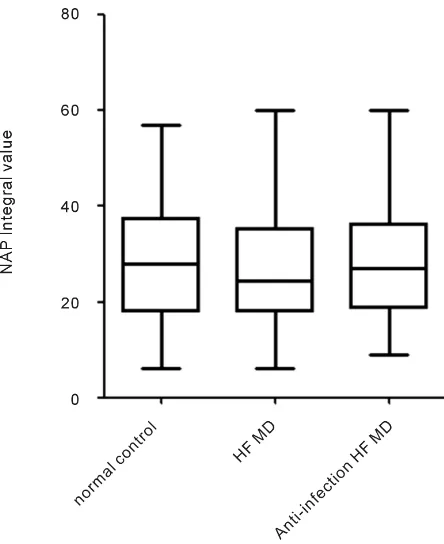
Among the 4 group patients, NAP positive rate of the normal control is (23.58 ± 11.89)% and integral value is 28.18 ± 13.82 (Figure 1); NAP positive rate of HFMD group is (22.8 ± 10.49)% and integral value is 26.92 ± 11.9; NAP positive rate of the HFMD co-infection group is (77.96 ± 8.99)% and integral value is 332.7 ± 58.42; NAP positive rate of Anti-infection HFMD group is (22.38 ± 10.54)% and integral value is 27.74 ± 12.16; Comparing the NAP positive rate of
Figure 1. NAP integral value in cases and controls.
HFMD concurrent infection group with the NAP positive rate of other three groups respectively, those are reference group, HFMD group and Anti-infection HFMD group, t value is 25.7964, 28.2326, 28.3696. The values of P are 0.0000; comparing the NAP integral value of HFMD co-infection with the NAP integral value of other three groups respectively, those are reference group, HFMD group and Anti-infection HFMD, t value is 35.8687, 36.2664, 36.1374. The values of P are 0.0000; there is no statistical significance to compare the NAP positive rate and the NAP integral value among the normal control, HFMD group and An-ti-infection HFMD. Abnormal lymphocytes and vacuoles degeneration occasio-nally be observed from the result of White blood cell morphology of both the normal control group and the HFMD group. Many abnormal lymphocytes, toxic particles cavitating cells and Dohle body can be observed from the result of White blood cell morphology of both HFMD concurrent infection group and Anti-infection HFMD group. The comparison result of the incidence of abnor-mal lymphocytes and vacuolar degeneration cells in White blood cell morphol-ogy between the normal control group and the HFMD group shows that the t is respectively 5.8005, 8.0225, the all P is respectively 0.0000.The comparison result of the incidence of abnormal lymphocytes and vacuolar degeneration cells in White blood cell morphology between the normal control group and shows that t is respectively 15.3897, 18.7609, the all P is respectively 0.0000.Contrast the in-cidence of abnormal lymphocytes and vacuolar degeneration cells in White blood cell morphology between the normal control group and the Anti-infection group we can see that t is respectively 2.5586, 1.8123, P is respectively 0.0060,
0.0365;Contrast the incidence of abnormal lymphocytes and vacuolar degenera-tion cells in White blood cell morphology between the HFMD group and the HFMD concurrent infection group, we can see that t is respectively 7.0166, 16.7142, the all P are respectively 0.0000. The comparison result of the incidence of abnormal lymphocytes and vacuolar degeneration cells in White blood cell morphology between the HFMD group and Anti-infection HFMD group shows that it is 4.3740 and 7.7346 respectively, the all P are respectively 0.0000.The comparison result of the incidence of abnormal lymphocytes ,toxic particles ca-vitating cells , vacuolar degeneration cells and Dohle body in White blood cell morphology between the HFMD concurrent infection group and the An-ti-infection HFMD group shows that t is respectively 16.0121, 28.0628, 18.5339, 6.0320, the all P are respectively 0.0000. Detail result is shown in below Table 2 and Table 3. White blood cell morphology is shown in below Figure 2.
[image:5.595.206.539.439.523.2]Figure 2. White blood cell morphology ((a) Toxic particles cavitating cells; (b) Abnormal lymphocytes; (c) Toxic particles).
Table 2. Comparison of NAP positive rate and integral value of 4 groups.
Group control group The normal The HFMD group HFMD concurrent infection group The anti-infection HFMD group
Positive
rate % 23.58 ± 11.89 22.8 ± 10.49 77.96 ± 8.99 22.38 ± 10.54 Integral
value 28.18 ± 13.82 26.92 ± 11.9 332.7 ± 58.42 27.74 ± 12.16
Table 3. The comparison of four groups’ WBC morphology.
Group The normal control group
The HFMD group
HFMD concurrent infection group
The anti-infection HFMD group
Abnormal
lymphocytes (%) 1.52 ± 1.93 4.04 ± 2.39 6.84 ± 1.50 2.36 ± 1.29
Toxic particles
cells (%) 0 0 40.14 ± 9.44 2.26 ± 1.41
Vacuolar
degeneration cells (%) 0.96 ± 1.43 3.10 ± 1.23 19.76 ± 6.94 1.40 ± 0.95
Durer
corpuscle (%) 0 0 2.90 ± 2.73 0.50 ± 0.68
[image:5.595.208.538.556.744.2]4. Discussion
HFMD (hand-foot-and-mouth disease) a kind of infectious disease caused by intestinal virus serious damage to children’s healthy life. According to the report, more than 20 intestinal virus strain can cause HFMD, among which Coxsackie A16 (CA16) and enterovirus 71 (EV 71) is the most common [13] [14] [15].
Children under the age of five are susceptible to infection. Who infected usually has symptoms such as low thermal pain, anorexia, oral pain, hands, foot and mouth with small herpes or ulcers. Most infected children whose immune system is strong can heal in a week, while the other few patients with complica-tions such as pulmonary edema, aseptic meningoencephalitis, myocarditis, etc. [16] [17] [18]. Some severe individual patient’s disease develops rapidly, causing heart failure and death. Currently there is no particularly effective medicine for the treatment, most of the clinical treatment can only symptomatic treatment [19] [20].
This paper is mainly study the changes of White blood cell morphology of those HFMD concurrent infection and the integral value and positive rate of NAP and analyze and contrast the differences with the normal control group, HFMD group and the Anti-infection HFMD group. The results showed that Neutrophil alkaline phosphatase positive rate and integral value of HFMD in-fected with complications is higher contrast the normal control group, HFMD group and the Anti-infection HFMD group. The comparison results of positive rate: t are 25.7964, 28.2326, 28.3696, P are all 0.0000; The comparison results of integral value: t are 35.8687, 36.2664, 36.1374, P are all 0.0000; he comparison results of the other groups is no statistical significance, all P are less than 0.05, P < 0.05. That is to say, positive rate and integral value of NAP of those infected HFMD with complications changes a lot and become normal after anti-infection. This suggests that clinic those infected HFMD with complications should do an-ti-infection treatment as soon as possible so that avoiding aggravation infection and endangering life. So it has very important clinical significance for those with HFMD should do examination in time to exclude the concurrent infection.
This study also shows that WBC morphological changes of patient who with HFMD concurrent infection changes more obviously than the one of the regular group, the HFMD group and Anti-infection HFMD group. Abnormal lympho-cytes and vacuoles degeneration occasionally be observed from the result of White blood cell morphology of both the reference group and the HFMD group. Many abnormal lymphocytes, toxic particles cavitating cells and Durer corpuscle can be observed from the result of White blood cell morphology of both HFMD concurrent infection group and the Anti-infection HFMD group. From compar-ison data of HFMD concurrent infection group and the others three groups, the regular group, the HFMD group and the Anti-infection HFMD, it can be found that the positive rate of abnormal lymphocytes, the t are respectively 15.3897, 7.0166, 16.0121, P are all 0.0000 and the positive rate of Vacuoles degeneration of cells, the t are respectively 18.7609, 16.7142, 18.5339, P are all 0.0000. Toxic
particles cavitating cells and Durer corpuscle was not found from test results of the WBC morphology of both the regular group and the HFMD group. From comparison data of HFMD concurrent infection group and the Anti-infection HFMD, it can be found that the positive rate of toxic particles cavitating cells and Durer corpuscle, the t are respectively 28.0628, 6.0320, P are all 0.0000. It is approved that changes of the leukocyte morphology of those HFMD concurrent infection is a very important measurement index for indicating and monitoring the status of WBC morphology. It enables doctor to timely intervention, so as to achieve therapeutic effect.
5. Conclusion
As mentioned above, it has very important clinical significance that taking the examination of leukocyte morphology and the positive and integral value of NAP as the diagnosis index and the prognosis monitoring for pediatric HFMD con-current infection. It is worth to promote in other hospitals.
6. The Limitation of the Study
The sample cases only include the patient in our hospital, and the study result just reflect the local situations not other places. A lot of manpower material re-sources is necessary if study the situation of other places. What’s more, due to selecting the sample cases take a long time and large span on the test time of all observation indexes, so there is some limitation in quality control.
Acknowledgements
In help and support of all colleagues and especially the hard work of all members of the team, this project can make it. Thanks for all colleagues who has made a great effort to this project. Wish them good health! Happy family! All the best!
Fund Project
Yulin Scientific research and technology development plan program Guangxi Zhuang Autonomous Region (No. Yushike 20171629).
References
[1] Huang, Y., Deng, T., Yu, S., et al. (2013) Effect of Meteorological Variables on the Incidence of Hand, Foot, and Mouth Disease in Children: A Time-Series Analysis in Guangzhou, China. BMC Infectious Diseases, 13, 134-134.
https://doi.org/10.1186/1471-2334-13-134
[2] Kashyap, R.R. and Kashyap, R.S. (2015) Hand, Foot and Mouth Disease—A Short Case Report. Journal of Clinical and Experimental Dentistry, 7, 336-338.
https://doi.org/10.4317/jced.52031
[3] Long, L., Xu, L., Xiao, Z., et al. (2016) Neurological Complications and Risk Factors of Cardiopulmonary Failure of EV-A71-Related Hand, Foot and Mouth Disease.
Scientific Reports, 6, 23444-23444. https://doi.org/10.1038/srep23444
[4] Keawcharoen, J. (2012) Hand, Foot, and Mouth Disease. Thai Journal of Veterinary
Medicine, 42, 225-257.
[5] Banta, J., Lenz, B., Pawlak, M., et al. (2016) Notes from the Field: Outbreak of Hand, Foot, and Mouth Disease Caused by Coxsackievirus A6 among Basic Military Trai-nees—Texas, 2015. Morbidity and Mortality Weekly Report, 65, 678-680.
https://doi.org/10.15585/mmwr.mm6526a3
[6] Feder, H.M., Bennett, N., Modlin, J.F., et al. (2014) Atypical Hand, Foot, and Mouth Disease: A Vesiculobullous Eruption Caused by Coxsackie Virus A6. Lancet Infec-tious Diseases, 14, 83-86. https://doi.org/10.1016/S1473-3099(13)70264-0
[7] Gaunt, E., Harvala, H., Osterback, R., et al. (2015) Genetic Characterization of Hu-man Coxsackievirus A6 Variants Associated with Atypical Hand, Foot and Mouth Disease: A Potential Role of Recombination in Emergence and Pathogenicity. Jour-nal of General Virology, 96, 1067-1079. https://doi.org/10.1099/vir.0.000062
[8] Vuorinen, T., Osterback, R., Kuisma, J., et al. (2014) Epididymitis Caused by Cox-sackievirus A6 in Association with Hand, Foot, and Mouth Disease. Journal of Clinical Microbiology, 52, 4412-4413. https://doi.org/10.1128/JCM.02441-14
[9] Li, T., Yang, Z., Liu, X., et al. (2014) Hand-Foot-and-Mouth Disease Epidemiologi-cal Status and Relationship with MeteorologiEpidemiologi-cal Variables in Guangzhou, Southern China, 2008-2012. Revista Do Instituto De Medicina Tropical De Sao Paulo, 56, 533-539. https://doi.org/10.1590/S0036-46652014000600014
[10] Huang, X., Wei, H., Wu, S., et al. (2015) Epidemiological and Etiological Characte-ristics of Hand, Foot, and Mouth Disease in Henan, China, 2008-2013. Scientific Reports, 5, 8904-8904. https://doi.org/10.1038/srep08904
[11] Han, J., Xu, S., Zhang, Y., et al. (2014) Hand, Foot, and Mouth Disease Outbreak Caused by Coxsackievirus A6, China, 2013. Journal of Infection, 69, 303-305. https://doi.org/10.1016/j.jinf.2014.03.015
[12] Yan, X., Zhang, Z.Z., Yang, Z.H., et al. (2015) Clinical and Etiological Characteris-tics of Atypical Hand-Foot-and-Mouth Disease in Children from Chongqing, Chi-na: A Retrospective Study. BioMed Research International, No. 8, 1-8.
[13] Lin, H., Sun, L., Lin, J., et al. (2014) Protective Effect of Exclusive Breastfeeding against Hand, Foot and Mouth Disease. BMC Infectious Diseases, 14, 645-645. https://doi.org/10.1186/s12879-014-0645-6
[14] Ventarola, D., Bordone, L., Silverberg, N.B., et al. (2015) Update on Hand-Foot-and-Mouth Disease. Clinics in Dermatology, 33, 340-346.
https://doi.org/10.1016/j.clindermatol.2014.12.011
[15] Zhang, S. and Zhao, J. (2015) Spatio-Temporal Epidemiology of Hand, Foot and Mouth Disease in Liaocheng City, North China. Experimental and Therapeutic Medicine, 9, 811-816.https://doi.org/10.3892/etm.2015.2207
[16] Wang, Z.L., Xia, A., Li, Y., et al. (2016) Socioeconomic Burden of Hand, Foot and Mouth Disease in Children in Shanghai, China. Epidemiology and Infection, 144, 138-143.https://doi.org/10.1017/S0950268815001569
[17] Li, Y., Zhang, J., Zhang, X., et al. (2014) Modeling and Preventive Measures of Hand, Foot and Mouth Disease (HFMD) in China. International Journal of Envi-ronmental Research and Public Health, 11, 3108-3117.
https://doi.org/10.3390/ijerph110303108
[18] Zhang, J., Kang, Y., Yang, Y., et al. (2015) Statistical Monitoring of the Hand, Foot and Mouth Disease in China. Biometrics, 71, 841-850.
https://doi.org/10.1111/biom.12301
[19] Zhang, X., Wang, H., Ding, S., et al. (2013) Prevalence of Enteroviruses in Children
with and without Hand, Foot, and Mouth Disease in China. BMC Infectious Dis-eases, 13, 606-606.https://doi.org/10.1186/1471-2334-13-606
[20] Mao, Q., Wang, Y., Bian, L., et al. (2016) EV71 Vaccine, a New Tool to Control Outbreaks of Hand, Foot and Mouth Disease (HFMD). Expert Review of Vaccines, 15, 599-606.https://doi.org/10.1586/14760584.2016.1138862