The chemokine receptor CXCR3 is expressed
on malignant B cells and mediates chemotaxis
Livio Trentin, … , Renato Zambello, Gianpietro Semenzato
J Clin Invest.
1999;
104(1)
:115-121.
https://doi.org/10.1172/JCI7335
.
B- and T-cell recirculation is crucial for the function of the immune system, with the control of
cell migration being mainly mediated by several chemokines and their receptors. In this
study, we investigated the expression and function of CXCR3 on normal and malignant B
cells from 65 patients with chronic lymphoproliferative disorders (CLDs). Although CXCR3
is lacking on CD5
+and CD5
–B cells from healthy subjects, it is expressed on leukemic B
lymphocytes from all (31/31) patients with chronic lymphocytic leukemia (CLL). The
presence of CXCR3 was heterogeneous in other B-cell disorders, being expressed in 2 of 7
patients with mantle cell lymphoma (MCL), 4 of 12 patients with hairy cell leukemia (HCL),
and 11 of 15 patients with other subtypes of non-Hodgkin’s lymphomas (NHLs). Chemotaxis
assay shows that normal B cells from healthy subjects do not migrate in response to
IFN-inducible protein 10 (IP-10) and IFN-
g
–induced monokine (Mig). In contrast, a definite
migration in response to IP-10 and Mig has been observed in all malignant B cells from
patients with CLL, but not in patients with HCL or MCL (1/7 cases tested). Neoplastic B cells
from other NHLs showed a heterogenous pattern. The migration elicited by IP-10 and Mig
was inhibited by blocking CXCR3. No effect of IP-10 and Mig chemokines was observed on
the cytosolic calcium concentration in […]
Article
Introduction
The superfamily of chemokines consists of an array of chemoattractant proteins that has been divided into 4 branches (C, CC, CXC, and CXXC) on the basis of the relative position of the cysteine residues in the mature protein (1–6). Structural variations of chemokines have been demonstrated to be associated with differences in their ability to regulate the trafficking of immune cells during hematopoiesis and inflammatory responses (1, 2, 7). Chemokines exert their attractant properties after binding to distinct membrane receptors. Because a sin-gle chemokine receptor binds several chemokines, it is often difficult to evaluate the activity of these structures in lymphocyte homing. For instance, IFN-inducible pro-tein 10 (IP-10) and IFN-γ–induced monokine (Mig) 2 CXC chemokines that are induced by IFN-γ (1, 4), bind the CXCR3 receptor and have been shown to be specifi-cally chemotactic for activated lymphocytes (8).
The recently cloned CXCR3 receptor cDNA (8) has been reported to be expressed on activated T lympho-cytes after in vitro stimulation, but it is usually lacking in, or present in only a small fraction of, resting T
lym-phocytes, B cells, monocytes, and granulocytes (9–14). It has been initially observed to mediate calcium changes and chemotaxis in response to IP-10 and Mig, but not to other chemokines (1, 4). Recently, IFN-inducible T-cell alpha chemoattractant (I-TAC) has also been observed to bind CXCR3 (13).
The mechanisms controlling malignant B-cell traf-ficking in the macro- and microenvironments are poor-ly understood. It is not clear why some disorders are preferentially confined to a limited number of organs, e.g., hairy cell leukemia (HCL), and why others, e.g., B-cell chronic lymphocytic leukemia (CLL), present with a wide diffusion to peripheral blood and other struc-tures at the onset of the disease. In this study, we inves-tigated the expression and chemotactic function of the CXCR3 receptor on normal B lymphocytes from healthy subjects and on malignant B cells from patients with different types of B-cell chronic lymphoprolifera-tive disorders (CLDs), including CD5+B-cell disorders,
e.g., CLL and mantle cell lymphoma (MCL), and CD5–
CLD, e.g., HCL and several subtypes of non-Hodgkin’s lymphomas (NHLs).
The chemokine receptor CXCR3 is expressed on
malignant B cells and mediates chemotaxis
Livio Trentin,
1Carlo Agostini,
1Monica Facco,
1Francesco Piazza,
1Alessandra Perin,
1Marta Siviero,
1Carmela Gurrieri,
1Silvia Galvan,
1Fausto Adami,
1Renato Zambello,
2and Gianpietro Semenzato
11Padua University School of Medicine, Department of Clinical and Experimental Medicine,
Clinical Immunology Branch, 35128 Padova, Italy
2Division of Hematology, Vicenza Hospital, 36100 Vicenza, Italy
Address correspondence to: Gianpietro Semenzato, Università di Padova, Dipartimento di Medicina Clinica e Sperimentale, Immunologia Clinica, Via Giustiniani 2, 35128 Padova, Italy. Phone: 39-049-821-2298;
Fax: 39-049-875-4179; E-mail: giansem@ux1.unipd.it.
Received for publication May 13, 1999, and accepted May 18, 1999.
B- and T-cell recirculation is crucial for the function of the immune system, with the control of cell migration being mainly mediated by several chemokines and their receptors. In this study, we inves-tigated the expression and function of CXCR3 on normal and malignant B cells from 65 patients with chronic lymphoproliferative disorders (CLDs). Although CXCR3 is lacking on CD5+and CD5–B cells
from healthy subjects, it is expressed on leukemic B lymphocytes from all (31/31) patients with chron-ic lymphocytchron-ic leukemia (CLL). The presence of CXCR3 was heterogeneous in other B-cell disorders, being expressed in 2 of 7 patients with mantle cell lymphoma (MCL), 4 of 12 patients with hairy cell leukemia (HCL), and 11 of 15 patients with other subtypes of non-Hodgkin’s lymphomas (NHLs). Chemotaxis assay shows that normal B cells from healthy subjects do not migrate in response to IFN-inducible protein 10 (IP-10) and IFN-γ–induced monokine (Mig). In contrast, a definite migration in response to IP-10 and Mig has been observed in all malignant B cells from patients with CLL, but not in patients with HCL or MCL (1/7 cases tested). Neoplastic B cells from other NHLs showed a het-erogenous pattern. The migration elicited by IP-10 and Mig was inhibited by blocking CXCR3. No effect of IP-10 and Mig chemokines was observed on the cytosolic calcium concentration in malig-nant B cells. The data reported here demonstrate that CXCR3 is expressed on maligmalig-nant B cells from CLDs, particularly in patients with CLL, and represents a fully functional receptor involved in chemo-taxis of malignant B lymphocytes.
Methods
Patient samples. Sixty-five patients with different B-cell malignancies had been studied at the time of diagno-sis. Thirty-one patients (16 men and 15 women, ages 48–78 years) with the diagnosis of B-CLL (15) were graded according to the Rai staging system (16) as fol-lows: stage 0 (3 cases), stage I (12 cases), stage II (10 cases), stage III (4 cases), and stage IV (2 cases); the total lymphocyte count ranged from 16,000 to 98,000/mm3.
Seven patients (6 men and 1 woman, ages 54–72 years) with the diagnosis of MCL (15) and with malig-nant B cells in the peripheral blood were studied.
Twelve patients with HCL (7 men and 5 women, ages 44–68 years) were studied. The diagnosis was established on the basis of clinical, morphological, cytochemical, his-tological, and immunological features (17). Fifteen patients (7 men and 8 women) with different histological entities of NHL (4 marginal zone, 8 lymphocytic, and 3 lymphoplasmacytic) in the leukemic phase were studied. Preparation of cell suspensions. PBMCs from patients with B-CLD were obtained from freshly heparinized blood samples by centrifugation on Ficoll-Hypaque (F/H) gradient (18). Normal B lymphocytes were obtained from 2 spleen specimens and from 6 tonsils after mechanic disruption (19). Mononuclear cells, recovered after centrifugation on F/H gradient, were washed 3 times with PBS and resuspended in endotox-in-free RPMI-1640 medium (Sigma Chemical Co., St. Louis, Missouri, USA) supplemented with 20 mM HEPES and L-glutamine, 100 U/mL penicillin, 100
µg/mL streptomycin, and 10% FCS (ICN Flow, Costa Mesa, California, USA) (19)
Cell samples with a percentage of monocytes and T and natural killer (NK) cells greater than 5% were fur-ther enriched in B lymphocytes by rosetting with sheep
red blood cells treated with neuroaminidase (Sigma Chemical Co.) and by removing residual CD3+, CD16+,
CD56+, and CD14+cells using magnetic separation
columns (Miltenyi Biotec, Bergisch Gladbach, Ger-many), as described previously (20). After the multi-step negative selection procedure, more than 98% of the resulting cell population was CD19+. The
purifi-cation of CD5+B cells was performed by a positive
selection using anti-CD5 mAb’s.
mAb’s and cytokines. CXCR3 receptor analysis was per-formed using 1C6 mAb (kindly provided by S. Qin, LeukoSite, Cambridge, Massachusetts, USA) (9). IP-10 and Mig chemokines were purchased from R&D Sys-tems Inc. (Minneapolis, Minnesota, USA).
For FACS analysis, 10–4cells were acquired and the
analysis was determined by overlaying the histograms of the samples stained with the different reagents. Cells were scored using a FACScan analyzer (Becton Dickinson Immunocytometry Systems, San Jose, Cal-ifornia, USA), and data were processed using the Cel-lQuest or Lysys software programs (Becton Dickin-son Immunocytometry Systems). Mean log fluorescence intensity (MFI) values were obtained by subtracting the MFI of the isotype control from the MFI of the positively stained sample. Furthermore, to evaluate whether the differences between the peaks of cells were statistically significant with respect to control, the Kolmogorov-Smirnov test for analysis of histograms was used, according to the CellQuest software user’s guide.
[image:3.612.80.522.48.254.2]Migration assay. Cell migration was measured in a 48-well modified Boyden chamber. The rhIP-10 and rhMig chemokines were diluted in RPMI-1640 medi-um at different concentrations (20 and 200 ng/mL for IP-10; 100 and 1,000 ng/mL for Mig) and were used to
Figure 1
Flow cytometry analysis of the expression of CXCR3 receptor on CD5+and CD5–B cells from healthy subjects and on malignant B
evaluate the chemotactic properties of B lym-phocytes from healthy donors and patients. The CXCR3–and CXCR3+cell lines (300-19; kindly
provided by B. Moser, Theodor-Kocher Institute, University of Bern, Bern, Switzerland) were used as negative and positive controls, respectively. Poly-vinylpyrrolidone-free polycarbonate mem-branes coated with fibronectin were used (3-µm pores for CLD; 5-µm pores for cell lines). A total of 30 µL of chemokines or control medium was added to the bottom wells, and 50 µL of 5.0 ×10⁶
cells/mL B cells or CXCR3–/CXCR3+cells
resus-pended in RPMI-1640 was added to the top wells. The chamber was incubated at 37°C with 5% CO2for 2 hours. The membranes were then
removed, washed with PBS on the upper side, and fixed and stained with Diff-Quick (Dade AG, Düdingen, Switzerland). Cells were counted at ×800 magnification in 3 fields per well. All assays were per-formed in triplicate. In blocking experiments, cell sus-pensions were preincubated before chemotaxis assay for 30 minutes at 4°C with anti-human CXCR3 mAb at the concentration of 20 µg/mL.
Cytosolic calcium. Changes in the intracellular calcium concentration ([Ca2+]i)were measured in 10 patients
with CLL with the fluorescent indicator fura-2/AM, as described previously (22). Briefly, 20 ×106cells were
incubated with 2 µM fura-2/AM at 37°C for 40 min-utes. After the loading procedure, aliquots of the cells (2 ×10⁶) were rapidly washed and resuspended in a
magnetically stirred thermostatted cuvette. Excitation and emission wavelengths were 340 and 500 nm, respectively; the excitation slit width was 5 nm, and the emission slit was 10 nm. The inhibitor of organic ion transport sulfinpyrazone was added in some experi-ments during [Ca2+]
imeasurements at a concentration
of 250 µM to prevent fura-2 release into the medium (23). Control experiments without sulfinpyrazone gave essentially the same results, except for a slowly increas-ing baseline due to fura-2 leakage (23).
Statistical analysis. Data are expressed as mean ± SEM, and comparisons between values were made using the Student’s ttest. P< 0.05 was considered as significant.
Results
Expression of CXCR3 chemokine receptor on normal and malignant B cells. The CXCR3 chemokine receptor was analyzed on normal B lymphocytes obtained from healthy donors and on tumor B cells recovered from patients with different B-cell malignancies (Table 1). Among different groups of disorders, high levels of CXCR3 expression were observed on B cells from CLL (172 ± 17 MFI) and NHL (151 ± 38 MFI) compared with normal CD5+and CD5–B lymphocytes (26 ± 3 and 35 ±
[image:4.612.279.535.78.163.2]17 MFI, respectively), with P< 0.001 and < 0.05, respec-tively. By contrast, only a few patients with HCL (4/12)
Table 1
Flow cytometry analysis of CXCR3 in B-cell CLDs and normal subjects
CD5+B lymphocytes CD5–B lymphocytes
Healthy CLL MCL Healthy HCL NHL
subjects subjects
MFIA 26 ± 3 172 ± 17 120 ± 50 35 ± 6 79 ± 32 151 ± 38
P P< 0.001 NS NS P< 0.05
No. positive 0/6 31/31 2/7 0/6 4/12 11/15 samples
AData are reported as mean ± SE of the MFI values for each group of subjects after
subtracting the MFI of isotype control mAb from the MFI of positively stained sam-ples. Comparisons between patients’ values and control subjects were made using the Student’s ttest. P< 0.05 was considered as significant. NS, not significant.
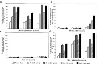
Figure 2
Chemotactic activity of CXCR3+and CXCR3–cell
lines and normal and malignant CD5+and CD5–
[image:4.612.59.333.488.733.2]and MCL (2/7) expressed CXCR3. Representative flow cytometry histograms obtained from subjects belong-ing to different groups are reported in Figure 1.
Cell migration assay. To characterize further the bio-logical properties of the CXCR3 chemokine receptor, normal and malignant B cells were assessed for their migration properties. Normal CD5+and CD5–B cells
from healthy subjects displayed very low or unde-tectable migration activity (Figure 2, b and c). Among CD5+B-cell malignancies, CLL leukemic cells from 13
of 13 patients tested exhibited a high migration activi-ty both at resting conditions and after triggering with IP-10 and Mig chemokines, whereas CD5+tumor B
cells from patients with MCL displayed low or unde-tectable migration. When CD5–malignant B cells were
considered (Figure 2c), hairy cells did not display any migration activity at resting conditions or after in vitro incubation with IP-10 and Mig chemokines. Further-more, tumor B lymphocytes recovered from subjects with NHL displayed a discrete chemotactic function, with the highest activity being usually observed in mar-ginal zone NHL.
Migration activity displayed by representative cases is shown in Figure 3. The test was performed with fresh-ly isolated B cells in the absence and presence of differ-ent concdiffer-entrations of IP-10 and Mig chemokines. Data reported here demonstrate that migration is mainly induced in CLL and NHL, whereas MCL and HCL tumor B cells are poorly induced to migrate. In addi-tion, among patients with NHL, the highest activity was demonstrated in 2 subjects with marginal zone B-cell lymphoma.
To verify further the role of CXCR3 in B-cell chemo-taxis, malignant B cells responsive to IP-10– and Mig-induced chemotaxis were preincubated with anti-CXCR3 mAb. The blocking of the anti-CXCR3 receptor determined a marked inhibition of both IP-10– and Mig-induced chemotaxis (Figure 4).
Cytosolic calcium measurement. We also examined the ability of IP-10 and Mig to stimulate functional respons-es in malignant B cells from patients with CLD through the evaluation of cytosolic calcium levels. The analysis was performed in 10 patients with CLL, and data relat-ed to 3 representative cases are reportrelat-ed in Figure 5. The assay was performed with both IP-10 and Mig at a 1 µM concentration. The data reported here demonstrate that no increase in [Ca2+]
i was observed after the addition of
IP-10 or Mig chemokines to malignant B cells. In con-trast, a significant increase was observed after addition of an anti-immunoglobulin antibody.
Discussion
The data reported here demonstrate that CXCR3 is constitutively expressed on malignant B cells obtained from patients with B-CLL, whereas its expression on other B-cell disorders is quite heterogeneous. Chemo-tactic in vitro studies showed that CXCR3 behaves as a functional receptor on CLL cells, as it transduces chemotactic activity after binding to relevant chemokines (IP-10 and Mig).
[image:5.612.112.492.52.304.2]Data from the literature have demonstrated that CXCR3 is commonly expressed on activated T cells and on a discrete proportion of resting T and NK cells (9, 11, 24). As far as normal B cells are concerned, only a
Figure 3
Chemotactic activity of representative patients with CD5+and CD5–B-lymphocyte malignancies to different concentrations of IP-10 (20 and
few peripheral blood B lymphocytes express the CXCR3 receptor (9). Our findings extend these obser-vations to the malignant counterpart of B cells and clearly indicate that CXCR3 is expressed on circulating B cells from patients with B-cell malignancies, in par-ticular from patients with CLL. The observation that CXCR3 is expressed on CLL leukemic cells suggests that this receptor might represent a hallmark of this disease. In fact, the expression of CXCR3 in other B-cell malignancies studied in this work is quite heteroge-neous, usually absent in HCL, and expressed at variable degrees of intensity in other NHLs. When compared with purified normal B cells, the differences in terms of MFI were statistically significant in CLL and NHL, but not in HCL and MCL.
Lymphocyte trafficking is a critical step of the sys-temic immunity (1–3). This process includes migration of T lymphocytes from the bone marrow and thymus to spleen and lymph nodes, as well as the migration of B lymphocytes in the lymph node microenvironment, i.e., from the mantle zones to germinal centers. Con-sidering the promiscuity of chemokines and their receptors, it is still difficult to understand how the cel-lular trafficking is regulated. Although the functions of some chemokines, e.g., stromal cell–derived factor-1 (SDF-1), are well characterized in terms of their ability to recruit B cells into the microenvironment of stroma cells (25), the role of IP-10 and Mig in B-cell trafficking is not defined. In addition to their chemotactic activi-ty for T lymphocytes, IP-10 and Mig share other prop-erties, including inhibition of neovascularization, inhi-bition of progenitor cells, and antitumor effects (4). For these reasons, the expression of CXCR3 may have a dual effect in these B-cell malignancies. Its presence may favor the trafficking of malignant B cells from one site to another site of disease involvement, thus favor-ing the spreadfavor-ing of the disease to peripheral blood, lymph nodes, bone marrow, and other organs. It could also be suggested that the interaction between CXCR3 and relevant chemokines might affect tumor growth by attracting T cells with antitumor functions. In this
view, a selection of T cells with a limited T cell–receptor repertoire has been observed in CLL and HCL (26, 27); these findings might be viewed as a possible conse-quence of an antitumor reaction.
The functional data on the CXCR3 receptor report-ed in this work deserve 2 considerations. The first comes from the demonstration that malignant B cells from patients with CLD are equipped with a chemo-tactic activity, not only in response to IP-10 and Mig chemokines but also in the absence of exogenous chemokine stimuli, indicating that these cells sponta-neously migrate in vitro. This effect was clearly detectable in patients with CLL and NHL. This find-ing may trigger speculation that other chemokines are likely to be involved in B-cell chemotaxis in patients with CLD. A second question deals with the evidence that although the triggering of the B-cell receptor by anti-immunoglobulin antibodies induced increases in the cytosolic calcium concentration, IP-10 and Mig binding to CXCR3 on malignant B cells did not. This latter observation might be a consequence of the den-sity of CXCR3 expression on malignant B cells or of the state of activation of these cells. In this regard, an increase in cytosolic calcium levels has not been detect-ed on resting human T lymphocytes, although the cells are equipped with surface CXCR3 (28). Calcium flux was observed only after in vitro preactivation of relevant cells (28). Alternatively, our findings could be the result of different in vitro experimental conditions to detect chemotaxis (i.e., migration assay in cells adhering to a substratum) and cytosolic calcium levels (i.e., calcium measurement in cell suspension) in the in vivo setting (29). This interpretation is consistent with recent evidence that calcium flux and chemotaxis are dissociated phenomena. In fact, calcium fluxes have been shown to be neither necessary nor sufficient for chemotaxis in standard assays using a variety of chemotactic factors (28).
[image:6.612.62.460.541.735.2]The evidence that CLL (a disease with a diffuse spreading) constitutively expresses CXCR3, whereas HCL (a disease usually limited to the spleen and bone
Figure 4
marrow) does not express CXCR3, indicates that this receptor plays a crucial role in the homing of malignant B cells. Although we did not find any correlation between the number of malignant B cells in the periph-eral blood and the intensity of the expression of CXCR3 on these cells, the highest levels of MFI were observed in patients with high numbers of hairy cells in the peripheral blood, compared with patients with only few circulating hairy cells. Furthermore, the evi-dence that patients with HCL have low or undetectable numbers of monocytes, which represent a relevant source of these chemokines, may further support the inability or the difficulty of hairy cells to spread to blood and lymph nodes. With respect to patients with NHL, our data demonstrate that some of them, e.g., marginal zone B-cell lymphomas with spleen involve-ment, bear CXCR3 receptor and show a strong migra-tion activity. This finding might be interpreted as a fur-ther demonstration that this receptor might correlate with the homing of malignant B cells and, in this par-ticular histotype, with the capability of these cells to migrate to circulation and different lymphoid organs. The complexity of this trafficking comes from the demonstration that most chemokines bind to 1 or more receptors and that a single receptor can bind several cytokines, usually either CC or CXC chemokines, but not both. In contrast, CXCR3 has been observed to bind, although with low affinity, CCR3 ligands that may affect CXCR3-mediated properties (24). Therefore, the num-ber of receptors on the cell surface, and its affinity and chemokine gradients at the site of malignant involve-ment, may play a key role in favoring the spread or exten-sion of the neoplastic process. In this context, it may be hypothesized that in the future the clinical use of recep-tor antagonists, e.g., eotaxin and other chemokines (30), might positively affect the spreading of the disease.
Acknowledgments
This work was supported by the Italian Association for Cancer Research (AIRC) and by the Ministero dell’ Uni-versitá e della Ricerca Scientifica e Technologica (MURST). The authors thank Tullio Pozzan and Cristi-na Fasolato for calcium flux evaluation and for review-ing the manuscript; Martin Donach for help in the preparation of the manuscript; S. Qin for providing the anti-CXCR3 mAb; and B. Moser for providing the 300-19 cell line.
1. Baggiolini, M., Dewald, B., and Moser, B. 1997. Human chemokines: an update. Annu. Rev. Immunol. 15:675–705.
2. Baggiolini, M. 1998. Chemokines and leukocyte traffic. Nature.
392:565–568.
3. Rollins, B. 1997. Chemokines. Blood. 90:909–928.
4. Farber, J.M. 1997. Mig and IP-10: CXC chemokines that target lympho-cytes. J. Leukoc. Biol. 61:246–257.
5. Yoshie, O., Imai, T., and Nomiyama, H. 1997. Novel lymphocyte-specif-ic CC chemokines and their receptors. J. Leukoc. Biol. 62:634–644. 6. Nelson, P.J., and Krensky, A.M. 1998. Chemokines, lymphocytes and
virus-es: what goes around, comes around. Curr. Opin. Immunol. 10:265–270. 7. Luster, A. 1998. Chemokines: chemotactic cytokines that mediate
inflammation. N. Engl. J. Med. 228:436–445.
8. Loetscher, M., et al. 1996. Chemokine receptor specific for IP10 and Mig: structure, function and expression in activated T-lymphocytes. J. Exp. Med. 184:963–969.
9. Qin, S., et al. 1998. The chemokine receptors CXCR3 and CCR5 mark subsets of T cells associated with certain inflammatory reactions. J. Clin. Invest. 101:746–754.
10. Piali, L., et al. 1998. The chemokine receptor CXCR3 mediates rapid and shear-resistant adhesion-induction of effector T lymphocytes by the chemokines IP10 and Mig. Eur. J. Immunol. 28:961–972.
11. Loetscher, M., Loetscher, P., Brass, N., Meese, E., and Moser, B. 1998. Lymphocyte-specific chemokine receptor CXCR3: regulation, chemokine binding and gene localization. Eur. J. Immunol. 28:3696–3705. 12. Sallusto, F., Lenig, D., MacKay, C.R., and Lanzavecchia, A. 1998. Flexible programs of chemokine receptor expression on human polarized helper 1 and 2 lymphocytes. J. Exp. Med. 187:875–883.
13. Cole, K.E., et al. 1998. Interferon-inducible T cell alpha chemoattractant (I-TAC): a novel non-ELR CXC chemokine with potent activity on acti-vated T cells through selective affinity binding to CXCR3. J. Exp. Med.
187:2009–2021.
[image:7.612.57.387.50.295.2]14. Kim, C.H., and Broxmeyer, H.E. 1999. Chemokines: signal lamps for traf-ficking of T and B cells for development and effector function. J. Leukoc. Biol. 65:6–15.
Figure 5
Effect of IP-10 and Mig on cytosolic calcium concentration in 3 representa-tive patients with CLL. Neither IP-10 nor Mig induced calcium mobilization in patients with leukemic CLL. A dis-crete calcium increase was observed after cross-linking surface membrane immunoglobulins. When indicated, the concentration of the stimuli was 1
µg/mL. At the end of each experiment, the calcium ionophore ionomycin (1 µM) was added to induce a massive calcium increase. The values are expressed as the ratio between the intensity of the fluorescence signals obtained by exciting at 340 and 380 nm. An increase in this ratio reflects an increase in [Ca2+]
i. The marginal
15. Harris, N.L., et al. 1994. A revised European-American classification of lymphoid neoplasms: a proposal from the international lymphoma study group. Blood. 84:1361–1392.
16. Rai, K.R., et al. 1975. Clinical staging of chronic lymphocytic leukemia. Blood. 46:219–234.
17. Trentin, L., et al. 1992. Expression and functional role of the p75 inter-leukin 2 receptor chain on leukemic hairy cells. Cancer Res. 52:5223–5228. 18. Trentin, L., et al. 1990. Mechanisms accounting for the defective natural killer activity in patients with hairy cell leukemia. Blood. 75:1525–1530. 19. Cerutti, A., et al. 1996. The CD5/CD72 receptor system is coexpressed with several functionally relevant counterstructures on human B cells and delivers a critical signaling activity. J. Immunol. 157:1854–1862. 20. Trentin, L., et al. 1997. B lymphocytes from patients with chronic
lym-phoproliferative disorders are equipped with different costimulatory molecules. Cancer Res. 57:4940–4947.
21. Trentin, L., et al. 1994. Expression and regulation of tumor necrosis fac-tor, interleukin-2, and hematopoietic growth factor receptors in B-cell chronic lymphocytic leukemia. Blood. 84:4249–4256.
22. Grynkiewicz, G., Poenie, M., and Tsien, R.Y. 1985. A new generation of Ca2+indicators with greatly improved fluorescence properties. J. Biol.
Chem. 260:3440–3450.
23. Di Virgilio, F., Steinberg, T.H., Swanson, J.A., and Silverstein, S.C. 1988.
Fura-2 secretion and sequestration in macrophages. A blocker of organ-ic anion transport reveals that these processes occur via a membrane transport system for organic anions. J. Immunol. 140:915–920. 24. Weng, Y., et al. 1998. Binding and functional properties of recombinant and
endogenous CXCR3 chemokine receptors. J. Leukoc. Biol. 273:18288–18291. 25. D’Apuzzo, M., et al. 1997. The chemokine SDF-1, stromal cell–derived factor 1, attracts early stage B cell precursors via the chemokine receptor CXCR4. Eur. J. Immunol. 27:1788–1793.
26. Farace, F., et al. 1994. T cell repertoire in patients with B chronic lym-phocytic leukemia. Evidence for multiple in vivo T cell clonal expan-sions. J. Immunol. 153:3281–3290.
27. Kluin-Nelemans, J.C., et al. 1996. Persistent clonal excess and skewed T-cell repertoire in T T-cells from patients with hairy T-cell leukemia. Blood.
87:3795–3802.
28. Rabin, R.L., et al. 1999. Chemokine receptor responses on T cells are achieved through regulation of both receptor expression and signaling. J. Immunol. 162:3840–3850.
29. Murgia, M., et al. 1994. Cytosolic free calcium concentration in the mito-genic stimulation of T lymphocytes by anti-CD3 monoclonal antibod-ies. Cell Calcium. 16:167–180.