Elevated blood pressure and enhanced
myocardial contractility in mice with severe
IGF-1 deficiency.
G Lembo, … , K R Chien, L Powell-Braxton
J Clin Invest.
1996;
98(11)
:2648-2655.
https://doi.org/10.1172/JCI119086
.
To circumvent the embryonic lethality of a complete deficiency in insulin-like growth factor 1
(IGF-1), we generated mice homozygous for a site-specific insertional event that created a
mutant IGF-1 allele (igf1m). These mice have IGF-1 levels 30% of wild type yet survive to
adulthood, thereby allowing physiological analysis of the phenotype. Miniaturized
catheterization technology revealed elevated conscious blood pressure in IGF-1(m/m) mice,
and measurements of left ventricular contractility were increased. Adenylyl cyclase activity
was enhanced in IGF-1(m/m) hearts, without an increase in beta-adrenergic receptor
density, suggesting that crosstalk between IGF-1 and beta-adrenergic signaling pathways
may mediate the increased contractility. The hypertrophic response of the left ventricular
myocardium in response to aortic constriction, however, was preserved in IGF-1(m/m) mice.
We conclude that chronic alterations in IGF-1 levels can selectively modulate blood
pressure and left ventricular function, while not affecting adaptive myocardial hypertrophy in
vivo.
Research Article
Find the latest version:
J. Clin. Invest.
© The American Society for Clinical Investigation, Inc. 0021-9738/96/12/2648/08 $2.00
Volume 98, Number 11, December 1996, 2648–2655
Elevated Blood Pressure and Enhanced Myocardial Contractility in Mice with
Severe IGF-1 Deficiency
Giuseppe Lembo,*§ Howard A. Rockman,* John J. Hunter,*§ Hope Steinmetz,** Walter J. Koch,¶ Lan Ma,i Morton P. Prinz,i
John Ross, Jr.,*§ Kenneth R. Chien,*‡§ and Lyn Powell-Braxton**
*Department of Medicine, ‡Center for Molecular Genetics, §American Heart Association-Bugher Foundation Center for Molecular
Biology, and iDepartment of Pharmacology, University of California, San Diego, La Jolla, California 92093; ¶Department of Surgery,
Duke University, Durham, North Carolina; and **Genentech, Inc., South San Francisco, California
Abstract
To circumvent the embryonic lethality of a complete defi-ciency in insulin-like growth factor 1 (IGF-1), we generated mice homozygous for a site-specific insertional event that created a mutant IGF-1 allele (igf1m). These mice have IGF-1
levels 30% of wild type yet survive to adulthood, thereby al-lowing physiological analysis of the phenotype. Miniatur-ized catheterization technology revealed elevated conscious blood pressure in IGF-1m/m mice, and measurements of left
ventricular contractility were increased. Adenylyl cyclase activity was enhanced in IGF-1m/m hearts, without an
in-crease in b-adrenergic receptor density, suggesting that crosstalk between IGF-1 and b-adrenergic signaling path-ways may mediate the increased contractility. The hyper-trophic response of the left ventricular myocardium in re-sponse to aortic constriction, however, was preserved in IGF-1m/m mice. We conclude that chronic alterations in
IGF-1 levels can selectively modulate blood pressure and left ventricular function, while not affecting adaptive myo-cardial hypertrophy in vivo. (J. Clin. Invest. 1996. 98:2648–
2655.) Key words: insulin-like growth factor I •
mutagene-sis, site-directed • myocardial contraction • hypertension •
adrenergic receptors • b receptors
Introduction
Insulin-like growth factor 1 (IGF-1) is a pleiotropic growth sig-nal produced by numerous tissues in response to growth hor-mone (GH)1 and local stimuli. In addition to its effects on
tis-sue growth, IGF-1 also influences function. Recently, we (1) and others (2, 3) reported the generation of mice (IGF-12/2) with a complete disruption of the igf1 gene. Our heterozygous
mice (IGF-11/2) exhibit decreased serum IGF-1 levels and are somewhat smaller than wild type (IGF-11/1) littermates; how-ever, the size reduction was proportional in most organs and all tissues appeared histologically normal. IGF-11/2 mice were fertile; however, when bred to homozygosity, more than 95% of the IGF-12/2 pups died at birth. Dead neonates were just over half the size of their littermates and did not breathe.
A number of experimental studies have suggested a poten-tial role of both GH and IGF-1 in cardiovascular physiology and function. However, the physiological role of IGF-1 in car-diovascular regulation is unclear, as no pharmacological inhib-itors of IGF-1 are available. The vasoactive effects of IGF-1 following in vivo administration suggest the possibility that IGF-1 may have a role in the control of blood pressure (4, 5), while the increase in mRNA for IGF-1 and its receptor in hearts subjected to pressure overload (6–8) implicate IGF-1 in adaptive cardiac growth. Recently, it has been shown that IGF-1 administration after experimental myocardial infarction in the rat produces hypertrophy of the noninfarcted myocytes with increased ventricular function in animals with large in-farcts (9); ventricular contractility is enhanced when either GH or IGF-1 is administered to normal rats (10), and a preliminary study of GH (sufficient to double IGF-1 levels) in humans with dilated cardiomyopathy demonstrated improved wall stress, hemodynamics, myocardial energy metabolism, and clinical status (11). An animal model of IGF-1 deficiency, coupled with miniaturized catheterization technology to assess in vivo cardiovascular physiological phenotypes (12–15), would pro-vide an appropriate vehicle to assess the need for IGF-1 in normal cardiovascular regulation. We describe a viable strain of mice with severe IGF-1 deficiency, who demonstrate chron-ically elevated blood pressure, enhanced cardiac contractility, and a preserved left ventricular hypertrophic response to pres-sure overload.
Methods
RNA isolation and RT-PCR. PolyA1 mRNA was isolated from 100 mg of each wild type and midi tissue using a mRNA STAT kit (TEL-TEST B, Inc., Friendswood, TX). cDNA was primed using an oligo-nucleotide homologous to exon 4 {I-CDNA 102: 59-CTG AGT CTT GGG CAT GTC AGT GTG G-39} (Perkin-Elmer Cetus Instru-ments, Emeryville, CA). PCR was carried out for 35 cycles (948C for 30 s, 658C for 30 s, 748C for 2 min), using I-CDNA 102 as the anti-sense primer and an oligonucleotide specific for the 39 end of exon 3 {I-CDNA 106: 59-CGT GTG TGG ACC GAG GGG CTT TTA C-39} as the sense primer. PCR products were separated on a 0.8% agarose gel, and transferred to a nylon membrane (Zetaprobe; Bio-Rad, Richmond, CA) using standard techniques. Blots were probed with
32P-end labeled oligonucleotides, specific for exon 4 {59 to I-CDNA
102 sequence above, I-CDNA 103: 59-GAC AGG CAT TGT GGA TGA GTG TTG C-39} or specific for the 59 end of exon 3 {I-CDNA 104: 59-CAC AGC TGG ACC AGA GAC CCT TTG-39}. Prehybrid-Address correspondence to Kenneth R. Chien, M.D., Ph.D.,
Direc-tor, American Heart Association-Bugher Foundation Center for Mo-lecular Biology, Department of Medicine, 0613-C, University of Cali-fornia, San Diego, 9500 Gilman Drive, La Jolla, CA 92093-0613. Phone: 619-534-2285; FAX: 619-534-8081; E-mail: [email protected]
G. Lembo’s present address is Department of Medicine, IRCCS Sanatrix, Pozzilli, Isermia, Italy.
Received for publication 24 January 1996 and accepted in revised form 2 October 1996.
ization and hybridization were performed in 3 3 SSC, 20 mM NaH2PO4, pH 7.0, 7% SDS, 10 3 Denhardt’s with 100 mg salmon
sperm DNA at 508C. The midi cDNA was not detectable by PCR when total RNA was used for RT; possibly, the presence of the extra 1.8 kb of neomycin cassette contributes to mRNA instability. The PCR reaction under the conditions used is not quantitative and was not designed to be representative of mRNA levels either between tis-sues or animals.
Measurements of pituitary GH levels. Frozen pituitaries were ho-mogenized in 0.5 ml lysis buffer (0.3 M sucrose, 10 mM Tris pH 8.0, 3 mM CaCl2, 2 mM MgCl2, 0.15% Triton X-100, 0.5 mM DTT in
DEPC-treated H2O) and layered over 0.25 ml cushion buffer (same
as lysis buffer, except 0.4 M sucrose) in microcentrifuge tubes. Sam-ples were centrifuged at 2,200 rpm for 10 min at 48 C, and the upper layer transferred to clean tubes. 0.1 ml of diluted homogenate was added to a well of an immunoplate (Nunc Inc., Naperville, IL) which had been precoated with 0.1 ml of 1:697 goat anti–rat antibody (lot 19898–54; Genentech Inc., South San Francisco, CA) for 15 h at 48C and blocked with 0.5% BSA. Samples were added in duplicate, along with controls and reference standard (NIH rat GH RP-2, AFP-3190 B), and incubated at room temperature for 2 h. Plates were then washed six times with PBS containing 0.05% Polysorbate 20 and incu-bated with 0.1 ml of 1:10 sheep anti–rat GH-HRP (Genentech lot 19898–15) at room temperature for 2 h, then washed as before. 0.1 ml of HRP substrate (2 component TMB Laboratories, Inc., Gaithers-burg, MD; Kirkegaard & Perry) was added to each well and plates in-cubated 25 min. Reaction was stopped by adding 0.1 ml 1M H3PO4
and ODs read on a plate reader at 450–650 nm.
Measurements of hormone levels. Mice were fasted overnight and blood collected retroorbitally. Serum was assayed for total IGF-1 levels by radioimmunoassay (16): to 50 ml serum, 0.7 ml cold acid/eth-anol (12.5% 2 N HCl, 87.5% EtOH) was added to separate binding proteins from IGF-1. After a 30-min incubation on ice, samples were spun 5 min in a microcentrifuge at 10,000 rpm, and 0.15 ml of the su-pernatant was neutralized with 0.03 ml 1 M Tris base. 0.1 ml of the neutralized supernatant was added to 0.4 ml assay buffer (1 3 PBS, 0.1% gelatin, 0.05% Polysorbate 20, 0.01% thiomerosal) and assayed for IGF-1 by RIA utilizing 125I–IGF-1 (20,000 cpm), rabbit anti–IGF-1
antibody, and goat anti–rabbit Immunobeads (BioRad International, Camarillo, CA). Insulin levels were measured using a RIA kit (Linco Research, St. Louis, MO), serum GH levels by immunoassay as de-scribed by Elias et al. (17), and glucose measured using a One Touch II monitor (Lifescan,Milpitas, CA).
Conscious blood pressure measurement. Mice were housed up to four per cage in a room in which temperature was controlled (20618C) and maintained on a 12-h light–dark cycle. Under anesthe-sia (ketamine 100 mg/kg i.p. plus xylazine 5 mg/kg i.p.), the femoral artery was isolated at the ventral junction of the right hindlimb with the abdomen. A flame-stretched PE50 catheter was inserted into the vessel through an arteriotomy and advanced into the abdominal aorta. The catheter containing heparinized 0.9% saline was securely sutured in place, plugged, and tunneled under the skin to exit through a small incision at the dorsal aspect of the neck. After recovery from anesthesia, animals were moved to individual cages. After 2 d recov-ery, mice were placed into a quiet environment and the catheter con-nected to a P23 Db strain gage transducer (Stratham Instruments, Hato Rey, PR). The signal was passed to a pressure processor for pul-satile blood pressure and electronically determined mean arterial pressure, and then to a biotach for beat-to-beat heart rate determina-tion (Gould Inc., Cleveland, OH). From the analogue tracings sys-tolic and diassys-tolic pressures were determined as well as computed mean arterial pressure, which was compared to the electronic mean and found to be indistinguishable. Measurements were obtained un-der conscious and unrestrained conditions, between 9 a.m. and 1 p.m., after an acclimation period of at least 30 min.
Left ventricular hemodynamics. Anesthetized, open chest left ven-tricular (LV) hemodynamics were performed as described (18, 19). Animals were anesthetized with a mixture of ketamine 100 mg/kg i.p.
and xylazine 5 mg/kg i.p., the trachea was intubated, and the animal connected to a volume-cycled ventilator. A carotid artery was cannu-lated with a flame-stretched PE 50 catheter connected to a modified P50 Statham transducer. The chest and pericardium were then opened and a 2F high-fidelity micromanometer catheter (Millar In-struments, Inc., Houston, TX) was inserted through the left atrium advanced across the mitral valve, and secured in the LV inflow tract. Hemodynamic measurements were recorded at baseline and 45 to 60 s after injection of graded bolus doses of isoproterenol. Continuous aortic pressure, LV systolic and diastolic pressures, and the first de-rivative of LV pressure (LV dP/dt) were recorded on an eight-chan-nel chart recorder and in digitized form on computer disk for beat av-eraging. 10 sequential beats were averaged for each measurement. The dose range of isoproterenol was previously determined to have an inotropic effect with minimal effect on HR (18, 19).
Acute and chronic aortic constriction. In separate experiments mice were instrumented as previously described (12, 13), and an additional thoracotomy was performed in the second intercostal space. The transverse aorta between right and left carotid arteries was isolated and a 7-0 nylon suture ligature was placed around the aorta, with the two ends of the suture left outside the chest. Hemodynamic recording was continuously acquired under basal conditions and during a 5 s acute transverse aortic occlusion produced by gently pulling on the suture ends. To study the effect of a transient increase of aortic and LV pressure on hemodynamic parameters, one beat just before occlu-sion and three beats after the occluocclu-sion were chosen for analysis. For studies of chronic transverse aortic constriction, the suture ligature was tied against either a 27 guage (wild type) or 27.5 gauge (IGF-1m/m)
needle, which was promptly removed to result in a significant pres-sure load on the LV. The chest was closed, mice were allowed to recover, and 7 d after transverse aortic constriction (TAC) simulta-neous left and right carotid pressures were measured under anesthe-sia to assess the degree of pressure load. Immediately following, the hearts were removed and chambers dissected and weighed.
Adenylyl cyclase activity and b-adrenergic receptor binding den-sity. Ventricles were homogenized in 5 ml of cold lysis buffer (5 mM Tris Cl, 5 mM EDTA) and spun at 500 g3 10 min to remove nuclei and cellular debris (18–20). The supernatant was passed over cheese cloth and membranes pelleted by centrifugation at 40,000 g3 15 min. Membranes were resuspended in 75 mM Tris Cl, 12.5 mM MgCl2, and
4 mM EDTA at concentration of 1 mg membrane protein/ml. 30 mg of membrane was incubated for 10 min at 378C in 50 ml of assay mix-ture containing 20 mM Tris Cl, 0.8 mM MgCl2, 2 mM EDTA, 0.12 mM
ATP, 0.05 mM GTP, 0.1 mM cAMP, 2.7 mM phosphoenolpyruvate, 0.05 IU/ml myokinase, 0.01 IU/ml pyruvate kinase, and [a-32P]cAMP
was isolated and cAMP quantitated (18).
Saturation ligand binding assays were performed on crude mem-brane preparations in 500 ml of binding buffer. Each reaction con-tained saturating quantities of the radioligand 125I-cyanopindolol.
Nonspecific binding was determined in the presence of 20 mM alpre-nolol. Binding assays were conducted at 378C for 60 min and termi-nated by rapid vacuum filtration over glass fiber filters, which were subsequently washed and counted in a gamma counter. Specific bind-ing was normalized to membrane protein and reported as mean fem-tomoles of receptor/mg (18–20).
Results
was not used during ES cell clone growth (21). The igf1 allele in this clone contained the entire construct including the neo-mycinr interrupted exon 3, the thymidine kinase gene, and
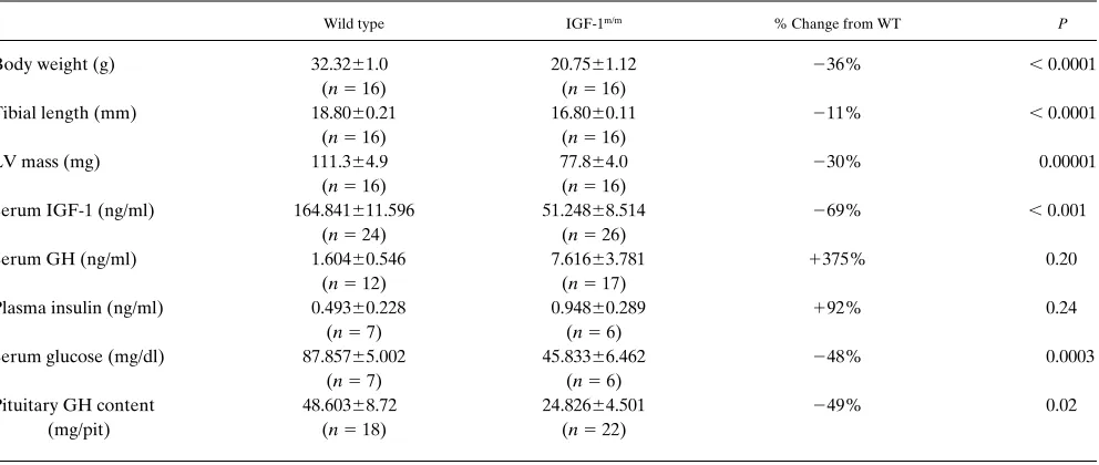
vec-tor sequences inserted 59 of the endogenous exon 3. The ge-nomic organization of the site-specific insertion allele was confirmed by diagnostic restriction digestion of DNA (BglII, KpnI, SacI, and BamHI each give unique fragments not seen in either the wild type or the null alleles), with southern blot-ting using probes from the targeblot-ting sequences (data not shown). Mice heterozygous for this allele exhibited reduced IGF-1 serum levels and body weights similar to those seen for mice heterozygous for the null allele. However, in contrast to the igf1 null mice, mice homozygous for this mutant gene were viable and fertile but profoundly growth retarded, being inter-mediate in size between wild type and IGF-12/2 (Table I); be-cause of their intermediate size, they have been identified as “midi” mice (IGF-1m/m), and had low but detectable IGF-1 in
their sera. If the mutant allele igf1m did not exhibit alternative
splicing, this would result in a null allele and no IGF-1 protein could be produced, as there are multiple stop codons in all three reading frames in the neomycinr cassette, which is
in-serted in an antisense orientation 13 amino acids after the start of the mature protein in exon 3. However, alternative splicing is known to occur with exon 5 in the wild type rodent igf1 gene, and we postulated that alternative splicing was occurring around the neomycinr-containing exon, resulting in the
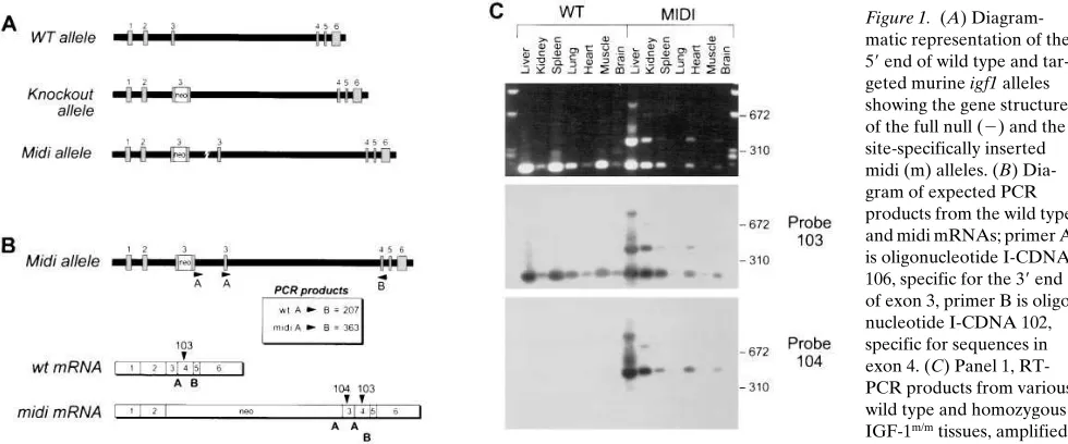
pro-duction of a small amount of wild type mRNA and protein. Exhaustive rtPCR was used to establish that wild type mRNA for IGF-1 was present in the IGF-1m/m mice (Fig. 1, B and C).
We were also able to detect the mutant mRNA in this manner. After injection of recombinant clone cells into C57BL/6J blastocysts, three founder chimeras were mated with C57BL/ 6J females to produce F1 heterozygous animals (129/SvX C57BL/6J). These animals were intercrossed to generate F2 homozygous, heterozygous, and wild type mice. Mating of the F2 homozygous animals to each other to produce F3
homozy-gote animals, and parallel mating of sibling F2 wild type ani-mals to each other to produce F3 wild type controls generated the animals used in this study. Large numbers of F2 animals were used to generate animals with as random a genetic back-ground as possible, and to avoid the possibility of genetic founder events.
Severely IGF-1 deficient young adult mice have reduced se-rum glucose. As seen in Table I, serum 1 levels in IGF-1m/m mice were 30% of wild type values. Plasma insulin levels
appeared to be slightly higher in these young adult IGF-1m/m
mice than in age-matched controls, although this difference did not reach statistical significance. Serum glucose was signifi-cantly lower in the IGF-1m/m mice than in age-matched controls,
however; insulin resistance, as determined by glucose toler-ance testing, was not present at this age (data not shown). Pitu-itary growth hormone content was significantly lower in IGF-1m/m mice than in wild type; although random serum growth
hormone levels do not accurately reflect the pulsatile secretion of GH, there was a trend for higher values in IGF-1m/m mice
compared with controls (Table I), suggesting that the reduced pituitary GH content is not indicative of decreased synthesis.
Cardiac chamber weights are reduced in proportion to body weight. The body weights of adult IGF-1m/m mice were
signifi-cantly reduced by 36% compared with their wild type counter-parts (Table I), comparably in males and females. Spleen and thymus were reduced proportionately to body weight, while brain, kidneys, and liver were less affected (L. Powell-Brayton, unpublished observations). Heart weight in adult IGF-1m/m
mice was likewise reduced, by 31.0% vs. wild type, due to pro-portionate decreases in all chambers. Tibial length was de-creased to a lesser extent in IGF-1m/m mice (10.6%, Table I); in
[image:4.612.58.548.60.264.2]our experience, tibial length correlates highly with body weight in normal young adult mice and shows much less vari-ability after maturity (H. Rockman and J. Hunter, unpublished observations), similar to previous results in adult rats (22). Thus, in attempting to normalize LV mass for body size, when
Figure 1. (A) Diagram-matic representation of the 59 end of wild type and tar-geted murine igf1 alleles showing the gene structure of the full null (2) and the site-specifically inserted midi (m) alleles. (B) Dia-gram of expected PCR products from the wild type and midi mRNAs; primer A is oligonucleotide I-CDNA 106, specific for the 39 end of exon 3, primer B is oligo-nucleotide I-CDNA 102, specific for sequences in exon 4. (C) Panel 1, RT-PCR products from various wild type and homozygous IGF-1m/m tissues, amplified
one uses body weight IGF-1m/m LVs are larger (3.7760.09 mg/g
vs. wild type 3.4360.09 mg/g, P 5 0.01), while using tibial length IGF-1m/m LVs are smaller (4.6260.22 mg/mm vs. wild
type 5.9560.29 mg/mm, P 5 0.001); therefore there is no con-sistent difference between chamber weights in severely IGF-1 deficient mice and controls beyond that explained by body size alone. There were no gross histological abnormalities in IGF-11/m or IGF-1m/m animals.
Severely IGF-1–deficient mice have elevated arterial blood pressure. Direct arterial blood pressure was measured in con-scious, unrestrained animals using an indwelling femoral ar-tery catheter no sooner than 48 h after catheter implantation. Heart rate in IGF-1m/m mice (3–4 mo of age) was not
signifi-cantly different from that in control animals (625616 bpm
[n5 11] vs. 597628 bpm [n 5 9]). However, there was a
signif-icant elevation of mean arterial pressure in the severely IGF-1 deficient IGF-1m/m mice compared with wild-type controls
(12561 mmHg vs. 11261 mmHg, P , 0.001; Fig. 2). Diastolic pressure also was higher (10063 vs. 9163 mmHg, P , 0.05) in IGF-1m/m mice, but the increase in systolic (14664 vs. 13564
mmHg, P 5 0.07) pressure attained only borderline statistical significance. Heterozygote animals had no detectable differ-ences in any parameter (data not shown). In separate experi-ments, conscious blood pressures in a small group (n 5 3) of IGF-12/2 (null) homozygotes (1), which survive to a limited ex-tent in a different genetic background (CD-1), were compared to those in age and sex-matched IGF-11/1 littermates (n 5 4). Mean arterial pressure in IGF-12/2 was higher than in wild type (122612 mmHg vs. 10761 mmHg), but this did not reach statistical significance due to the small sample size. Thus, de-spite homeostatic mechanisms involved in blood pressure con-trol, the arterial pressure was higher in IGF-1m/m mice than in
wild type animals, clearly suggesting a role of IGF-1–depen-dent pathways in the control of basal blood pressure; the trend toward higher blood pressure in IGF-1 null mice in the CD-1 background confirms a specific effect of IGF-1 deficiency on blood pressure across different genetic substrates.
Cardiac contractility is increased in IGF-1m/m mice. The
im-portance of growth hormone in the regulation of myocardial contractile force has recently been demonstrated in GH-defi-cient dwarf rats (23), raising the possibility that IGF-1, a prom-inent endocrine mediator of GH effects, may be involved in cardiac contractility. To examine whether IGF-1 signaling pathways are necessary for the maintenance of normal cardiac function, we measured both basal and isoproterenol-stimu-lated cardiac contractility in IGF-1 deficient mice and wild type controls. Cardiac catheterization and in vivo hemody-namic measurements were performed in anesthetized mice (13) with continuous recording of heart rate (HR), aortic pres-sure, LV dP/dt, and the first derivative of LV dP/dt at baseline and after progressive doses of isoproterenol (18). Before tho-racotomy, mean aortic pressure was significantly increased in anesthetized IGF-1m/m mice compared to wild type (8163
mmHg [n 5 4] vs. 7163 [n 5 4] mmHg, P , 0.05), confirming that differences obtained in conscious animals remain present under anesthesia. After thoracotomy, basal LV systolic pres-sure was similarly elevated in IGF-1m/m mice (87.065.9 mmHg
[n 5 12] vs. 70.261.4 mmHg [n 5 13], P , 0.0001) (Fig. 3 A). The heart rate was slightly but significantly higher in IGF-1m/m
mice at control and with lower doses of isoproterenol, but it was identical to that in wild type animals at high doses (Fig. 3 B). Although a trend toward higher LV end-diastolic pressure was present in the IGF-1m/m group compared with wild type,
this difference did not reach statistical significance. Surpris-ingly, the maximum first derivative of the LV pressure (LV dP/ dtmax), an index of myocardial inotropic state, was significantly
increased in the IGF-1m/m mice compared to wild type controls
(63956684 mmHg/s vs. 40236277 mmHg/s, respectively, P , 0.0001, Fig. 3 C). Similarly, LV dP/dtmin, an index of relaxation,
was also enhanced in the IGF-1 deficient mice (248406648 mmHg/s vs. 231236212 mmHg/s, P , 0.0008, Fig. 3 D); none of these differences was seen in heterozygote animals. These data indicate that a severe in vivo deficiency of IGF-1 is associ-ated with a significant increase in LV inotropic state.
LV dP/dtmax has previously been demonstrated to be
[image:5.612.61.557.72.282.2]de-pendent on preload but relatively indede-pendent of isolated Table I. Principal Characteristics of IGF-1m/m Mice and Age-matched Wild Type Controls
Wild type IGF-1m/m % Change from WT P
Body weight (g) 32.3261.0 20.7561.12 236% , 0.0001
(n5 16) (n5 16)
Tibial length (mm) 18.8060.21 16.8060.11 211% , 0.0001
(n5 16) (n5 16)
LV mass (mg) 111.364.9 77.864.0 230% 0.00001
(n5 16) (n5 16)
Serum IGF-1 (ng/ml) 164.841611.596 51.24868.514 269% , 0.001 (n5 24) (n5 26)
Serum GH (ng/ml) 1.60460.546 7.61663.781 1375% 0.20
(n5 12) (n5 17)
Plasma insulin (ng/ml) 0.49360.228 0.94860.289 192% 0.24
(n5 7) (n5 6)
Serum glucose (mg/dl) 87.85765.002 45.83366.462 248% 0.0003
(n5 7) (n5 6)
Pituitary GH content 48.60368.72 24.82664.501 249% 0.02
(mg/pit) (n5 18) (n5 22)
changes in the LV systolic pressure except when a low aortic diastolic pressure prevents the development of LV dP/dtmax
(24). To exclude the possibility that low diastolic pressure was a factor in the wild type mice and that enhanced LV dP/dtmax
was secondary to a hemodynamic effect of increased arterial pressure in IGF-1 deficient mice, we measured LV dP/dtmax
under conditions of acutely increased LV systolic pressure in-duced by thoracic aortic constriction in wild type mice. As shown in Fig. 4, in initial beats at pressures encompassing the range of LV pressure previously observed in IGF-1m/m mice,
no difference in LV dP/dtmax was recorded. Thus, the enhanced
contractility in the IGF-1m/m animals does not appear to be
sec-ondary to the increase in blood pressure per se.
To exclude an increase in b-adrenergic receptor density as the etiology, binding assays demonstrated that the cardiac b receptor density was comparable between IGF-1m/m mice and
wild type (30.1463.15 vs. 31.0460.83 fmol/mg protein, P 5 NS, n 5 5 in each group). However, basal adenylyl cyclase activity was enhanced in the IGF-1m/m mice compared to controls (9.166
1.61 [n 5 10] vs. 4.8860.69 [n 5 7] pmol cAMP/mg protein per min, respectively, P 5 0.05). In the absence of an appreciable change under basal conditions in two primary regulators of LV performance that are sensitive to catecholamine levels, i.e., the heart rate and the LV end-diastolic pressure, it is suggested that enhanced coupling of b-adrenergic receptors to down-stream signaling pathways rather than increased circulating
catecholamines may contribute to the increased myocardial contractility reflected in augmented LV dP/dtmax. These results
[image:6.612.59.292.59.377.2]are in contrast to those obtained in a genetic rat model of GH deficiency, the primary regulator of IGF-1 biosynthesis, in which increased basal cardiac contractility has been reported in the setting of increased b-adrenergic receptor density (23). Figure 2. (Top) Individual measurements of mean arterial pressure
in male and female IGF-1m/m mice and their wild type controls. *P,
0.01, and ‡P, 0.05, compared to sex-matched wild type, Student’s t
[image:6.612.313.459.62.546.2]test. (Bottom) Examples of recordings of pulsatile arterial pressure in IGF-1m/m and wild type animals.
Figure 3. Hemodynamic variables are shown at baseline and after graded doses of isoproterenol. (A) peak LV systolic pressure, (B) heart rate LV end-diastolic pressure, (C) LV dP/dtmax (D) LV dP/
dtmin. Data are reported as mean6SEM. Open and filled circles
repre-sent wild type (n5 13, 6 male and 7 female) and IGF-1m/m mice (n5
12, 5 male and 7 female), respectively. Statistical analysis was per-formed using repeated measures analysis of variance with post hoc (Scheffe’s) analysis. *P, 0.001 vs. wild type; ‡P, 0.05 vs. wild type.
Hypertrophic response to hemodynamic load is preserved in severe IGF-1 deficiency. IGF-1 has been shown to induce hy-pertrophy of neonatal rat cardiac myocytes in vitro (25) and in the normal rat left ventricle and the infarcted heart in vivo (9); others have not found a significant hypertrophic effect of IGF-1 on ventricular myocytes in vitro, however (26). mRNA for both IGF-1 and its receptor are upregulated in the ventricle of animals subjected to pressure overload (6–8) or infarction (27), raising the possibility that IGF-1 may be an important au-tocrine/paracrine mediator of adaptive cardiac hypertrophy. To determine whether normal levels of IGF-1 are required for the hypertrophic response to pressure overload, severely IGF-1 deficient IGF-1m/m mice and nontransgenic controls
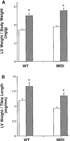
un-derwent transverse aortic constriction or sham operation using previously established techniques (13). As shown in Fig. 5, when normalized for either body weight or tibial length, LV mass in the IGF-1m/m mice increased to a comparable extent
(147% vs. 148%, normalized for body weight) to wild type animals; the range of transstenotic pressure gradients was comparable between the two groups. Thus, the adaptive hy-pertrophic response of the left ventricle is unimpaired in the IGF-1m/m mice, documenting selectivity of the effects of IGF-1
deficiency on contractility and resting blood pressure.
Discussion
The identification of a line of mice harboring a site-specific in-sertion in the IGF-1 gene has allowed us to study the effects in adult animals of severe IGF-1 deficiency, a condition in which complete absence of IGF-1 is lethal at birth. At present, no pharmacological inhibitors specific for IGF-1 signaling are available, so this model is the first to provide insights into the in vivo physiological role of IGF-1 on basal cardiovascular function. These animals exhibit a functional phenotype which is distinct from that seen in GH deficient dwarf rats, in which both GH and IGF-1 are reduced.
Blood pressure. Essential hypertension is a polygenic dis-order that is a major contributor to morbidity and mortality in human cardiovascular disease. Recent advances have led to the identification of a large number of candidate genes which might regulate basal blood pressure; however, to date, the
genes which have demonstrated linkage with hypertension are those in the renin–angiotensin pathway (28) and in adrenal steroid biosynthesis (29). Mice transgenic for additional copies of the angiotensinogen gene have a gene-dosage dependent in-crease in blood pressure (30), while genetic deficiency of the vasoactive peptide ANP, generated by targeted disruption of the gene, has produced a murine model of salt-sensitive hyper-tension (31). It has been noted in this gene-targeted model that the number of copies of the Ren gene (two per haploid type inherited from the 129/Sv strain, one per haploid geno-type inherited from the C57BL/6J strain) directly affects the blood pressure in individual animals (32). Our observations likewise suggest a strong influence of genetic modifiers on blood pressure. In IGF-1m/m animals in a CD-1 background
(with reduction in IGF-1 levels comparable to that seen in 129/
Sv 3 C57BL/6J) there is little difference in blood pressures
from wild type (data not shown); in that background, however, IGF-1 null animals can survive, and in such mice (with no de-tectable IGF-1) there seems to be an increase in blood pres-sure comparable to that in IGF-1m/m mice in the 129/Sv 3
C57BL/6J background. These data indicate that the sensitivity of blood pressure to serum IGF-1 levels varies depending on the genetic background.
[image:7.612.316.459.372.660.2]Igf1 could therefore be considered a candidate gene for blood pressure regulation, since it (like angiotensin II) has both growth-promoting properties for vascular smooth muscle
Figure 4. LV systolic pressure (open circles) and LV dP/dtmax (filled
[image:7.612.56.204.544.650.2]circles) before and for four beats after the creation of acute pressure overload with sudden transverse aortic constriction (TAC) in control (wild type) mice (n5 10). Data are reported as mean6SEM and in-clude both male and female mice. Statistical analysis was performed using repeated measures analysis of variance with post hoc (Scheffe’s) analysis. *P, 0.0001 vs. basal (before acute TAC). Num-bers represent LVEDP.
Figure 5. Response of IGF-1m/m mice and wild type controls to
trans-verse aortic constriction (TAC). (A) LV weight normalized for body weight; (B) LV weight normalized for tibia length. Open bars denote sham-operated animals (wild type, n5 16; IGF-1m/m, n5 16), hatched
bars denote TAC (wild type, n5 15; IGF-1m/m, n5 14). WT, wild
cells (33) as well as potent vasodilator effects when adminis-tered in vivo (4, 5), suggesting a possible role for IGF-1 in the physiological regulation of vascular tone. IGF-1 produces vas-cular effects qualitatively similar to insulin, although at tenfold lower doses, indicating that the hemodynamic actions of insu-lin may be mediated through the IGF-1 receptor (34). In light of recent evidence that insulin and IGF-1 converge on a com-mon signaling pathway (35), it is intriguing to note that resis-tance to the vasorelaxant effects of insulin, which has been re-ported in several pathological circumstances (including obesity (36), noninsulin dependent diabetes mellitus (37), and essential hypertension (38)), has been hypothesized to be a critical de-terminant of the increased blood pressure existing in each of these disease states (39). The increase in mean arterial pres-sure in IGF-1m/m transgenic mice, with parallel elevations of
systolic and diastolic pressures, suggests a role for IGF-1 in maintaining the set point of arterial tone and vascular resis-tance. This finding is consonant with previous data on the va-sodilator effect of IGF-1 infusion (4, 40, 41) which suggest that the effect on arterial pressure is at the level of the arteriolar re-sistance vessels, rather than an indirect effect on autonomic tone (the latter is also less likely, since no significant difference was seen in heart rate in conscious midi mice vs. wild type). Modulatory effects on sympathetic tone, as well as the possi-bility of an increase in cardiac output, cannot be excluded as contributory factors, however. The observation of enhanced adenylyl cyclase activity in cardiac myocyte extracts cannot readily be extrapolated as a mechanism for increased blood pressure; indeed, if also present in vascular smooth muscle one might anticipate a vasorelaxant effect from this pathway. Other potential contributors to the increase in blood pressure include plasma insulin and growth hormone (although trans-genic mice overexpressing GH (42) and rats receiving GH over 5 wk (43) have no elevation in blood pressure; increases in renal plasma flow in response to GH may in fact be medi-ated by IGF-1 (44) GH deficient rats have lower mean arterial pressure than control Lewis rats (23) although recorded pres-sures in both groups are higher (13267 vs. 16368 mmHg, re-spectively) than in other rat strains (41). No relation has been found between body weight and mean arterial pressure in mice (H. Rockman, unpublished observations), so the lower body weight in the midi mice is unlikely to play a substantial role. The observation that IGF-1 deficiency produces elevated arte-rial pressure, regardless of mechanism, strongly suggests that IGF-1 signaling pathways contribute to blood pressure regula-tion, and that components of this pathway (IGF-1, its receptor, and the intracellular effector molecules under its control) are candidate genes for hypertension.
Left ventricular contractility. The finding of increased car-diac contractility was rather surprising, since our hypothesis had been that IGF-1 was essential for normal cardiac function. That this was not simply due to increased afterload due to in-creased arterial pressure with a secondary effect on LV
dP/dt-max was shown by the lack of responsiveness of LV dP/dtmax to
acute increases in arterial pressure. In contrast to a defined transgenic model of increased contractility which is known to be due to increased b-adrenergic signaling (18), and in contrast to GH-deficient rats which have a similar phenotype (23), binding assays indicate that b receptor number is not increased in the IGF-1m/m mice. In the setting of increased adenylyl
cy-clase activity, the pattern of hemodynamic response is similar to that seen in mice transgenic for an inhibitor of the b
adren-ergic receptor kinase (bARK) (19); as in this model, the con-tractile performance of the IGF-1m/m mice can be further
stimulated by isoproterenol, indicating that the additional re-cruitment of cardiac performance by b-adrenergic agonists is preserved in IGF-1 deficiency. The possibility of cross-talk be-tween intracellular signaling pathways has been suggested for insulin and b adrenergic signaling in vitro (45); the present data support this hypothesis, and raise the possibilities that such interaction may be mediated through the IGF-1 receptor and involve the level or activity of the b adrenergic receptor kinase. This does not preclude additional increases in intrinsic cardiac myocyte contractility due to altered isoforms of con-tractile proteins such as a-actin and myosin heavy chain, of cal-cium regulatory proteins such as the troponins or phospholam-ban (which has also been implicated in the control of cardiac contractility in a transgenic model [46]), or by other increases in intracellular calcium or myofilament calcium sensitivity. The observation that b adrenergic coupling is enhanced with-out an increase in receptor density raises several interesting possibilities in light of the previous observations in GH-cient rats. In the latter, presumptively also with secondary defi-ciency in IGF-1, a contribution of enhanced b adrenergic cou-pling to the increased contractility was not excluded. Thus GH and IGF-1 deficiencies synergistically enhance b adrenergic signaling by increases in b adrenergic receptor number and coupling, respectively; it is tempting to speculate that GH and IGF-1 excess might therefore inhibit b adrenergic signaling.
Cardiac hypertrophy. The lack of effect of IGF-1 defi-ciency on the adaptive hypertrophic response of the myocar-dium to the load imposed by transverse aortic constriction was unexpected. In light of evidence that IGF-1 produces a hyper-trophic phenotype in cultured neonatal ventricular myocytes (25) as well as in vivo (9) and that IGF-1 mRNA and that for its receptor are upregulated in the ventricular myocardium fol-lowing hemodynamic loading (6–8), it appeared likely that the hypertrophic response in vivo in the midi mice would at least be blunted. Instead, no effect was found, suggesting that either IGF-1 is not necessary for cardiac myocyte hypertrophy (or that other growth signals can compensate) or that the low lev-els of IGF-1 in the midi mice are sufficient (threshold effect).
mice should provide a model system in which to unravel these IGF-1 dependent pathways.
Acknowledgments
We thank Wes Wong and Ross Clark for assaying pituitary GH, and Richard Latvala for technical assistance.
This work was supported by grants from the National Heart, Lung, and Blood Institute (HL-36139, HL-45069, and HL-46345) and American Heart Association (91-022170) to K.R. Chien, HL-53773 to J. Ross, and HL-35018 to M.P. Prinz.
References
1. Powell-Braxton, L., P. Hollingshead, C. Warburton, M. Dowd, S. Pitts-Meek, D. Dalton, N. Gillett, and T.A. Stewart. 1993. IGF-I is required for nor-mal embryonic growth in mice. Genes Dev. 7:2609–2617.
2. Liu, J.P., J. Baker, A.S. Perkins, E.J. Robertson, and A. Efstratiadis. 1993. Mice carrying null mutations of the genes encoding insulin-like growth factor I (Igf-1) and type 1 IGF receptor (Igf1r). Cell. 75:59–72.
3. Baker, J., J.P. Liu, E.J. Robertson, and A. Efstratiadis. 1993. Role of in-sulin-like growth factors in embryonic and postnatal growth. Cell. 75:73–82.
4. Copeland, K.C., and K.S. Nair. 1994. Recombinant human insulin-like growth factor-I increases forearm blood flow. J. Clin. Endocrinol. Metab. 79: 230–232.
5. Fryburg, D.A. 1994. Insulin-like growth factor I exerts growth hormone-and insulin-like actions on human muscle protein metabolism. Am. J. Physiol.
267:E331–E336.
6. Wahlander, H., J. Isgaard, E. Jennische, and P. Friberg. 1992. Left ven-tricular insulin-like growth factor I increases in early renal hypertension. Hy-pertension. 19:25–32.
7. Czerwinski, S.M., J. Novakofski, and P.J. Bechtel. 1993. Is insulin-like growth factor gene expression modulated during cardiac hypertrophy? Med. Sci. Sports. Exercise. 25:495–500.
8. Donohue, T.J., L.D. Dworkin, M.N. Lango, K. Fliegner, R.P. Lango, J.A. Benstein, W.R. Slater, and V.M. Catanese. 1994. Induction of myocardial insu-lin-like growth factor-I gene expression in left ventricular hypertrophy. Circula-tion. 89:799–809.
9. Duerr, R.L., S. Huang, H.R. Miraliakbar, R. Clark, K.R. Chien, and J. Ross, Jr. 1995. Insulin-like growth factor-1 enhances ventricular hypertrophy and function during the onset of experimental cardiac failure. J. Clin. Invest. 95: 619–627.
10. Cittadini, A., H. Stromer, S.E. Katz, R. Clark, A.C. Moses, J.P. Morgan, and P.S. Douglas. 1996. Differential cardiac effects of growth hormone and in-sulin-like growth factor-1 in the rat. A combined in vivo and in vitro evaluation.
Circulation. 93:800–809.
11. Fazio, S., D. Sabatini, B. Capaldo, C. Vigorito, A. Giordano, R. Guida, F. Pardo, B. Biondi, and L. Sacca. 1996. A preliminary study of growth hormone in the treatment of dilated cardiomyopathy. N. Engl. J. Med. 334:809–814.
12. Rockman, H.A., R.S. Ross, A.N. Harris, K.U. Knowlton, M.E. Stein-helper, L.J. Field, J. Ross, Jr., and K.R. Chien. 1991. Segregation of atrial-spe-cific and inducible expression of an atrial natriuretic factor transgene in an in vivo murine model of cardiac hypertrophy. Proc. Natl. Acad. Sci. USA. 88: 8277–8281.
13. Rockman, H.A., S. Ono, R.S. Ross, L.R. Jones, M. Karimi, V. Bhar-gava, J.J. Ross, and K.R. Chien. 1994. Molecular and physiological alterations in murine ventricular dysfunction. Proc. Natl. Acad. Sci. USA. 91:2694–2698.
14. Chien, K.R. 1993. Molecular advances in cardiovascular biology. Science (Wash. DC). 260:916–917.
15. Chien, K.R. 1995. Cardiac muscle diseases in genetically engineered mice: evolution of molecular physiology. Am. J. Physiol. 269:H755–H766.
16. Lieberman, S.A., J. Bukar, S.A. Chen, A.C. Celniker, P.G. Compton, J. Cook, J. Albu, A.J. Perlman, and A.R. Hoffman. 1992. Effects of recombinant human insulin-like growth factor-I (rhIGF-I) on total and free IGF-I concentra-tions, IGF-binding proteins, and glycemic response in humans. J. Clin. Endo-crinol. Metab. 75:30–36.
17. Elias, K.A., G.S. Ingle, J.P. Burnier, R.G. Hammonds, R.S. McDowell, T.E. Rawson, T.C. Somers, M.S. Stanley, and M.J. Cronin. 1995. In vitro char-acterization of four novel classes of growth hormone-releasing peptide. Endo-crinology. 136:5694–5699.
18. Milano, C.A., L.F. Allen, H.A. Rockman, P.C. Dolber, T.R. McMinn, K.R. Chien, T.D. Johnson, R.A. Bond, and R.J. Lefkowitz. 1994. Enhanced myo-cardial function in transgenic mice overexpressing the beta 2-adrenergic recep-tor. Science (Wash. DC). 264:582–586.
19. Koch, W.J., H.A. Rockman, P. Samama, R.A. Hamilton, R.A. Bond, C.A. Milano, and R.J. Lefkowitz. 1995. Cardiac function in mice overexpressing the beta-adrenergic receptor kinase or a beta ARK inhibitor. Science (Wash. DC). 268:1350–1353.
20. Bond, R.A., P. Leff, T.D. Johnson, C.A. Milano, H.A. Rockman, T.R. McMinn, S. Apparsundaram, M.F. Hyek, T.P. Kenakin, L.F. Allen et al., 1995. Physiological effects of inverse agonists in transgenic mice with myocardial overexpression of the beta 2-adrenoceptor. Nature (Lond.). 374:272–276.
21. Hasty, P., J. Rivera-Perez, C. Chang, and A. Bradley. 1991. Target fre-quency and integration pattern for insertion and replacement vectors in embry-onic stem cells. Mol. Cell. Biol. 11:4509–4517.
22. Yin, F.C., H.A. Spurgeon, K. Rakusan, M.L. Weisfeldt, and E.G. Lakatta. 1982. Use of tibial length to quantify cardiac hypertrophy: application in the aging rat. Am. J. Physiol. 243:H941–H947.
23. Brown, L., B. Wyse, and C. Sernia. 1993. Adrenoceptor-mediated car-diac and vascular responses in genetically growth hormone-deficient rats. Bio-chem. Pharmacol. 45:2223–2229.
24. Ross, J. Jr., J.W. Covell, E.H. Sonnenblick, and E. Braunwald. 1966. Contractile state of the heart characterized by force-velocity relations in vari-ably afterloaded and isovolumic beats. Circ. Res. 18:149–163.
25. Ito, H., M. Hiroe, Y. Hirata, M. Tsujino, S. Adachi, M. Shichiri, A. Koike, A. Nogami, and F. Marumo. 1993. Insulin-like growth factor-I induces hypertrophy with enhanced expression of muscle specific genes in cultured rat cardiomyocytes. Circulation. 87:1715–1721.
26. Kajstura, J., W. Cheng, K. Reiss, and P. Anversa. 1994. The IGF-1-IGF-1 receptor system modulates myocyte proliferation but not myocyte cellular hy-pertrophy in vitro. Exp. Cell. Res. 215:273–283.
27. Reiss, K., L.G. Meggs, P. Li, G. Olivetti, J.M. Capasso, and P. Anversa. 1994. Upregulation of IGF1, IGF1-receptor, and late growth related genes in ven-tricular myocytes acutely after infarction in rats. J. Cell. Physiol. 158:160–168.
28. Kurtz, T.W. 1994. Genetic models of hypertension. Lancet. 344:167–168. 29. Cicila, G.T., J.P. Rapp, J.M. Wang, E. St. Lezin, S.C. Ng, and T.W. Kurtz. 1993. Linkage of 11 beta-hydroxylase mutations with altered steroid bio-synthesis and blood pressure in the Dahl rat. Nat. Genet. 3:346–353.
30. Kim, H.S., J.H. Krege, K.D. Kluckman, J.R. Hagaman, J.B. Hodgin, C.F. Best, J.C. Jennette, T.M. Coffman, N. Maeda, and O. Smithies. 1995. Ge-netic control of blood pressure and the angiotensinogen locus. Proc. Natl. Acad. Sci. USA. 92:2735–2739.
31. John, S.W., J.H. Krege, P.M. Oliver, J.R. Hagaman, J.B. Hodgin, S.C. Pang, T.G. Flynn, and O. Smithies. 1995. Genetic decreases in atrial natriuretic peptide and salt-sensitive hypertension. Science (Wash. DC). 267:679–681.
32. Smithies, O., and N. Maeda. 1995. Gene targeting approaches to com-plex genetic diseases: atherosclerosis and essential hypertension. Proc. Natl. Acad. Sci. USA. 92:5266–5272.
33. Hwang, D.L., L.J. Latus, and A. Lev-Ran. 1992. Effects of platelet-con-tained growth factors (PDGF, EGF, IGF-I, and TGF-beta) on DNA synthesis in porcine aortic smooth muscle cells in culture. Exp. Cell. Res. 200:358–360.
34. Wu, H.Y., Y.Y. Jeng, C.J. Yue, K.Y. Chyu, W.A. Hsueh, and T.M. Chan. 1994. Endothelial-dependent vascular effects of insulin and insulin-like growth factor I in the perfused rat mesenteric artery and aortic ring. Diabetes.
43:1027–1032.
35. White, M.F., and C.R. Kahn. 1994. The insulin signaling system. J. Biol. Chem. 269:1–4.
36. Laakso, M., S.V. Edelman, G. Brechtel, and A.D. Baron. 1990. De-creased effect of insulin to stimulate skeletal muscle blood flow in obese man. A novel mechanism for insulin resistance. J. Clin. Invest. 85:1844–1852.
37. Laakso, M., S.V. Edelman, G. Brechtel, and A.D. Baron. 1992. Im-paired insulin-mediated skeletal muscle blood flow in patients with NIDDM.
Diabetes. 41:1076–1083.
38. Feldman, R.D., and G.S. Bierbrier. 1993. Insulin-mediated vasodilation: impairment with increased blood pressure and body mass. Lancet. 342:707–709. 39. Anderson, E.A., and A.L. Mark. 1993. The vasodilator action of insulin. Implications for the insulin hypothesis of hypertension. Hypertension. 21:136– 141.
40. Haylor, J., I. Singh, and A.M. el Nahas. 1991. Nitric oxide synthesis in-hibitor prevents vasodilation by insu8lin-like growth factor I. Kidney Int. 39: 333–335.
41. Hirschberg, R., J.D. Kopple, R.C. Blantz, and B.J. Tucker. 1991. Effects of recombinant human insulin-like growth factor I on glomerular dynamics in the rat. J. Clin. Invest. 87:1200–1206.
42. Dilley, R.J., and S.M. Schwartz. 1989. Vascular remodeling in the growth hormone transgenic mouse. Circ. Res. 65:1233–1240.
43. Harrap, S.B., F. Macpherson, V.G. Wilson, D.L. Davies, O.P. Isaksson, B. Folkow, and A.F. Lever. 1988. Failure of chronic administration of growth hormone to affect blood pressure, vascular reactivity and sodium metabolism in normal rats. J. Hypertens. (Suppl.) 6:S170–S172.
44. Hirschberg, R., and J.D. Kopple. 1989. Effects of growth hormone and IGF-I on renal function. Kidney Int. (Suppl.):S20–S26.
45. Hadcock, J.R., J.D. Port, M.S. Gelman, and C.C. Malbon. 1992. Cross-talk between tyrosine kinase and G-protein-linked receptors. Phosphorylation of beta 2-adrenergic receptors in response to insulin. J. Biol. Chem. 267:26017–26022.