R E S E A R C H A R T I C L E
Open Access
Effect of training and sudden detraining on the
patellar tendon and its enthesis in rats
Antonio Frizziero
1, Milena Fini
2*, Francesca Salamanna
2, Arsenio Veicsteinas
3, Nicola Maffulli
4, Marina Marini
1Abstract
Background:Different conditions may alter tendon characteristics. Clinical evidence suggests that tendon injuries are more frequent in athletes that change type, intensity and duration of training. Aim of the study was the assessment of training and especially detraining on the patellar tendon (PT) and its enthesis.
Methods:27 male adult Sprague-Dawley rats were divided into 3 groups: 20 rats were trained on a treadmill for 10 weeks. Of these, 10 rats were euthanized immediately after training (trained group), and 10 were caged without exercise for 4 weeks before being euthanized (de-trained group). The remaining 7 rats were used as controls (untrained rats). PT insertion, structure (collagen fiber organization and proteoglycan, PG, content), PT thickness, enthesis area, and subchondral bone volume at the enthesis were measured by histomorphometry and microtomography.
Results:Both PG content and collagen fiber organization were significantly lower in untrained and detrained animals than in trained ones (p < 0.05 andp< 0.0001). In the detrained group, fiber organization and PG content were worse than that of the untrained groups and the untrained group showed a significantly higher score than the detrained group (p< 0.05). In the trained group, the PT was significantly thicker than in untrained group (p< 0.05). No significant differences in the enthesis area and subchondral bone volume among the three groups were seen.
Conclusions:Moderate exercise exerts a protective effect on the PT structure while sudden discontinuation of physical activity has a negative effect on tendons. The present results suggest that after a period of sudden de-training (such as after an injury) physical activity should be restarted with caution and with appropriate rehabilitation programs.
Background
Tendon is a highly organized connective tissue, capable of resisting high tensile strength while transmitting forces from muscle to bone. The region where a tendon, liga-ment or joint capsule attaches to bone is the enthesis. In the context of a tendon, the enthesis is a fibrous or fibro-cartilagineous zone of tendon attachment to bone that ensures that the contractile forces produced by the mus-cle contraction are transmitted to the skeleton [1].
Tendons consist of a densely packed well organized con-nective tissue dominated by collagen (60-85% of tendon dry weight and mainly collagen I) arranged into fibrils, fibers, bundles, and fascicles, embedded in extracellular
matrix (ECM) proteins, including proteoglycans (PG). Mechanical stimuli have different impact on the expres-sion of PG in tendons [2-6]. For example, mechanical tension induces the synthesis of decorin, whereas the pro-duction of the large PG aggrecan is stimulated by com-pression [7].
A variety of conditions, including immobilization, training, aging and medications, may influence tendon characteristics [2,8-11]. Immobilization results in pro-found reduction in the mechanical properties of tendon. Most noticeably, there is a decrease in tendon strength and an increase in collagen turnover [12]. With exercise, the turnover of mature collagen and collagen cross-links increases [13,14], large diameter fibrils are formed with increased packing density of fibrils [15] and increased tendon stiffness [16]. Exercise also leads to changes in PG content [17]. Strenuous exercise in mature rodents or * Correspondence: [email protected]
2
Laboratory of Pre-clinic and Surgical Studies, Rizzoli Orthopaedic Institute, Bologna, Italy
Full list of author information is available at the end of the article
chickens leads to thickening of collagen fibrils and increase in the galactosamine-containing glycosaminogly-cans (GAGs) [18]. In contrast, immature tendons respond to exercise with higher collagen turnover, reduced maturation of collagen [19] and changes in hya-luronan concentration [20]. Kubo et al observed that tendon adaptation to detraining is faster than the one to resistance training [21], but to the present author knowl-edge, in comparison with training or immobilization, the effects of sudden detraining on tendons have not been deeply investigated.
In fact, there is a paucity of data examining the impact that cessation of training may have on tendon. In prac-tice, we do not fully understand how tendons respond to a period of training followed by sudden detraining. On the other site, clinical evidence suggests that tendon injuries are more frequent in athletes that change type, intensity and duration of training. Thus, the aim of the present research was the study of PT and its enthesis after training and especially following sudden disconti-nuation of activity. We evaluated the histological charac-teristics of the patellar tendon (PT) and its enthesis in trained, detrained, and untrained rats. Because both the magnitude and the type of load transmission across the joint surface are likely to change with exercise and with tendon health status we measured subchondral bone thickness, bone area and bone volume. Also bone volume, as an indicator of bone adaption, was evaluated by microtomography at the level of the subchondral bone connected to the enthesis.
Methods
Animal and training protocol
The animal study was performed according to the Ita-lian Law by Decree on Animal Experimentation (N. 116/92), and was approved by the Health Authority of Milan (Italy) Ethical Committee for the Use of Labora-tory Animals. Training was carried out according to the American Physiological Society guidelines for exercising rodents on treadmills [22].
A total of 27 male albino Sprague-Dawley rats aged 9 weeks, between 250 g to 350 g, were placed in indivi-dual standard cages and fed a standard diet without limitations; room temperature was kept at 21 ± 2°C; 12 hrs of light were automatically alternated with 12 hrs of dark. After 1 week of acclimatization, 20 rats were randomly chosen to run on a six-lane rodent treadmill 1 hr a day, three times a week. The speed was gradually increased to reach 25 m/min in 5 weeks, which corre-sponds to ~ 60% VO2max, and was then maintained constant for a further 5 weeks [20]. Seven control untrained animals were placed on a non-moving tread-mill during the training session and were euthanized at 19 weeks of age (untrained group). At the end of the
10-week training, under general anesthesia 10 trained rats were randomly chosen for immediate euthanasia (trained group) whereas 10 were caged without exercise for a further 4 weeks before being euthanized (detrained group).
The rats were periodically examined by a veterinarian. Food consumption and body weight were evaluated three times a week.
Histological studies
The right and left knee joints were explanted and fixed in 4% buffered paraformaldehyde for 48 hours for un-decalcified bone processing. The samples were then dehydrated by placing them in graded series of increas-ing percentage of alcohol with one step in 50% alcohol, one step in 75% alcohol, two steps in 95% alcohol, and two steps in 100% alcohol, for 48 hours per each step. Finally, they were embedded in polymethyl-methacrylate (Merck, Shuchardt, Hohenbrunn, Germany). Blocks were sectioned in the sagittal plane. A series of sections 200 ± 100μm thick, spaced about 300μm apart because of the thickness of the microtome diamond saw, were obtained with a Leica 1600 diamond saw microtome (Leica SpA, Milan, Italy). The sections were then auto-matically thinned (EXAKT Cutting and Grinding Systems, GmbH & Co., Norderstedt, Germany) to 40 ± 10μm with different abrasive papers (EXAKT Abrasive Disc, GmbH & Co., Norderstedt, Germany), from 1200 to 300 grit in steps of 15 minutes each. A total of 27 samples (10 from trained, 10 from detrained, and 7 from control animals) were processed and analyzed. They were stained with Toluidine Blu, Acid Fuchsin, Fast Green, and processed for routine histological and histomorphometrical analyses by using a transmission and polarized light AxioSkop Microscope (Carl Zeiss GmbH, Jena, Germany). Computerized image analysis was performed with Axio Vision 4.6 (Carl Zeiss GmbH, Jena, Germany). Three sections per each sample were analyzed by two blinded investigators.
Histology and Histomorphometry
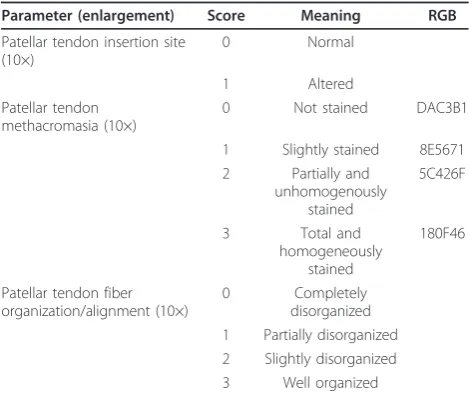
beneath tendon was normal [23]. For the evaluation of PT metachromasia, the score evaluated the intensity of staining by Toluidine Blue O. This metachromatic dye binds to the ECM proteoglycans (PGs) staining the PT matrix blue [24];“blue” was defined, on the red-green-blue (RGB) scale [25,26], as (R = 24; G = 15; B = 70; hexadecimal RGB value = 180F469). The analyses were carried out at 10× magnification. The method provides discrete numbers to which we assign the following scores: 0 for unstained tendons (70 ± 10% of pixels hav-ing hexadecimal RGB value = DAC3B1: R = 218; G = 195; B = 177), 1 for slightly stained tendons (70 ± 10% of pixels having hexadecimal RGB value = 8E5671: R = 142; G = 86; B = 113), 2 for partially and inhomogen-eously stained tendons (70 ± 10% of pixels having hexa-decimal RGB value = 5C426F: R = 92; G = 66; B = 111) and 3 for tendons with total and homogeneous staining (70 ± 10% of pixels having hexadecimal RGB value = 180F46: R = 24; G = 15; B = 70) (Table 1).
Another parameter that was evaluated was the collagen PT fiber organization and alignment [23]. In normal con-ditions, these fibers are arranged in closely aligned bun-dles. The analysis was carried out at 10× magnification. The score for the fiber bundle evaluation was 0 for a com-plete disorganization of the fibers, with bundles lacking any alignment through their whole length, 1 for partial disorganization, with bundles non-aligned in more than half of the tendon length, 2 for slight disorganization, with bundles non-aligned in less than half of the tendon length and 3 for tendons with closely aligned bundles through their whole length. The score received by each sample was then averaged within the experimental group (trained, detrained and untrained), so that each group was assigned a total score between 0 and 7, with 0 corresponding to
altered insertion site, unstained and not organized tendon, and 7 corresponding to a normal insertion site, stained and structurally well organized tendon.
Enthesis area (μm2) and PT thickness (μm) were also measured. The enthesis area was evaluated at a magnification of 10×. PT thickness was measured at a magnification of 2.5×, with 10 measurements performed along the length of the tendon. To avoid biases due to subjectivity, the histological evaluations and measure-ments were performed by two experienced and blinded investigators. Possible discrepancies were resolved by sharing the final results.
Microcomputer Tomography (Micro-CT) Analyses
After dehydration in graded series of alcohols and before embedding in poly-methyl-methacrylate, 4 samples for each group were randomly chosen for analysis by Micro-CT SkyScan 1172 (μCT, SKYSCAN, Kartuizersweg 3B 2550 Kontich, Belgium). The system consisted of a micro focus X-ray source, a routable specimen holder and a detector system equipped with a 4000 × 2624 pixel CCD camera. The samples were wrapped in parafilm, to pre-vent drying, and scanned. Each X-ray projection was acquired in a 12-bit gray level image, 2000×1150 pixel, 10.97μm image pixel sizes, with an aluminum filter and for a time of 35 minutes. After acquisition, projection images, 935 for the entire sample, were reconstructed (NRecon 1.4.4), and all the sections were cut along a longitudinal plane (CTAn 1.7) (Figure 1a,b). Then, a region of interest (ROI) of subchondral bone at the level of the enthesis was selected within the volume of the bone area where the PT inserts. This step was repeated for different sections arranged along the volume of the bone, and subsequently the ROI was interpolated. Bone volume and bone area were measured. To compare the same number of sections for each sample, the ROI data-set of every examined sample was reduced to the 70 most central sections. Subsequently, the images inside the selected ROI were binarized considering the bone as white, and the quantitative 2D and 3D parameters were analyzed.
Statistical Analysis
Statistical evaluation was performed using the software package SPSS/PC+ Statistics TM 12.1 (SPSS Inc., Chicago, IL USA). After verifying the normal distribu-tion of data, one-way ANOVA followed by the Scheffé
post hocmulti comparison tests was used to analyze his-tomorphometric differences between groups.
Results
Histology and Histomorphometry
[image:3.595.56.291.110.308.2]Histomorphometry data are reported in Table 2. In all groups, the PT tendon insertion was normal with
Table 1 Semi-quantitative score for the evaluation of patellar tendon
Parameter (enlargement) Score Meaning RGB
Patellar tendon insertion site (10×)
0 Normal
1 Altered
Patellar tendon methacromasia (10×)
0 Not stained DAC3B1
1 Slightly stained 8E5671 2 Partially and
unhomogenously stained
5C426F
3 Total and
homogeneously stained
180F46
Patellar tendon fiber organization/alignment (10×)
0 Completely
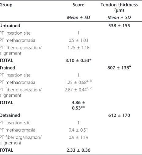
collagen fiber organization continued into fibrocartilagi-nous and mineralized zones with healthy bone beneath the tendon. The untrained group yielded a total score of 3.10 ± 0.53, and PT displayed the most reduced staining, although, remarkably, one single untrained control scored 3. The total score was 4.86 ± 0.53 for the trained group and 2.33 ± 0.36 for the detrained group, respec-tively. Comparison ANOVA showed that in the trained group, the total score was significantly higher than in the detrained and in the untrained groups (p< 0.0005). The untrained group showed a significantly higher score than the detrained group (p< 0.05).
More in detail, histomorphometry showed that both PG content and collagen fiber organization were signifi-cantly lower in untrained and detrained animals than in trained ones (p< 0.05 andp< 0.0001).
Collagen fibers were partially disorganized in untrained rat tendons (mean score: 1.75 ± 1.18) (Figure 2a). On the other hand, the samples of the trained group displayed closely aligned bundles; collagen fibers were parallel and closely packed together and with PGs neatly arranged (mean score: 2.87 ± 0.44) (Figure 2b). Therefore, 9 ten-dons of the trained group received a score of 3, and one sample scored 1. In the detrained group, fiber organiza-tion was worse than that of both the trained and the untrained groups, with disorganization of the collagen bundles (Figure 2c); 9 tendons scored 0-1 and only one sample which scored 3 (mean score: 0.9 ± 1.19).
Polarized microscopy, showed that even the smallest visible collagen fibril was birifrangent, which indicates the presence of elongated submicroscopic units oriented in the direction of the fiber axis. Such characteristic is particularly conspicuous in the trained tendon (Figure 3).
In the untrained and detrained groups, collagen fibers slightly stained with toluidine blue O (means score: 0.5 ± 1.03 and 0.4 ± 0.51, respectively), while in the trained group collagen fibers were homogeneously stained and therefore with a physiologic amount of PGs (mean score: 1.25 ± 0.68) (Figure 4a,b,c).
Histomorphometric measurements showed no signifi-cant differences in the enthesis area among the three groups (untrained group: 2.7 ± 0.9μm2; trained group: 2.3 ± 0.4 μm2; detrained group: 2.6 ± 0.5μm2). In the trained group, the PT was significantly thicker than in untrained group (p< 0.05), while no significant differ-ences were observed between the trained and detrained groups, and detrained and untrained groups (Table 2).
Microtomography
[image:4.595.59.540.88.219.2]Microtomographic analysis showed no significant differ-ences in subchondral bone volume among the three
Table 2 Histomorphometric scores of PT in Untrained, Trained and Detrained rats
Group Score Tendon thickness
(μm)
Mean ± SD Mean ± SD
Untrained 538 ± 155
PT insertion site 1
PT methacromasia 0.5 ± 1.03
PT fiber organization/ alignement
1.75 ± 1.18
TOTAL 3.10 ± 0.53*
Trained 807 ± 138a
PT insertion site 1
PT methacromasia 1.25 ± 0.68a, b
PT fiber organization/ alignement
2.87 ± 0.44a, c
TOTAL 4.86 ±
0.53**
Detrained 612 ± 170
PT insertion site 1
PT methacromasia 0.4 ± 0.51
PT fiber organization/ alignement
0.9 ± 1.19
TOTAL 2.33 ± 0.36
*p< 0.05 (untrainedvsdetrained); **p< 0.0005 (trainedvsdetrained; trained
vsuntrained).
a
p< 0.05 (trainedvsuntrained);b
p< 0.05 (trainedvsdetrained);c
[image:4.595.58.290.442.695.2]p< 0.0001 (trainedvsdetrained).
groups (untrained group: 3.08 ± 0.2 mm3; trained group: 2.37 ± 0.15 mm3; detrained group: 2.72 ± 0.3 mm3).
Discussion
Tendon is not a static tissue, rather it adapts itself according to the level, direction and frequency of the applied load through a process of remodeling probably mediated by tenocytes [27,28].
It is well known that running activity improves PT and its enthesis [29,30]. On the contrary there is a pau-city of data examining the impact that a sudden detrain-ing may have on tendons.
We studied several structural features of the PT in trained, de-trained and control untrained rats by histolo-gical, histomorphometric and microtomographic ana-lyses using a semi-quantitative score which took into account several aspects of tendon morphology/structure and by measuring enthesis area, PT thickness and sub-chondral bone volume and collagen fiber arrangement and stainability. On the other hand, it was not possible to evaluate other parameters, since, in order to evaluate the enthesis area and to maintain the integrity at the
tendon-bone interface, the joints were embedded in methacrylate and then processed for non decalcified his-tology (without undergoing the decalcification proce-dure). Therefore, this procedure did not allow the use of some quantitative analyses and the adoption of com-monly used scores for the semiquantitative evaluation of tendinopathy, as those suggested by Movin and Bonar [31]. Thus it was not possible to measure cellularity, cel-lular activity, biochemical properties and vascularity of tendon.
Tendon thickness was also measured because this parameter changes with mechanical loading [32].
The training-induced modifications that we observed in the PT are in agreement with published data showing that exercise increases mature collagen replacement and fibril diameter and density. It has been also described that in exercising rodents the diameter of collagen fibrils increases, together with the galactosamine content in GAGs [16,18]. Exercise modifies PG content, as evi-denced by Toluidine Blue staining of calcified sections [16]. Moreover, tendon thickness changes with mechani-cal loading [32]. These modifications may be associated with moderate rather than intense exercise and may have a protective effect [7,10]. In fact, in vitrorepetitive application of high tensile stresses to tenocytes stimu-lates collagen synthesis, pro-inflammatory cytokines and gene expression of mediators, such as cyclooxygenase 2 (Cox-2), prostaglandin E2 (PGE2), matrix metalloprotei-nase 1 (MMP-1) [32,33]. Low tensile stresses have the opposite effects, with a reduction in pro-inflammatory mediators. It is still unclear how these findings can be applied to the management of tendinopathy [34,35]. Tensile and compressive forces exert a different influ-ence on the expression of PGs in tendons. Tension induces decorin syntheses, and compression stimulates the production of aggrecan [9,11].
[image:5.595.60.537.88.181.2]In detrained rats, fiber re-arrangement might arise from a sudden interruption of training which induces a decrease in the synthetic activities of the tenocytes. A sudden build-up of MMPs associated with training interruption might be also involved. In fact, it may be
[image:5.595.58.291.513.684.2]Figure 3 Fibril birefringence observed with polarized microscopy in patellar tendon from the trained group. CF: collagen fibers. Magnification 10×.
advanced the hypothesis that after the cessation of phy-sical activity, PG synthesis is reduced, while MMPs, that had been synthesized during the training phase to help the fiber remodeling processes, persist and contribute to the disruption of bundle organization.
Subchondral bone undergoes functional adaptation in response to exercise, because both the magnitude and the type of load across the joint surface are likely to change with exercise. Casting immobilization and exer-cise are accompanied by remodeling of bone at the insertion of tendon and ligament even if it is still unclear if or how this process alters the enthesis [28]. Therefore, training was expected to alter bone remodel-ing, quantity and mechanical properties [34,35]. In our study, no significant differences in the enthesis area and adjacent subchondral bone volume were observed among the groups. It is possible that lack to significant differences is due to the magnitude and type of load. In fact, in this study, rats did not undergo high intensity training, but only moderate exercise which did not seem to affect subchondral bone volume at the enthesis. On the other hand, training significantly increased PT thick-ness in trained rats compared to sedentary untrained ones. Moderate physical activity might have a protective effect on tendon structures, however, a sudden interrup-tion of such activity may, at least in the short term, influence negatively tendon morphology.
The observed negative effect of sudden detraining on PT requires further investigation also because of the limitations of the present study and the novelty of the observations.
Conclusions
Moderate prolonged physical activity exerts a protective effect on the tendon structure and morphology and induces an increase of PGs. Discontinuing such activity has the opposite effects, and, in the short term, disrupts intra-tendinous tendon morphology.
The present results suggest that after a period of sudden de-training (such as after an injury) physical activity
should be restarted with caution and with appropriate rehabilitation programs because cessation of activity cause modifications of PT collagen organization and PG content.
List of abbreviations
PT: Patellar tendon; PGs: proteoglycans; ECM: extracellular matrix; GAGs: glycosaminoglycans; Cox-2: cyclooxygenase 2; PGE2: prostaglandin E2; MMP-1: matrix metalloproteinase 1; MMPs: matrix metalloproteinases; RGB: hexadecimal values of Red, Green, Blue.
Acknowledgements
This work was partially supported by the Project“Regione Emilia Romagna Programma di Ricerca Regione-Università 2007-2009 and by the Italian Ministry of University and Research, grant PRIN 2007Y4WF3T.
Author details
1Department of Histology, Embryology and Applied Biology, University of
Bologna, Italy.2Laboratory of Pre-clinic and Surgical Studies, Rizzoli Orthopaedic Institute, Bologna, Italy.3Department of Sport Science, Nutrition
and Health, University of Milan, and Center of Sport Medicine, Don Gnocchi Foundation, Milan, Italy.4Queen Mary University of London, Barts and The
London School of Medicine and Dentistry, Center for Sports and Exercise Medicine London, United Kingdom.
Authors’contributions
AF has conceived the study, has participated in its design and was involved in drafting the manuscript. MF participated in the design of the study, performed the statistical and microtomographical analysis and was involved in drafting the manuscript. FS carried out the histological and
histomorphometrical measures and was involved in drafting the manuscript. AV performed the animal study and was involved in drafting the manuscript. NM was involved in discussing the histological and histomorphometrical analyses and participated in drafting the manuscript. MM developed the experimental model design and the animal training and participated in drafting the manuscript. All authors read and approved the final manuscript.
Competing interests
The authors declare that they have no competing interests.
Received: 2 August 2010 Accepted: 19 January 2011 Published: 19 January 2011
References
1. Kirkendall DT, Garrett WE:Function and biomechanics of tendons.Scand J Med Sci Sports1997,7:62-66.
2. Elliott DH:Structure and function of mammalian tendon.Biol Rev1965,
40:392-421.
3. Józsa L, Kannus P:Overuse injuries of tendons.InHuman tendons: anatomy, physiology and pathology.Edited by: Józsa L, Kannus P. Champaign IL: Human Kinetics; 1997:164-253.
[image:6.595.66.538.89.180.2]4. Riley G:The pathogenesis of tendinopathy. A molecular perspective. Rheumatology2004,43:131-142.
5. Vogel HG:Mechanical and chemical properties of various connective tissue organs in rats as influenced by non-steroidal anti-rheumatic drugs.Connect Tissue Res1977,5:91-95.
6. Vogel KG, Meyers AB:Proteins in the tensile region of adult bovine deep flexor tendon.Clin Orthop Relat Res1999,367:344-355.
7. Baldock C, Koster AJ, Ziese U, Rock MJ, Sherratt MJ, Kadler KE, Shuttleworth A, Kielty CM:The supramolecular organization of fibrillin-rich microfibrils.J Cell Biol2001,152:1045-1056.
8. Banes AJ, Horesovsky G, Larson C, Tsuzaki M, Judex S, Archambault J, Zernicke R, Herzog W, Kelly S, Miller L:Mechanical load stimulates expression of novel genes in vivo and in vitro in avian flexor tendon cells.Osteoarthritis Cartilage1999,7:141-153.
9. Robbins JR, Vogel KG:Regional expression of mRNA for proteoglycans and collagen in tendon.Eur J Cell Biol1994,64:264-270.
10. Robbins JR, Evanko SP, Vogel KG:Mechanical loadingand TGF-beta, regulate proteoglycan synthesis in tendon.Arch Biochem Biophys1997,
342:203-211.
11. Watanabe H, Yamada Y, Kimata K:Roles of aggrecan, a large chondroitin sulfate proteoglycan, in cartilage structure and function.J Biochem1998,
124:687-693.
12. Ingelmark Bo E:The structure of tendon at various age and under different functional conditions. II. An electron-microscopic investigation of Achilles tendons from white rats.Acta Anatomica1948,6:193-225. 13. Curwin SL, Vailas AC, Wood J:Immature tendon adaptation to strenuous
exercise.J Appl Physiol1988,65:2297-2301.
14. Nielsen HM, Skalicky M, Viidik A:Influence of physical exercise on aging rats. III. Life-long exercise modifies the aging changes of the mechanical properties of limb, muscle, tendons.Mech Ageing Dev1998,
100:243-260.
15. Buchanan CI, Marsh RL:Effects of long-term exercise on the biomechanical properties of the Achilles tendon of the guinea fowl. J Appl Physiol2001,90:164-171.
16. Yoon JH, Brooks R Jr, Kim YH, Terada M, Halper J:Proteoglycans in chicken gastrocnemius tendons change with exercise.Arch Biochem Biophys2003,
412:279-286.
17. Michna H, Hartmann G:Adaptation of tendon collagen to exercise.Int Orth1989,13:161-165.
18. Vailas AC, Zernicke RF, Matsuda J, Peller D:Regional biochemical and morphological characteristics of rat knee meniscus.Comp Biochem Physiol B1985,82(2):283-5.
19. Gillard GC, Merrilees MJ, Bell-Booth PG, Reilly HC, Flint MH:The proteoglycan content and the axial periodicity of collagen in tendon. Biochem J1977,163:145-151.
20. Wisloff U, Helgerud J, Kemi OJ, Ellingsen O:Intensity-controlled treadmill running in rats: VO2max and cardiac hypertrophy.Am J Physiol Heart Circ Physiol2001,280(3):1301-1310.
21. Kubo K, Ikebukuro T, Yata H, Tsunoda N, Kanehisa H:Time course of changes in muscle and tendon properties during strength training and detraining.J Strength Cond Res2010,24(2):322-331.
22. Kregel KC, Allen DL, Booth FW,et al:Resource book for the design of animal exercise protocols.American Physiological Society Bethesda2006, 1-80.
23. Cooper RR, Misol S:Tendon and ligament insertion. A light and electron microscopic study.J Bone Joint Surg Am1970,52(1):1-20.
24. Buckwalter A, Mankin HJ, Grodzinsky AJ:Articular cartilage and osteoarthritis.Instr Course Lecture2005,54:465-480.
25. Fermin CD, Degraw S:Colour thresholding in video imaging.J. Anat1995,
186:469-81.
26. Fassina L, Visai L, Benazzo F,et al:Effects of electromagnetic stimulation on calcified matrix production by SAOS-2 cells over a polyurethane porous scaffold.Tissue Eng2006,12(7):1985-99.
27. Sharma P, Maffulli N:Basic biology of tendon injury and healing.Surgeon
2005,3(5):309-16.
28. Sharma P, Maffulli N:Tendon Injury and Tendinopathy: Healing and Repair.J Bone Joint Surg Am2005,87:187-202.
29. Viidik A, Skalicky M:Voluntary exercise and mild food restriction effectively retard the collagen biomarker of aging.Aging Clin Exp Res
2003,15(6):475-81.
30. Clark J, Stechschulte DJ:The interface between bone and tendon at an insertion site: a study of the quadriceps tendon insertion.J Anat1998,
192(4):605-16.
31. Maffulli N, Longo UG, Franceschi F, Rabitti C, Denaro V:Movin and Bonar scores assess the same characteristics of tendon histology.Clin Orthop Relat Res2008,466:1605-1611.
32. Grigg NL, Wearing SC, Smeathers JE:Eccentric calf muscle exercise produces a greater acute reduction in Achilles tendon thickness than concentric exercise.Br J Sports Med2009,43(4):280-3.
33. Yang G, Im HJ, Wang JH:Repetitive mechanical stretching modulates IL-1 beta induced Cox-2, MMP-1 expression, and PGE2 production in human patellar tendon fibroblast.Gene2005,19(363):166-72.
34. Murray RC, Vedi S, Birch HL, Lakhani KH, Goodship AE:Subchondral bone thickness, hardness and remodelling are influenced by short-term exercise in a site-specific manner.J Orthop Res2001,19:1035-1042. 35. Wang JH, Li Z, Yang G, Khan M:Repetitively stretched tendon fibroblast
produce inflammatory mediators.Clin Orthop Relat Res2004,442:243-50.
Pre-publication history
The pre-publication history for this paper can be accessed here: http://www.biomedcentral.com/1471-2474/12/20/prepub
doi:10.1186/1471-2474-12-20
Cite this article as:Frizzieroet al.:Effect of training and sudden detraining on the patellar tendon and its enthesis in rats.BMC Musculoskeletal Disorders201112:20.
Submit your next manuscript to BioMed Central and take full advantage of:
• Convenient online submission
• Thorough peer review
• No space constraints or color figure charges
• Immediate publication on acceptance
• Inclusion in PubMed, CAS, Scopus and Google Scholar
• Research which is freely available for redistribution