Open Access
Study protocol
Prolonged conservative treatment or 'early' surgery in sciatica
caused by a lumbar disc herniation: rationale and design of a
randomized trial [ISRCT 26872154]
Wilco C Peul*
1,2, Hans C van Houwelingen
1, Wilbert B van der Hout
3,
Ronald Brand
1, Just AH Eekhof
4, Joseph ThJ Tans
5, Ralph TWM Thomeer
1and Bart W Koes
6Address: 1Department of Neurosurgery, Leiden University Medical Center, PO Box 9600, 2300 RC Leiden, The Netherlands, 2Department of
Medical Statistics, Leiden University Medical Center, Leiden, The Netherlands, 3Department of Medical Decision Analysis, Leiden University
Medical Center, Leiden, The Netherlands, 4Department of General Practice, Leiden University Medical Center, Leiden, The Netherlands, 5Department of Neurology, Medical Center Haaglanden, The Hague, The Netherlands and 6Department of General Practice, University Medical
Center Rotterdam (Erasmus MC), PO Box 1736 Rotterdam, The Netherlands
Email: Wilco C Peul* - [email protected]; Hans C van Houwelingen - [email protected]; Wilbert B van der Hout - [email protected]; Ronald Brand - [email protected]; Just AH Eekhof - [email protected];
Joseph ThJ Tans - [email protected]; Ralph TWM Thomeer - [email protected]; Bart W Koes - [email protected] * Corresponding author
Abstract
Background: The design of a randomized multicenter trial is presented on the effectiveness of a prolonged conservative treatment strategy compared with surgery in patients with persisting intense sciatica (lumbosacral radicular syndrome).
Methods/design: Patients presenting themselves to their general practitioner with disabling sciatica lasting less than twelve weeks are referred to the neurology outpatient department of one of the participating hospitals. After confirmation of the diagnosis and surgical indication MRI scanning is performed. If a distinct disc herniation is discerned which in addition covers the clinically expected site the patient is eligible for randomization. Depending on the outcome of the randomization scheme the patient will either be submitted to prolonged conservative care or surgery. Surgery will be carried out according to the guidelines and between six and twelve weeks after onset of complaints. The experimental therapy consists of a prolonged conservative treatment under supervision of the general practitioner, which may be followed by surgical intervention in case of persisting or progressive disability. The main primary outcome measure is the disease specific disability of daily functioning. Other primary outcome measures are perceived recovery and intensity of legpain. Secondary outcome measures encompass severity of complaints, quality of life, medical consumption, absenteeism, costs and preference. The main research question will be answered at 12 months after randomization. The total follow-up period covers two years.
Discussion: Evidence is lacking concerning the optimal treatment of lumbar disc induced sciatica. This pragmatic randomized trial, focusses on the 'timing' of intervention, and will contribute to the decision of the general practictioner and neurologist, regarding referral of patients for surgery.
Published: 11 February 2005
BMC Musculoskeletal Disorders 2005, 6:8 doi:10.1186/1471-2474-6-8
Received: 10 January 2005 Accepted: 11 February 2005
This article is available from: http://www.biomedcentral.com/1471-2474/6/8
© 2005 Peul et al; licensee BioMed Central Ltd.
Background
One of the greatest advantages of publishing the design of a randomized controlled trial (RCT) before results are available is the accessibility to criticism of the methodo-logical quality irrespective of the results. Firstly the scien-tific reader must be enabled to search for epidemiological shortcomings when the results differ from the expected outcome as compared to results in line with one's expec-tations. Secondly, it is possible to more extensively elabo-rate the background and rationale of the research question, the study population, the chosen treatments and outcome measures, as compared to publications describing the trial results. Thirdly, but not less important, publishing the design of a RCT is instrumented in prevent-ing publication bias in subsequent meta-analyses. Studies with non-significant results are less likely to be published than those with significant results [1,2]. It is a considera-ble loss for data pooling that unpublished trial results are omitted. After pre-publishing the study design even unpublished data can be used in a systematic review, since these can be required from the study group. This article describes the rationale and parallel group design of a RCT in which the optimal timing of disc surgery for sciatica will be investigated.
The lumbosacral radicular syndrome (LSRS or LRS; also called sciatica) is typically characterized by radiating pain in the dermatome of a lumbar or sacral spinal nerve root. Occasionally more than one root is involved. Contained in the syndrome pain may be accompanied with lumbar fixation, reflex abnormalities motor and sensory distur-bances. In diagnosis includes stenosis of the spinal and/or root canal, infection, multiple sclerosis, autoimmune or metabolic neuropathy, and tumour. This study will be restricted to herniations at the lowest three lumbar disc levels, since these represent the most common sites. In the vast majority of cases LSRS is the result of a herniated disc. In the Netherlands annually between 60,000 and 75,000 new cases of LSRS are diagnosed by the General Practi-tioner (GP) [3]. The presumed direct medical costs of treatment of LSRS are € 133 million each year [4]. Most of these costs are attributable to in-hospital treatment; only a small portion is incurred by GP's or physiotherapists (€ 3.2 million). In a study, performed in 1988, more than 11.000 patients were operated in the Netherlands and this frequency did not change in the past years [4,5]. The com-bined direct and indirect costs are estimated to be € 1,2 billion per year [6]. The indirect costs are considerable due to the high rate of production loss caused by sciatica.
The natural history of LSRS is in general favourable. In 60–80 percent of patients, the leg pain decreased or disap-peared within 6–12 weeks after onset [7-9,51]. These patients no longer experienced problems at work or in their private lives after three months. The minority with
lasting complaints beyond three months further decreases with time. At one year only a small proportion of herni-ated discs continues to produce discomfort and disability. At present it is not possible to identify these latter groups of patients in an early stage of their disease by means of intensity of pain, neurological deficit, root irritation signs, or diagnostic imaging. For this reason it is not helpful to perform early diagnostic imaging (CT or MRI), unless a disease entity different from disc herniation is considered. After the indication for surgery has been set diagnostic imaging is helpful in defining the exact site of disc herni-ation and its anatomical relherni-ationship with the nerve root involved.
Since the first publication on lumbar disc surgery by Mix-ter and Barr [17] many studies have demonstrated the suc-cess of surgery for the treatment of LSRS. Unfortunately only a few prospective studies investigated the difference in outcome between surgical and conservative care [7,8,18-22]. The published treatment results vary as much as the frequency of reported complications and the recur-rence rate.
The only study, which compared surgery with conserva-tive care directly in a RCT, was performed by Weber more than 20 years ago [7,8]. He found better results for surgery at one-year follow-up. At four and ten years follow-up the results of surgical and conservative care no longer differed. Being the only published RCT comparing surgical and conservative care, this study regrettably carries some important methodological flaws in both design and out-come measures when compared to today's epidemiologi-cal standard rules [23]. One of the main shortcomings is the exclusion of patients, who do have an indication for surgery because of "intolerable" pain. Those are the cur-rent patients who ask for surgery and are not comparable to the randomized population of Weber. Therefore it is impossible to extrapolate and generalize these results to the treatment policy of today.
Whether particular demographic findings, symptoms, physical signs and/or MRI findings either separately or combined do have prognostic value has not been investi-gated scientifically yet. It would be of great value if one were able to identify early in the course of the disease those patients who will have an unfavourable outcome without surgery.
In spite of the known favourable natural course the surgi-cal rate in the Netherlands is quite high [10]. We perform six times as many lumbar discectomies compared to Scot-land, four times the number in England and two times the number in Sweden. In the latter study comparing 12 West-ern countries the United States is the only country where more operations are performed for the indication LSRS. There are no substantial differences in the incidence of this disease in the countries mentioned that can explain the difference in surgical rates. There is no indication [6] that the surgical rate has changed under influence of the consensus reports [11,12,11]. Actually change was not likely to occur because the published guidelines were rep-resentative for daily practice and normal care before 1996 in the Netherlands. With respect to the indications for and timing of surgery no evidence in the literature is available to either support or contradict these guidelines. These guidelines were produced after agreement between all medical (sub-) disciplines involved in the care for patients with LSRS. Our high surgical rate, as contradictory as it may seem, may reflect good clinical practice.
Because of the observation that most people recover from their complaints in the first 6–8 weeks [9,51] this period of persistent radicular leg pain is considered a good indi-cation for surgery in the Netherlands. Although there is consensus that surgery is only offered in case of persistent pain, the timing of this treatment seems to depend on local production capacity and patient and doctor prefer-ences rather than on evidence-based practice. This lack of evidence for the timing of surgery after the 6–8 week period explains the large variations in daily practice. Exact data on the problems associated with surgery, such as sur-gical failure, recurrent disc herniation and adverse effects are limited. This is one of the reasons that in some regions surgery will only be carried out after a period of 3–6 months of LSRS. [14].
It is not known whether the relative high rate of disc sur-gery in the Netherlands is cost-effective or not, compared to other countries [15,16].
In summary, consensus is missing on the preferred timing of disc surgery, due to insufficient evidence that a pro-longed conservative care strategy is effective. More insight is needed into the potential short-term effects of a relative early surgery strategy, as compared to an extended
wait-and-see period. In particular the effects on the return to work or resumption of previous daily activities as well as the complications of both strategies have not yet been investigated.
The main goal of this comparative study is to investigate whether the completion of a 6–12 weeks period of lasting radicular pain constitutes a solid indication for surgery and is superior to prolonged conservative care. A second-ary goal is to identify possible subgroups of patients who will substantially benefit from one of the proposed treat-ment strategies. The cost-effectiveness results will be a trade-off between a quicker relief of leg pain in the surgery group versus the advantage of lower costs and avoiding the negative effects of surgery in the conservatively treated group. The difference in disease related quality of life depends on the duration of persisting pain and disability after randomization in the prolonged conservative care group.
This study to investigate this scientific gap in our under-standing of the effectiveness of surgery for LSRS is in line with a recommendation by the Dutch Health Council in 1999 to the Minister of Health [4] and the current Cochrane Review [15,16].
The results of this trial will lead to a more rational use of the existing guidelines if the hypothesis is rejected. If the latter is accepted and prolongation of the conservative treatment policy is more cost-effective than surgery after 6–12 weeks, the current guidelines for the timing of sur-gery need correction.
Methods/design
To answer the main research question the investigators propose to conduct a multi-centre comparative rand-omized clinical trial with parallel group design. The main research question will be answered after a follow-up of six months (Figure 1). The complete follow-up will last two years. The multi-centre design is necessary to collect enough patients in two years. The Medical Ethics Com-mittee of all participating hospitals approved the study protocol.
Patients
Flow chart of the Sciatica Trial
Figure 1
Flow chart of the Sciatica Trial
GP Referral Patients with < 6-10 weeks LSRS
Neurologist: Inclusion Criteria LSRS for Sciatica Trial
Research Nurse: Baseline measurements and Informed Consent
Randomization
Approval Neurosurgeon
Clinical signs related to nerve root compression
Surgery in 2 weeks
Conservative Care with possible
late surgery
Follow-up 2-year
Follow-up 2-year
Randomization will start after at least 6 weeks persistent disabling pain in the dermatome of the leg served by the L4, L5 or S1 root. All 1100 GP's involved will be informed about this study and receive information about develop-ments and the results of the trial. They will refer patients within the first 6–12 weeks after onset sciatica.
During the first visit to the neurological outpatient clinic the patient's history will be taken and a standardized neu-rological examination will be performed. During this visit the neurologist will inform the patient on the cause and course of a lumbosacral radicular syndrome and convey the doubt regarding the timing of surgery for this condi-tion. The study will be explained to the patient and in case of a positive reaction an appointment is made to meet one of the research nurses as soon as possible.
Preferably the study MRI scans will be performed after informed consent during the first visit to the research nurse. Because the patient needs some time to consider participation a second visit will be planned at least two days after the first visit to the outpatient clinic. The research nurse will give all extra information needed to understand the trial and will ask the patient if he/she agrees to be randomized. Informed by the radiologist and surgeon, the research nurse will only randomize the patient during the third visit if the MRI confirms the pres-ence of unilateral disc herniation and the patient is eligi-ble according to the inclusion and exclusion criteria. The patient will not be aware of detailed MRI data. The radiol-ogist and neurosurgeon independently using a standard-ized Case Record Form (CRF) will register the MRI findings. The MRI will be performed according to a
stand-ardized protocol and including Gadolinium series for the intended subgroup analysis.
Treatment allocation
Patients will randomly be allocated to either surgery within 1–2 weeks or prolonged conservative treatment by their GP. Patients, their doctors and research nurses can obviously not be blinded for the allocated treatment. Blinding of the outcome measurements is not possible, due to the fact that mainly self-reported outcomes are used. A randomization list is prepared for every participat-ing hospital. Permuted blocks of random number patients are formed to ensure near-equal distribution of patients over the two randomization arms in the hospitals. Using random number tables generates the random sequence of the permuted blocks. The data manager, who is not involved in the selection and allocation of patients will prepare coded, sealed envelopes containing the treatment allocation. During the second patient visit the research nurse will open the envelope together with the patient and appointments will be made for the allocated treat-ment, either surgery or referral back to the GP, to ensure that treatment is started as soon as possible after randomi-zation. This will be done after checking all the criteria and especially the persistence of pain with disability in daily functioning. A letter about the allocated treatment arm informs all caregivers. Although the principal investigator will not include and operate upon trial patients he may be biased with a preference for surgery, which could theoret-ically influence analysis. Therefore the principal investiga-tor is blinded for the allocated treatment. As he is not involved in treatment of the study population blinding during later analysis is only possible after blinding during the randomization and follow-up period.
Interventions
After randomization two groups of patients will exist.
Group A; the surgically treated patients and group B; the conservatively managed patients.
Surgical treatment
(A) will be performed in the conventional manner with microscope or loupe magnification. The investigators pre-fer the standard surgical approach because the other (min-imally invasive) surgical approaches have limited indications, are not more cost-effective, and have a long learning curve. During the transflaval approach care is undertaken to minimize bony removal and on the other hand to prevent overstretching of the compromised nerve root. In addition to removal of herniated disc material as much as possible nuclear material will be removed with pituitary forceps, curettes and rongeurs in order to prevent recurrence. The participating treating doctors are 2 ortho-paedic- and 12 neurosurgeons with large experience in the
Table 1: Selection criteria for trial eligibility
Inclusion criteria: • Age 18–65 yr.
• Persistent radicular pain in the L4, L5 or S1 dermatome with or without mild neurological deficit
• Severe disabling leg pain of 6–12 weeks duration • Evidence of a unilateral disc herniation confirmed on MRI • Sufficient knowledge of Dutch language
• Informed consent
Exclusion criteria:
• Cauda equina syndrome or severe paresis (MRC<3) • Complaints of a lumbosacral radicular syndrome in the same dermatome within the past 12 months
• A history of unilateral disc surgery on the same level • Spinal canal stenosis
• Degenerative or lytic spondylolisthesis • Pregnancy
• "Severe life-threatening" or psychiatric illness
standard approach with loupe magnification or micro-scope. A standardized CRF will register the findings of the surgeon and the herniated disc material will be investi-gated histologically for granular infiltration.
Surgery will take place as soon as possible and within a maximum of two weeks after randomization. Hospital admission will be 2–7 days, including the day of surgery. During the immediate post-operative period the patients will be mobilised with the help of a physiotherapist. At home guidance is confirmed by their own physiothera-pist. The frequency will be 2 times a week for 8 weeks.
Conservative management
(B) will be conducted by the general practitioner (GP) or neurologist when necessary. The GP will provide ample information about the favourable prognosis of LSRS. The treatment of LSRS is aimed primarily at pain relief and maintenance/restoration of normal day-to-day activities. Unfortunately, the effect of giving information and coun-selling has not been studied specifically among LSRS patients. However, various studies have evaluated the effect of such support for people suffering from other pain syndromes [24]. Inferences can reasonably be made from the findings of these studies. Hence, it may be assumed that adequate and unambiguous information about what is wrong (the nature of the condition) and what the patient can expect (the prognosis), together with trustwor-thy counselling can reduce the anxiety and uncertainty felt by the patients and thus ease the pain [12]. The GP's will encourage the patients to continue with normal day-to-day activities in so far as possible. When necessary analge-sic medication can be prescribed according to the guide-lines. The GP will advise the patients to stay active and if possible return to work and/or their leisure activities.
After the first consultation the GP will make a follow-up schedule. During the next visit the patient and doctor will look at the changes since the first visit to determine whether there is any improvement in the ability to per-form normal activities. The doctor will check the efficacy of the prescribed pain medication and may adjust the dose or sort of analgesics according to the NHG guide-lines. In these guidelines paracetamol is the first choice. If not effective, NSAID's (ibuprofen, diclofenac or naproxen) are to be prescribed. Only in the event of severe disabling pain morphine may be given for a restricted period of time. By preference all analgesics should be taken at fixed times of the day rather than on a 'if neces-sary' basis. If the GP and the patient conclude that there is considerable kinesiophobia because of the fear that the radicular or low back pain will increase, the help of a physiotherapist can be recommended. Guided by the GP (and physiotherapist) the patient will upgrade his or her activities according to the agreed time schedule [25,26].
The guide will be time, not the intensity of the pain. The GP will be free in her/his choice of prescription of medi-cation and referral to physiotherapists. The research nurse will register the conservative management strategy after communication with the responsible GP. In case of pro-gressive neurological deficit or worsening intolerable pain the GP can refer the patient back to the research nurse or neurosurgeon. If, six months after randomization, the patient has still not improved or suffers from intermittent LSRS, surgical treatment will be offered. Some patients will ask for surgery earlier because of worsening drug resistant leg pain. In these cases and in the case of a pro-gressive neurological deficit, surgery will be performed in consultation with the patient. If after maximum conserva-tive treatment and counselling the patient is still not able to cope with the functional disability surgery can be requested. If surgery in these cases is not offered by the study-group the patient does have the right to have a sec-ond opinion with an undependable neurosurgeon of another university hospital.
Outcome assessment
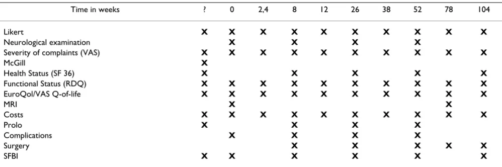
In the LSRS the most common complaints are pain and disability to perform normal daily activities. We will use below described validated outcome parameters, which will be assessed by means of questionnaires. Patients are not informed about their earlier scores. Follow-up exami-nations by the research nurse will take place 8, 26 and 52 weeks after randomization and the patients will keep a diary (table 2). In between at 2, 4, 12, 38, and 78 and after 104 weeks the main questionnaire (primary outcome measures) will be filled in at home and send to the data centre.
Primary outcome measures
1) Roland Disability Questionnaire for Sciatica.' This ill-ness-specific 23-item functional assessment questionnaire is frequently used for low back pain and sciatica [38,39]. Scores range from 0 to 23, reflecting a simple unweighted sums of items endorsed by the respondent. Patients with high scores at baseline do have a severe disabling LSRS. To define recovery a difference of at least 11 points from baseline has to be seen [38,17]. The Roland Question-naire for Sciatica has a documented high level of internal consistency; construct validity, and responsiveness [38,39]. It is the main primary outcome measure in this trial.
rank their five most important functional disabilities in daily live (work, hobby), which they can use in their own evaluation overall and in separate items.
3) VAS pain in the leg. This parameter will measure the experienced intensity of pain in the leg during the week before visiting the research nurse. Pain will be assessed on a horizontal 100 mm scale varying from 0 mm, 'no pain in the leg', to 100 mm, 'the worst pain ever'. Patients do not see the results of earlier assessments and will score the pain experienced at the visit. [28-32].
Secondary outcome measures
[image:7.612.55.554.99.265.2]1) EuroQol classification system and VAS rating personal health. A cost-utility analysis will be performed using QALY's based on the EuroQol questionnaire, which has been validated in many studies and is easy to fill out [41,42,51]. The EuroQol will be measured twice a week during the first four weeks and at all follow-up moments. Patients describe their general health status using the EuroQol classification system, consisting of 5 questions on mobility, self care, usual activities, pain/discomfort, and anxiety/depression [44]. From the EQ-5D classifica-tion system, the EQ-5D utility index will be calculated [43]. This utility measure reflects how the general public values the health status described by the patient, which is preferred for economic evaluations from a societal per-spective. Patients also rated their personal health using a visual analog scale (VAS) ranging from worst imaginable health to best imaginable health.
2) Short-Form 36 (SF-36). Quality of life was also assessed using the RAND-36 questionnaire. This is a generic health status questionnaire, which can easily be filled out at home. The questionnaire consists of 36 items on physical and social functioning has 8 domains; 1) physical func-tioning, 2) physical restrictions, 3) emotional restrictions,
4) social functioning, 5) somatic pain, 6) general mental health, 7) vitality, 8) general health perception. This ques-tionnaire has been used frequently and was validated in studies on low back pathology and surgery [33-37]. From the RAND-36, the SF-6D utility index was calculated. Like the EQ-5D, this SF-6D reflects the general public's valua-tion of the health described by the patient. The SF-6D is a recent instrument that has not been used much yet, but it richer classification system could make it a more sensitive utility measure than the EuroQol measure.
3) Sciatica Frequency and Bothersome Index (SFBI). This is a scale from 0 to 6, which can assess the frequency (0 = not at all to 6 = always) and bothersomeness (0 = not bothersome to 6 = extreme bothersome) of back and leg symptoms. The sum of the results of four symptom ques-tions yields both indexes, ranging from 0 to 24: leg pain; numbness and/or tingling in the leg; weakness in the leg or foot; pain in the back or leg while sitting. [17].
5) PROLO-scale. This scale measures the evaluation of the research nurse of the functional-economic status of the patients. This parameter has been used in studies on the difference in functional outcome between different tech-niques of lumbar spine fusion [40].
6) VAS pain in the back. This parameter measures the intensity of the pain in the back experienced during the week before visiting the research nurse. Assessment will be based on a horizontal 100 mm scale varying from 0 mm, 'no pain in the back', to 100 mm, 'the worst pain ever'. Patients do not see the results of earlier assessments and will score their pain during the visit. This parameter is included because a lot of patients with LSRS also have back pain in varying intensities, which can change after surgery or conservative treatment.
Table 2: Data collection and outcome measures
Time in weeks ? 0 2,4 8 12 26 38 52 78 104
Likert X X X X X X X X X X
Neurological examination X X X X
Severity of complaints (VAS) X X X X X X X X X X
McGill X
Health Status (SF 36) X X X X X
Functional Status (RDQ) X X X X X X X X X X
EuroQol/VAS Q-of-life X X X X X X X X X X
MRI X X
Costs X X X X X X X X X X
Prolo X X X X
Complications X X X X
Surgery X X X X X
Other outcome measures
1) Costs. The societal costs during the first year will be estimated in accordance with the recent pharmacoeco-nomic guideline [47,48]. The costs of hospital admission and surgery will be based on an integral top-down cost analysis in three large regional participating hospitals (aggregated according to the total number of patients per department). From this institutional analysis, the con-stant costs per admission and the variable costs per admis-sion day will be estimated. From these constant and variable costs, the individual costs of hospital admission and surgery for all patients can be estimated, using the duration of the hospitalization. In the study an MRI is per-formed in all cases. The costs of this MRI will only be cal-culated for patients undergoing surgery, because in the normal situation MRI would only be performed when a surgical indication exists.
Patients will register other health care needs in a diary (including physiotherapy, visits to GP's and specialists, nursing care and medication). Each diary covers a period of 3 months and will be discussed with the patient during the follow-up visits to the research nurse. The volume of health care will be assessed using standard prices [48].
In the diary the patient will also register direct non-medi-cal costs (including time costs, travel expenses and domes-tic help). To estimate productivity costs the patients will also report absenteeism in the diary. At the follow-up vis-its, the research nurse will register the work situation, work efficiency and gross wages. Absenteeism will be val-ued according to the friction-cost method.
2) Incidence of (re-) surgery. One of the goals of the policy for group B is to avoid surgery while achieving at least the same effects. The surgical rate is therefore an indication of the success or failure of this policy. The incidence of re-operation at the same disc level in group A will be an indi-cation of the failure rate for surgery.
3) Side-effects or complications that are ascribed to the treatment are recorded by the patients, their treating phy-sicians and the research nurses.
4) MRI findings. The results of the differences between the baseline MRI and the MRI made 52 weeks after randomi-zation are important secondary outcome measures. The difference in size of the disc herniation (in mm), nerve root compression, and amount of scar tissue will be regis-tered. Failures of surgery can be recognized by inadequate disc removal or decompression of the nerve. The data will be gathered, using a standardized CRF, which will be filled out by the local radiologist, orthopaedic- or neurosurgeon and (neuro-) radiologist
Sample size
The result of this study is based on the short-term success of surgical intervention and will be a trade-off between a quicker relief of leg pain versus an advantage in cost-effec-tiveness for conservatively managed patients. The sample size is calculated on the basis of the Roland Disability Questionnaire for Sciatica averaged during the 12 months follow-up period. The numbers used for this sample-size are drawn from the Maine Lumbar Spine Study 1 year and recently published 5-year results [19,55]. The difference in the Roland score between the surgical- and non-surgical group in this study did not change between 3 and 12 months follow-up as shown in their study [19] and can be averaged over the first year. The main aim of this study is to measure the short-term functional difference at 12 months follow-up. Surgical treatment is considered better when the post treatment change is at least 4 points more when compared to the conservative treatment arm [38] and constant over time. Considering this constant differ-ence and a mean standard deviation =10 over the first year [55] 140 patients per treatment arm are needed to reach a power (1-β) of 0,90 with α = 0.05 (two-sided). To answer the main research question 280 patients are needed for analysis with at least 12 months follow-up. The aim is to enrol 300 (150 per arm) patients in the study, including 8 % loss to follow-up after 1 year. The total number of oper-ated patients each year in all participating hospitals exceeds 1400. With this number of patients also a clini-cally important difference in median time to recovery of two months can be detected by survival analysis. Although the time to recovery is the main issue, the prob-lem of recurrent complaints is still not solved in the differ-ent approaches of survival and proportional hazard analysis.
Statistical and cost analysis
in the same group who had not and with patients in the surgery group. To compare the actual treatment sec instead of strategies an explorative analysis will be per-formed in subgroups off all patients who actually received surgery and who did not receive surgery in both groups. All patients who withdraw from the study are included in the analysis until the time of withdrawal.
The result of this study will be a trade off between the dis-advantages of surgery (hospitalisation, reduced quality of life and costs) versus the possible advantages (earlier relief of pain and return to work). For that reason recovery, measured as an 11 point difference in score when com-pared to baseline (Roland Disability Questionnaire for Sciatica), is the clinically most relevant patient outcome. Quality of Life (SF-36) and perceived recovery are impor-tant to compare the reduced quality of life from surgery to the possibly prolonged pain from conservative therapy and also to be able to compare cost-effectiveness with that of other spine interventions. The EuroQol is important to obtain cost-utility ratio's that can be compared with those of a wide range of other interventions. Utilities are obtained from the descriptive classification system of the EuroQol, using the model described by Dolan [43,53]. Conservative treatment may decrease costs compared to surgery but possibly at the expense of delayed effective-ness. In an incremental cost-effectiveness analysis, societal
costs during the first year will be compared to the primary outcome measure (Roland Disability Questionnaire for Sciatica, averaged over the first year), Quality of Life (SF-36, during the first year) and perceived recovery (7-points Likert scale). Cost-effectiveness analyses with these effec-tiveness measures have been conducted before, allowing comparison with other spine interventions.
Finally, to answer the second research question explora-tive analyses are conducted to investigate whether the treatment effect after two, six and twelve months varies in specific subgroups of patients (Table 3).
Using logistic regression for success rate and linear regres-sion for severity of the disability, each prognostic indica-tor is checked for interaction with treatment. If the interaction term is significant, a stratified analysis will be performed.
Discussion
In this article the rationale and design of a pragmatic RCT on the cost-effectiveness of timing of disc surgery for LSRS is described. The only randomized trial [7] so far on this subject only included patients where the caregiver was in doubt about the surgical indication. Patients with severe disabling pain were not randomized [8]. The Sciatica Trial is directed to those patients with a clear surgical indication according to current usual care. The study is pragmatic because it acknowledges that sometimes it may not be possible to postpone surgery for every conservative care patient until 6 months after allocation and that some patients will recover before surgery is performed in the surgical group. In these cases we consider it unethical to hold on to the randomized treatment. Because of the Intent-to-Treat analysis these cases will be analysed in their own allocated randomization arm and will not cause methodological problems because it is two healthcare strategies that are compared, as opposed to two treat-ments. The objective of this trial is to provide evidence on the preferred timing of disc surgery for sciatica. A pro-longed conservative treatment strategy is compared to the international guideline advise of surgery after 6–8 weeks LSRS. The intended size of the study population is suffi-ciently large to detect short and long term differences between both strategies.
Abbreviations
GP = General Practitioner
LSRS = Lumbosacral Radicular Syndrome
RCT = Randomized Controlled Trial
VAS = Visual Analogue Scale
Table 3: Selected prognostic variables for subgroup analysis
Demographic Variables
• Age < 39 years versus > 39 years, • Intellectual versus physical demanding job,
Anamnestic and Neurological Variables • Acute start LSRS versus slow start, • History of backpain versus no history,
• Influence of coughing, sneezing on complaints versus no influence, • Difficulty to put on shoes and/or socks versus no difficulty, • Straight leg raising ≤ 30 degrees versus > 30 degrees, • Positive crossed straight leg raising sign versus negative sign, • VAS-pain > 70 versus < 69 mm,
• Tingling/numbness in pain area versus no tingling (9), • Pain leg worse by sitting versus no worsening (9), • McGill affective high score versus low score,
Radiological Variables
• MRI disc sequester versus contained disc herniation, • MRI circumferential gadolinium enhancement versus no enhancement of disc herniation,
• Mediolateral versus median and lateral disc herniation, • High versus low height of disc level (height 9 mm),
Miscellaneous Variables
Competing Interests
The author(s) declare that they have no competing interests.
Author's contributions
WP designed the study is responsible for the protocol. HH is responsible for the calculation of the sample size and contributed to the design of analysis. WH is responsible for the design of the cost-effectiveness analysis. RB has contributed in the case record forms and is responsible for the database ProMIse. JE contributed to the involvement of the GP's. JT structured the ideas about the diagnostics of LSRS and intake by neurologists. RT is the neurosurgical supervisor of WP. BK is the epidemiological supervisor of WP. All authors participated in the trial design and coor-dination. All authors read and approved the final manuscript.
Acknowledgements
The Sciatica Trial is funded by ZonMW/NOW. Furthermore we do want to thank the six researchnurses and datamanagers of the Sciatica Research Team for their work in making the start of this trial possible in the partici-pating hospitals.
References
1. Egger M, Smith GD: Bias in location and selection of studies.BMJ
1998, 316:61-6.
2. Thornton A, Lee P: Publication bias in meta-analysis: its causes and consequences.J Clin Epidemiol 2000, 53:207-16.
3. Van de Velden J, de Bakker DH: Basisrapport: Morbiditeit in de huisartsenpraktijk.Utrecht Nivel 1990.
4. Van den Bosch JH, Kardaun JW: Ziekten van het zenuwstelsel in Nederland.Den Haag: SDU/uitgeverij 1993.
5. SIG Zorginformatie Utrecht Gegevens uit de landelijke registratie van Nederlandse Ziekenhuizen; 1996.
6. Van Tulder, Koes BW, Bouter LM: A cost-of-illness study of back pain in the Netherlands.Pain 1995, 62:233-40.
7. Weber H: Lumbar Disc Herniation. A controlled prospective study with ten years of observation.Spine 1983, 8:131-140. 8. Weber H: Lumbar Disc Herniation. A prospective study of
prognostic factors including a controlled trial.J of Oslo City Hospital 1978, 28:33-64.
9. Vroomen PC: The diagnosis and conservative treatment of Sciatica.Thesis Maastricht 1998.
10. Cherkin DC, Deyo RA: An international comparison of back surgery rates.Spine 1994, 19:1201-1206.
11. Smeele IJ, van den Hooghen JM, Mens JM: E.a. NHG-standaard Lumbosacraal Radiculair Syndroom. Huisarts Wet 1996,
39:78-89.
12. Stam J: Consensus over diagnostiek en behandeling van het lumbosacraal radiculair syndroom.Ned Tijdschr Geneesk 1996,
140:2621-27.
13. CBO Consensus Het lumbosacraal radiculair syndroom.
1996.
14. Gezondheidsraad 1999/18.Diagnostiek en behandeling van het lum-bosacraal radiculair syndroom .
15. Gibson JN, Grant IC, Waddell G: The Cochrane Review of Sur-gery for Lumbar Disc Prolapse and Degenerative Lumbar Spondylosis.Spine 1999, 24:1820-32.
16. Gibson JN, Grant IC, Waddell G: Surgery for lumbar disc pro-lapse (Cochrane Review). In Cochrane Library Issue 4 Oxford: Update Software; 2000.
17. Mixter WJ, Barr J: Rupture of the intervertebral disc with involvement of the spinal canal.N Engl J Med 1934, 211:210-15. 18. Spanfort EV: Lumbar disc herniation: a computer aided analy-sis of 2504 operations.Acta Orthopaedica Scandinavica 1972:142.
19. Atlas SJ, Deyo RA: The Maine Lumbar Spine Study, part II. 1-Year Outcomes of Surgical and Nonsurgical Management of Sciatica.Spine 1996, 15:1777-1786.
20. Hakelius A: Prognosis in Sciatica: A clinical follow-up of surgi-cal and nonsurgisurgi-cal treatment.Acta Orthop Scand 1970:1-76. 21. Malter AD, Deyo RA: 5-Year Reoperation Rates After Different
Types of Lumbar Spine Surgery.Spine 1998, 23:814-820. 22. Saal JA: Natural history and non-operative treatmenet of
lum-bar disc herniation.Spine 1996:2S-9S.
23. Bessette L, Liang MH: Classics in Spine. Surgery literature revisited.Spine 1996, 21:259-263.
24. Malter AD, Deyo RA: Cost-Effectiveness of lumbar discectomy for the treatment of herniated intervertebral disc.Spine 1996,
21:1050-1055.
25. Saal JA: Saal JS The natural history of lumbar intervertebral disc extrusions treated non-operatively.Spine 1989, 15:683-6. 26. Gatchel , Turk DC: Psychological approaches to
painmanage-ment: a practitioner's handbook. The Guilford Press; 1996. 27. Main CJ, Wood PLR, Hollis S: The distress and risk assesment
method.Spine 1992, 17:42-52.
28. Fordyce WE: Behavioral methods for chronic pain and illness.
St Louis: CV Mosby Company; 1976.
29. Deyo RA: Outcome measures for low back pain research. A proposal for standardized use.Spine 1998, 23:2003-2013. 30. Scott J, Huskisson EC: Graphic representation of pain.Pain 1976,
2:175-184.
31. Huskisson : Measurement of pain.Lancet 1974:1127-1131. 32. Joyce CRB, Zutshi DW: Comparison of fixed interval and Visual
Analogue Scales for rating Chronic Pain.Eur J Clin Pharmacol
1975, 8:415-420.
33. Melzack R, Katz J: Pain measurement in persons in pain. Text-book of Pain .
34. Collins SL, Moore A: The visual analogue pain intensity scale: what is moderate pain in millimetres?Pain 1997, 72:95-97. 35. Ware JE, Sherbourne C: The MOS 36-item short-form survey
(SF 36): Conceptual framework and item selection.Med Care
1992, 30:473-483.
36. Van der Zee K, Sanderman R: De psychometrische kwaliteiten van de MOS 36-item Short Form Health Survey (SF-36) in een Nederlandse populatie. T Soc Gezondheidsz 1993,
71:183-191.
37. Brazier JE, Harper R: Validating the SF-36 health survey questionnaire.BMJ 1992, 305:160-64.
38. Aaronson NK, Acquadro C: International quality of life asses-ment (IQOLA) project.Q Of Life Research 1992, 1:349-51. 39. Stansfeld SA, Roberts R: Assessing the validity of the SF-36
Gen-eral Health Survey.Q Of Life Research 1997, 6:217-24.
40. Patrick DL, Deyo RA: Assessing health related quality of life in patients with sciatica.Spine 1995, 20:1899-909.
41. Gommans I, Koes BW: Validity and responsiveness of the Dutch adaptation of the Roland Disability Questionnaire. In
Low Back Pain Edited by: Tulder MW, Koes BW, Bouter LM. EMGO; 1996:57-70.
42. Prolo DJ, Oklund SA: Toward uniformity in evaluating results of lumbar spine operations.Spine 1986, 11:601-606.
43. Dolan PP: Modeling valuations for EuroQol health states.Med Care 1997, 35:1095-1108.
44. EuroQol Group: A new facility for the measurement of health-related quality of life.Health Policy 1990, 16:199-208.
45. Matsubara Y: Serial changes on MRI in lumbar disc herniations treated conservatively.Neuroradiology 1995, 37:378-83. 46. Bozzao A, Galluci M: MRI imaging assessment of natural history
in patiënts treated without surgery.Radiology 1992, 185:135-41. 47. College voor zorgverzekeringen: Richtlijnen voor
farmaco-econ-omisch onderzoek.CvZ, afdeling FO/G&s 1999.
48. Oostenbrink JB, Koopmanschap MA, Rutten FF: Handleiding voor kostenonderzoek, methoden en richtlijnprijzen voor econo-mische evaluaties in de gezondheidszorg.CvZ 2000.
49. Jansen SJT, Stiggelbout AM: Unstable preferences: a shift in val-uation or an effect of the elicitation procedure?Med Decis Making 2000, 20:62-71.
50. Tubach F, Beaute J: Natural History and prognostic indicators of sciatica.J of Clin Epidemiology 2004, 57:174-179.
Publish with BioMed Central and every scientist can read your work free of charge "BioMed Central will be the most significant development for disseminating the results of biomedical researc h in our lifetime."
Sir Paul Nurse, Cancer Research UK
Your research papers will be:
available free of charge to the entire biomedical community
peer reviewed and published immediately upon acceptance
cited in PubMed and archived on PubMed Central
yours — you keep the copyright
Submit your manuscript here:
http://www.biomedcentral.com/info/publishing_adv.asp
BioMedcentral
controlled study of bedrest and physiotherapy for acute sciatica.J Neurosurg (Spine 1) 2002, 96:45-49.
52. Stiggelbout AM, Eijkemans MJC, Kiebert GM, Kievit J, Leer JWH, De Haes JCJM: The "utility" of the Visual Analog Scale in medical decision-making and technology assessment: is it an alterna-tive to the Time Trade-Off?Int J Technol Assessm Health Care 1996,
12:291-8.
53. Dolan P: Modelling valuations for health states: the effect of duration.Health Policy 1996, 38:189-203.
54. Stolk EA, Busschbach JJ, Caffa M, Meuleman EJ, Rutten FF: Cost util-ity analysis of sildenafil compared with papaverine-phen-tolamine injections.BMJ 2000, 29(320):1165-8.
55. Atlas SJ, Keller RB, Deyo RA: Surgical and non-surgical Manage-ment of Sciatica Secondary to a Lumbar Disc Herniation. Five-Year Outcomes from the Maine Lumbar Spine Study. Spine 2001, 15(26(10)):1179-1187.
Pre-publication history
The pre-publication history for this paper can be accessed here: