Hepatorenal Syndrome
Tyree H. Kiser
Department of Clinical Pharmacy, University of Colorado Skaggs School of Pharmacy and Pharmaceutical Sciences, Anschutz Medical Campus, Aurora, USA.
Ema
Received November 13th, 2013; revised December 10th, 2013; accepted January 5th, 2014
Copyright © 2014 Tyree H. Kiser. This is an open access article distributed under the Creative Commons Attribution License, which permits unrestricted use, distribution, and reproduction in any medium, provided the original work is properly cited. In accordance of the Creative Commons Attribution License all Copyrights © 2014 are reserved for SCIRP and the owner of the intellectual property Tyree H. Kiser. All Copyright © 2014 are guarded by law and by SCIRP as a guardian.
ABSTRACT
Hepatorenal syndrome (HRS) is the most serious hepatorenal disorder and one of the most difficult to treat. To date, the best treatment options are those that reverse the mechanisms underlying HRS: portal hypertension, splanchnic vasodilation, and/or renal vasoconstriction. Therefore, liver transplantation is the preferred definitive treatment option. The role of other therapies is predominantly to prolong survival sufficiently to allow patients to undergo transplantation. Terlipressin with the addition of adjunctive albumin volume expansion is the pre-ferred pharmacologic therapy for the treatment of patients with HRS. Norepinephrine and vasopressin are ac-ceptable alternatives in countries where terlipressin is not yet available. For patients with Type II HRS, mido-drine plus octreotide appears to be an effective pharmacologic regimen that can be administered outside of an intensive care unit setting. Regardless of chosen vasoconstrictor therapy, careful monitoring is needed to ensure tissue ischemia and severe adverse effects do not occur. Artificial hepatic support devices, renal replacement therapy, and transjugular intrahepatic portosystemic shunt (TIPS) are non-pharmacologic options for patients with HRS. However, hepatic support devices and renal replacement therapies have not yet demonstrated im-proved outcomes and TIPS is difficult to be employed in patients with Type I HRS due to contraindications in the majority of patients. Despite advances in our understanding of hepatorenal syndrome, the disease is still as-sociated with significant morbidity, mortality, and costs. More evidence is urgently needed to help improve pa-tient outcomes in this difficult-to-treat population.
KEYWORDS
Hepatorenal Disorder; Cirrhosis; Portal Hypertension; Transplantation; Vasopressors
1. Introduction
Hepatorenal syndrome (HRS) is the most significant he-patorenal disorder affecting patients with advanced cirr-hosis. If not treated, patients with HRS Type I survive only a median of 2 weeks and 95% of patients die within the first 30 days after onset. The median survival time is 4 to 6 months in patients with Type II HRS [1]. Patients with advanced cirrhosis and ascites are at high risk for HRS, with 18% of patients developing hepatorenal syn-drome (HRS) within 1 year and up to 39% developing HRS by 5 years [2-4].
HRS is a unique renal dysfunction, because it is a functional renal failure that occurs in the absence of pa-renchymal kidney disease. Patients with cirrhosis have
tention to increase intravascular blood volume. Cardiac output is increased in response to reduced effective ar-terial volume resulting in tachycardia and low systemic blood pressure. Ultimately, this cascade of events causes a shift in the renal autoregulation curve, making renal
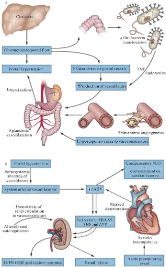
[image:2.595.134.460.155.681.2]perfusion much more sensitive to changes in mean ar-terial pressure. As cirrhosis progresses, further renal va-soconstriction and sodium retention occurs as the splan-chnic and systemic vasodilation worsens leading to the development of a functional renal failure (Figure1) [5,6].
Frequently, the development of HRS is precipitated by an acute event. In patients with cirrhosis and ascites, bacterial infections appear to be the most important risk factor for the development of HRS. Infection results in circulatory dysfunction by releasing various cytokines, such as tumor necrosis factor and interleukin 6 in the splanchnic vasculature [5,6]. These inflammatory cyto-kines activate endothelial and inducible nitric oxide syn-thases increasing the production of nitric oxide [6]. As many 33% of patients who develop spontaneous bacterial peritonitis will develop HRS [7]. Other HRS triggering events include severe alcoholic hepatitis, hypovolemia as the result of excess diuresis or gastrointestinal losses (bleeding or diarrhea), large volume shifts between in- travascular and extravascular compartments, and use of medications that affect afferent or efferent arteriole con-striction or vasodilation in the kidney (e.g., nonsteroidal anti-inflammatory drugs [NSAIDs], angiotensin-con- verting enzyme inhibitors [ACE-Is], angiotensin II re-ceptor blockers [ARBs]) [5]. Additionally, cirrhotic car-diomyopathy resulting in the inability to maintain the re- quired cardiac reserve may be an important contributor to HRS.
2. Diagnosis of HRS
The assessment of HRS is daunting, because of the dif-ficulties in providing a definitive diagnosis and the poor overall response rate to currently available therapies. The diagnosis of HRS is made by excluding all other possible causes of renal failure and utilizing revised criteria pub-lished by the International Ascites Club in 2007 (Table1) [8,9]. These diagnostic criteria have been widely ac-cepted; however, they may be difficult to apply in the acute care setting. The major struggle within the diag-nostic criteria is the difficulty in fulfilling all of the di-agnostic criteria in patients who have a presentation sug-gestive of HRS. One of the major limitations in the clin-ical setting is the ability to rule out renal failure caused by other factors, because many patients with HRS physi-ology have bacterial infections with or without shock, are receiving diuretic therapy prior to their AKI, or may be receiving medications or undergoing procedures that are detrimental to renal blood flow or kidney function. Addi-tionally, patients with prolonged Type I HRS may even-tually develop acute tubular necrosis due to intense renal arteriole vasoconstrition. As a result of these limitations, several renal biomarkers are being studied to help deci-pher HRS from other causes of AKI in patients with cirrhosis; but further studies are required before they can be applied in clinical practice [10-12].
After establishing the diagnosis, HRS can be divided into one of two forms: Type I and Type II. Type I HRS is diagnosed when there is a doubling in SCr to a val-ue >2.5 mg/dL in a period of less than 2 weeks. Type I
Table 1. Diagnostic criteria for hepatorenal syndrome.
Diagnostic Criteria for Hepatorenal Syndrome
Cirrhosis with ascites
Serum creatinine (SCr) >1.5 mg/dL (>133 umol/L)
No improvement in serum creatinine levels (decrease to of ≤1.5 mg/dL) after at least 2 days with diuretic withdrawal (if on diuretics) and volume expansion with 20% to 25% albumin. The recommended
dose of albumin is 1 g/kg of body weight per day, up to a maximum of 100 g/day.
Absence of shock
No current or recent treatment with nephrotoxic medications
Absence of parenchymal kidney disease as defined by proteinuria <500 mg/day, no microhematuria (<50 red blood cells per high
power field), and normal renal ultrasonography)
Classification of Hepatorenal Syndrome
Type I — doubling in SCr to a value > 2.5 mg/dL in a period of less than 2 weeks
Type II — stable or more slowly progressive renal dysfunction (SCr > 1.5 mg/dL) not meeting the criteria for Type I HRS
Adapted from Solerno, et al. [9] and Gines, et al. [5].
HRS usually develops as a result of a triggering factor that causes acute deterioration of hepatic function to-gether with other organ dysfunctions. The most common triggers for Type I HRS are bacterial infections and se-vere alcoholic hepatitis. In contrast, Type II HRS occurs in patients with refractory ascites and involves renal dysfunction (SCr > 1.5 mg/dL) that is more slowly pro-gressive and does not meet the criteria for Type I HRS. It is common for patients with Type II HRS to eventually develop Type I HRS as the result of a precipitating event [4,12].
3. Prevention of HRS
liter of ascites removed) is necessary to avoid large vo-lume shifts from the intravascular space [13,14]. Addi-tionally, antibiotic prophylaxis in patients with a ga-strointestinal bleed can reduce the incidence of sponta-neous bacterial peritonitis and renal failure and pentox-ifylline therapy may reduce the incidence of HRS in pa-tients with acute alcoholic hepatitis [15,16].
4. Treatment of HRS
Liver transplantation is the optimal and definitive therapy for patients with HRS because it cures the underlying organ dysfunction responsible for the pathophysiologic pathway to HRS [17-19]. Liver transplantation drastical-ly improves mortality for patients with HRS, resulting in 5-year survival rates similar to patients without HRS who underwent liver transplantation (67.1% versus 70.1%, respectively; P = NS) [18]. Renal dysfunction can often be reversible and, therefore, patients with HRS are not frequently listed for combined liver-kidney transplants (LKTx); however, recommendations have been made to consider LKTx in HRS patients who have received he-modialysis (HD) for >8 weeks [14], with some groups advocating for a requirement of >12 weeks of HD prior to transplantation before consideration of LKTx [12].
Treatment of HRS with the intent to improve renal function and prolong survival long enough to allow for liver transplantation is the ultimate goal of pharmacolog-ic therapy for HRS. Vasoconstrpharmacolog-ictors are the mainstay of therapy for HRS due to their ability to improve the he-modynamic instability that is responsible for the de-creased renal perfusion pressure. The addition of albumin therapy to vasoconstrictors may further improve renal blood flow, glomerular filtration, and ultimately response rates to vasoconstrictor therapy [20]. If albumin is uti-lized, the admixture should provide a high concentration of albumin and the typical dose is 1 g/kg (up to 100 g) on day 1 or 2, then 25 to 50 g/day of 25% albumin (or 20 to 40 g/day of 20% albumin) thereafter [4]. Albumin thera-py is continued, along with vasoconstrictor therathera-py, until a complete response in SCr is realized or until futility of therapy is determined. The dose and duration of albumin therapy should be dictated by volume status; as albumin is initially effective at improving intravascular volume, but will eventually result in third space volume expan-sion. Volume status should be assessed by hemodynamic monitoring, although the optimal method for evaluating volume status is controversial and likely includes the interpretation of several possible measurements, includ-ing heart rate, mean arterial pressure (MAP), central ve- nous pressure, pulse pressure variation, stroke volume variation, echocardiography, urine output, ascites, and edema.
Terlipressin is a unique vasopressin analogue with po-tential advantages that make it the preferred
vasocon-strictor for patients with HRS. The effects of vasopressin and vasopressin analogues on the V1 receptor are the predominate mechanism for treating the underlying splanchnic vasodilation present in those with HRS. There are a large number of V1 receptors in the splanchnic vasculature, making this area especially sensitive to the vasoconstrictive effects [21]. Vasoconstriction of the splanchnic vascular beds is believed to reverse HRS by increasing effective arterial blood volume, thereby sup-pressing activation of the renin-angiotensin-aldosterone system (RAAS) and the sympathetic nervous system, reversing compensatory renal vasoconstriction and ulti-mately increasing renal perfusion. Terlipressin reduces portal vein pressure and increases MAP in patients with cirrhosis and splanchnic vasodilation. It improves splan-chnic blood flow and down-regulates the excessive salt and water retention that leads to ascitic fluid accumula-tion [22]. These attributes have made terlipressin one of the most widely used agents for the treatment of Type I HRS outside of North America.
Although the number of prospective controlled trials is small, terlipressin has been one of the most studied
vaso-pressor agents for the treatment of HRS (Table2) [22].
A comprehensive review of the terlipressin literature for HRS through January 2012 can be found in the Cochrane Database [23]. Combined analysis of 6 prospective stu-dies demonstrates that terlipressin treatment improves renal function and mortality for patients with HRS. HRS reversal (reversal or complete response is defined as a decrease in SCr to a value ≤1.5 mg/dL) occurs in 25% to 50% of patients treated with terlipressin. Relapse rates after stopping therapy do occur and retreatment with va-soconstrictor therapy may be necessary. Adverse effects related to terlipressin include tachycardia, arrhythmias, chest pain, diarrhea, abdominal pain, bronchospasm, and peripheral ischemia [24]. Serious ischemic adverse events have required discontinuation of terlipressin ther-apy in a small percentage of patients (e.g., nonfatal myo- cardial infarction, livedo reticularis, and cyanosis of the fingers) [25]. The phase III Multi-Center Randomized, Placebo-Controlled, Double-Blind Study to Confirm the Reversal of Hepatorenal Syndrome Type 1 With Lucas-sin (TerlipresLucas-sin) (REVERSE) trial [ClinicalTrials. gov identifier NCT01143246] with provide further evidence evaluating terlipressin therapy for the management of patients with HRS.
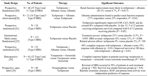
Table 2. Selected clinical studies of vasoconstrictors in the treatment of HRS.
Study Design No. of Patients Therapy Significant Outcomes
Prospective, Randomized [40]
N = 46 (35 Type I and 11 Type II HRS)
Terlipressin + Albumin versus Albumin
Renal function improvement more likely in terlipressin + albumin (43.5% versus 8.7%, P = 0.017)
Prospective, observational[20]
N = 21 (16 Type I and 5 Type II HRS)
Terlipressin + Albumin versus Terlipressin
Albumin administration found to predict renal function response (77% responders versus 25% responders, P = 0.03)
Prospective, randomized [41]
N = 24
(Type I HRS) Terlipressin versus Placebo
Terlipressin significantly improved UOP, CrCl, MAP, and de-creased SCr compared with placebo. At day 15, 5 of 12 patients
receiving terlipressin survived compared with 0 of 12 patients receiving placebo (P < 0.05).
International, Multi-center, Randomized [25]
N = 112
(Type I HRS) Terlipressin versus Placebo
Treatment success: terlipressin 25% versus placebo 12.5%, P = 0.093. HRS reversal: terlipressin 34% versus 13%, P = 0.008. Related adverse effects: terlipressin 9% versus placebo 2%, P = NS
Prospective, randomized [24]
N = 52 (Type I HRS)
Terlipressin + Albumin versus Albumin
80% complete response with terlipressin + albumin versus 19% response with albumin (p < 0.01). Improved survival at 180 days
with terlipressin + albumin (p < 0.01)
Retrospective [26]
N = 43 (32 Type I and 11 Type II HRS)
Vasopressin + Octreotide versus Vasopressin versus
Octreotide
Complete response higher in patients receiving vasopressin or vasopressin + octreotide versus octreotide monotherapy (P = 0.01)
Prospective, open label [29]
N = 40 (Type I HRS)
Norepinephrine versus Terlipressin
Reversal of HRS occurred in 50% of patients in each treatment group (p = NS). Survival was similar between groups (p = 0.8). Baseline creatinine clearance, MAP, and plasma renin activity were
independent predictors of response.
UOP = urine output; CrCl = creatinine clearance; MAP = mean arterial pressure; SCr = serum creatinine; Adapted from Kiser et al. [22].
(Table3) [4]. It may take 2 to 3 days for a response in SCr to be observed, so early dosing titration decisions should focus on achieving a MAP increase of 10 mm Hg, UOP improvement, and avoidance of ischemic adverse effects. Therapy should be discontinued if patients dem-onstrate no response in SCr by day 4 of therapy, despite adequate titration and an increase in MAP, because a response to therapy at this point is unlikely.
In countries where terlipressin is not commercially available, other vasoconstrictor treatment options (e.g., vasopressin or norepinephrine) must still be considered. Vasopressin is considered a reasonable alternative to terlipressin therapy because of its effects on the V1 re-ceptor; however, it is less selective than terlipressin and must be administered by a continuous infusion because of its shorter half-life. Evidence for vasopressin use in HRS comes from a retrospective study that evaluated 43 patients who had received vasopressin and/or octreotide for treatment of HRS. Response in SCr (SCR < 1.5 mg/dL) occured in 41% of the patients that received va-sopressin therapy. Therapy with vava-sopressin, either alone or in combination with octreotide, was an independent predictor of renal function recovery (odds ratio [OR] 6.4; 95% confidence interval [CI] 1.3 - 31.8). The mean va-sopressin dose in patients that responded to therapy was 0.23 ± 0.19 units/min, which is significantly higher than typically utilized in shock syndromes [26]. Although patients with cirrhosis and HRS appear to be more tole-rant to higher doses of vasopressin, caution and careful monitoring of serum lactate levels and the monitoring of
extremities for ischemia should be maintained for pa-tients receiving vasopressin doses >0.1 units/min as ad-verse effects related to vasopressin are ischemic in nature and dose dependent [27].
Norepinephrine’s alpha-adrenergic agonist activity makes it a potent vasoconstrictor of both the venous and arterial vasculature. Similar to terlipressin, in patients with HRS, norepinephrine effectively improves UOP, sodium excretion, serum sodium concentration, creati-nine clearance (CrCl), MAP, plasma renin activity, and aldosterone activity. In small comparative studies, nopinephrine has demonstrated a similar rate of HRS re-versal and patient survival when compared with terli-pressin [28,29]. Adverse effects between norepinephrine and terlipressin are similar, with reversible cardiac and digital ischemia being the most common adverse events [29]. The cost of norepinephrine therapy is also signifi-cantly lower than terlipressin (107 ± 31 versus 1536 ± 40 Euros, P < 0.0001), making it an attractive alternative therapy [28]. However, the possibility of utilizing terli-pressin in patients outside of a monitored hospital setting may reduce the overall difference in cost between treat-ment options.
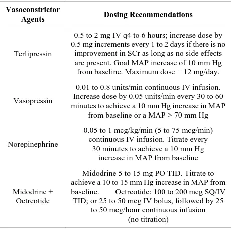
Table 3. Dosage and administration of vasoconstrictor me-dications for HRS.
Vasoconstrictor
Agents Dosing Recommendations
Terlipressin
0.5 to 2 mg IV q4 to 6 hours; increase dose by 0.5 mg increments every 1 to 2 days if there is no
improvement in SCr as long as no side effects are present. Goal MAP increase of 10 mm Hg from baseline. Maximum dose = 12 mg/day.
Vasopressin
0.01 to 0.8 units/min continuous IV infusion. Increase dose by 0.05 units/min every 30 to 60 minutes to achieve a 10 mm Hg increase in MAP
from baseline or a MAP > 70 mm Hg
Norepinephrine
0.05 to 1 mcg/kg/min (5 to 75 mcg/min) continuous IV infusion. Titrate every
30 minutes to achieve a 10 mm Hg increase in MAP from baseline
Midodrine + Octreotide
Midodrine 5 to 15 mg PO TID. Titrate to achieve a 10 to 15 mm Hg increase in MAP from baseline. Octreotide: 100 to 200 mcgSQ/IV TID; or 25 to 50 mcg IV bolus, followed by 25
to 50 mcg/hour continuous infusion (no titration)
SCr = serum creatinine; MAP = mean arterial pressure; IV = intravenous; 1) Adjunctive albumin administration is recommended: 1 g/kg (up to 100 g) on day 1 or 2, then 25 to 50 g/day of 25% albumin (or 20 to 40 g/day of 20% albumin) thereafter. 2) Therapy should be discontinued after 4 days if no response in SCr is observed, despite adequate dosage titration, because the likelihood of a response to therapy is low. 3) All patients should be monitored for signs of ischemia (i.e., visual evaluation of digits, distal pulses, abdominal pain, serum lactate, and/or troponin) at least every 12 hours and after any dosing titration. 4) In patients that demonstrate a complete response to therapy, dosage reduction or vaso-constrictor discontinuation should be attempted by day 14 of therapy to determine the sustainability of the response. Restarting therapy may be necessary if a relapse occurs; Adapted from Nadim et al. [4]
usual midodrine dosage range for the treatment of HRS is 5 to 15 mg orally TID. If goal MAP cannot be achieved and there is no response in UOP or SCr, despite titration to 15 mg orally TID, consideration of switching to a more potent intravenous vasoconstrictor may be neces-sary [4].
TIPS procedures can significantly decrease the porto-systemic pressure gradient. This leads to decreased plasma renin and sympathetic activity potentially im-proving or reversing HRS physiology [31]. Insertion of the shunt within 4 to 6 weeks of HRS onset may improve renal function recovery and survival [32]. Improvement in renal function after TIPS may take several weeks, so the use of other therapies for treating HRS is commonly required until the effect of TIPS placement is realized. TIPS can be beneficial for patients with both Type I and Type II HRS; however, many patients with Type I HRS cannot safely undergo the procedure due to their ad-vanced liver disease or other contraindications. Patients with lower bilirubin and those with Type II HRS are more likely to have prolonged survival post TIPS [32].
Artificial liver support therapies have been evaluated
for the treatment of HRS; including molecular adsorbent recirculating system (MARS), Prometheus, single pass albumin dialysis (SPAD), and single pass albumin ex-tended dialysis (SPAED) [33-37]. These extracorporeal systems provide combined hepatic and renal support by removing water-soluble and albumin-bound toxins re-sulting in improved serum bilirubin, creatinine, and other laboratory measurements. Unfortunately, these artificial support systems do not provide sustained responses in kidney function after discontinuation and laboratory val-ues commonly return to pretreatment levels after discon-tinuation [33]. In addition to the inability to produce meaningful outcomes, other challenges to artificial liver support systems include hypotension, blood loss each time the circuit is replaced, and the frequent need for anticoagulant administration into the extracorporeal cir-cuit to prevent clotting of the circir-cuit. Therefore, use of these systems for the management of patients with HRS are not recommended at this time [4,6].
Few studies have evaluated renal replacement therapy (RRT) for the treatment of HRS [38,39]. The use of RRT can improve short-term survival for patients with HRS and may be helpful with bridging patients to transplant or treating patients who have an acute reversible cause of hepatic decompensation. Use by patients who are not transplant candidates and those without an acute reversi-ble component is unlikely to change a patient’s disease course and merely results in resource overutilization and substantial costs to the health care system [39]. Therefore, the initiation of continuous or intermittent RRT for pa-tients with HRS is generally reserved until a significant indication for dialysis arises (e.g., severe hyperkalemia, metabolic acidosis, or volume overload). Individual pa-tient selection, according to the severity of illness, Child- Pugh and MELD scores, and the potential for liver trans-plantation should all be considered prior to the initiation of RRT.
5. Conclusions
HRS is the most significant disease within the spectrum of hepatorenal disorders and is associated with a substan-tial mortality rate. HRS physiology is characterized by splanchnic arterial vasodilation causing reduced effective arterial volume, renal vasoconstriction as a result of ac-tivation of the sympathetic nervous system and the RAAS, reduced cardiac output as the result of cirrhotic cardiomyopathy, and release of vasoactive mediators that affect renal blood flow and glomerular microcirculatory hemodynamics. Patients with Type 1 HRS have a more acute rise in their SCr values and shorter survival times as compared to patients with Type II HRS.
The goal of all other pharmacologic and nonpharmaco- logic therapies is to prolong survival time enough to al- low for liver transplantation. Several small studies have attempted to evaluate various pharmacologic agents for the treatment of HRS, but only vasoconstrictors com- bined with albumin therapy have emerged as a preferred initial treatment option. Terlipressin, in combination with albumin volume expansion, is the preferred pharmaco- logic therapy for the management of patients with HRS. Norepinephrine and vasopressin are alternatives if terli- pressin is unavailable. Midodrine with octreotide appears to be an effective pharmacologic regimen in patients with Type II HRS and those who require an alternative to intravenous therapy. Although TIPS is effective for both Type I and II HRS, it is less commonly employed in Type I HRS patients due to the presence of contraindica- tions to the procedure. Artificial hepatic support devices and renal replacement therapy are effective for correcting abnormal laboratory values, but have not demonstrated the ability to reverse HRS. The decision to deliver these therapies should be limited to patients who have an indi- cation for dialysis and are high on the liver transplant list.
Substantial advances in understanding the pathophysi- ology and management of HRS has improved the recog- nition and treatment of HRS patients. However, several questions still remain regarding how best to optimize current treatment options. Given the substantial morbidi- ty, mortality, and cost associated with HRS management; more studies are urgently needed to help improve patient outcomes in this difficult population.
REFERENCES
[1] P. Gines, M. Guevara, V. Arroyo and J. Rodes., “Hepato- renal Syndrome,” The Lancet, Vol. 362, No. 9398, 2003, pp. 1819-1827.
[2] A. Gines, A. Escorsell, P. Gines, et al., “Incidence, Pre- dictive Factors, and Prognosis of the Hepatorenal Syn- drome in Cirrhosis with Ascites,” Gastroenterology, Vol. 105, No. 1, 1993, pp. 229-236.
[3] F. Wong, M. K. Nadim, J. A. Kellum, F. Salerno, R. Bel- lomo, A. Gerbes, P. Angeli, R. Moreau, A. Davenport, R. Jalan, C. Ronco, Y. Genyk and V. Arroyo, “Working Par- ty Proposal for a Revised Classification System of Renal Dysfunction in Patients with Cirrhosis,” Gut, Vol. 60, No. 5, 2011, pp. 702-709.
[4] M. K. Nadim, J. A. Kellum, A. Davenport, F. Wong, C. Davis, N. Pannu, A. Tolwani, R. Bellomo, Y. S Genyk and The ADQI Workgroup, “Hepatorenal Syndrome: The 8th International Consensus Conference of the Acute Di- alysis Quality Initiative (ADQI) Group,” Critical Care, Vol. 16, No. 1, 2012, p. R23.
[5] P. Gines and R. W. Schrier, “Renal Failure in Cirrhosis,”
The New England Journal of Medicine, Vol. 361, No. 13, 2009, pp. 1279-1290.
[6] F. Wong, “Recent Advances in our Understanding of He- patorenal Syndrome,” Nature Reviews Gastroenterology and Hepatology, Vol. 9, No. 7, 2012, pp. 382-391.
[7] P. Sort, M. Navasa, V. Arroyo, X. Aldeguer, R. Planas, L. Ruiz-del-Arbol, L. Castells, V. Vargas, G. Soriano, M. Guevara, P. Ginès and J. Rodés, “Effect of Intravenous Albumin on Renal Impairment and Mortality in Patients with Cirrhosis and Spontaneous Bacterial Peritonitis,” New England Journal of Medicine, Vol. 341, 1999, pp. 403-409.
[8] V. Arroyo, P. Gines, A. L. Gerbes, F. J. Dudley, P. Genti- lini, G. Laffi, T. B. Reynolds, H. Ring-Larsen and J. Schölmerich, “Definition and Diagnostic Criteria of Re- fractory Ascites and Hepatorenal Syndrome in Cirrhosis. International Ascites Club,” Hepatology, Vol. 23, No. 1, 1996, pp. 164-176.
[9] F Salerno, A Gerbes, P. Gines, et al., “Diagnosis, Preven- tion and Treatment of Hepatorenal Syndrome in Cirrho- sis,” Gut, Vol. 56, 2007, pp. 1310-1318.
[10] E. Sola, A. Cardenas and P. Gines, “Results of Pre-trans- plant Treatment of Hepatorenal Syndrome with Terlipres- sin,” Current Opinion in Organ Transplantation, Vol. 18, No. 3, 2013, pp. 265-270.
[11] C. Fagundes, M. N. Pepin, M. Guevara, et al., “Urinary Neutrophil Gelatinase-Associated Lipocalin as Biomarker in the Differential Diagnosis of Impairment of Kidney Function in Cirrhosis,” Journal of Hepatology, Vol. 57, No. 2, 2012, pp. 267-273.
[12] European Association for the Study of the Liver, “EASL Clinical Practice Guidelines on the Management of As- cites, Spontaneous Bacterial Peritonitis, and Hepatorenal Syndrome in Cirrhosis,” Journal of Hepatology, Vol. 53, No. 3, 2010, pp. 397-417.
[13] K. P. Moore, F. Wong, P. Gines, et al., “The Management of Ascites in Cirrhosis: Report on the Consensus Confe- rence of the International Ascites Club,” Hepatology, Vol. 38, No. 1, 2003, pp. 258-266.
[14] B. A. Runyon, “Introduction to the Revised American Association for the Study of Liver Diseases Practice Gui- deline Management of Adult Patients with Ascites Due to Cirrhosis 2012,” Hepatology, Vol. 57, 2013 pp. 1651- 1653.
[15] J. Fernandez, M. Navasa, R. Planas, et al., “Primary Prophylaxis of Spontaneous Bacterial Peritonitis Delays Hepatorenal Syndrome and Improves Survival in Cirrho- sis,” Gastroenterology, Vol. 133, No. 3, 2007, pp. 818-
824.
Does not Decrease Short-Term Mortality But Does Re- duce Complications in Patients with Advanced Cirrhosis,” Gastroenterology, Vol. 138, No. 5, 2010, pp. 1755-1762. [17] T. A. Gonwa, C. A. Morris, R. M. Goldstein, et al., “Long-
Term Survival and Renal Function Following Liver Transplantation in Patients with and without Hepatorenal Syndrome—Experience in 300 patients,” Transplantation, Vol. 51No. 2, 1991, pp. 428-430.
[18] D. R. Jeyarajah, T. A. Gonwa, M. McBride, et al., “He- patorenal Syndrome: Combined Liver Kidney Transplants versus Isolated Liver Transplant,” Transplantation, Vol. 64, No. 12, 1997, ppl. 1760-1765.
[19] R. Ruiz, H. Kunitake, A. H. Wilkinson, et al., “Long- Term Analysis of Combined Liver and Kidney Trans- plantation at a Single Center,” JAMA Surgery, Vol. 141, No. 8, 2006, pp. 735-741; Discussion 41-42.
[20] R. Ortega, P. Gines, J. Uriz, et al., “Terlipressin Therapy with and without Albumin for Patients with Hepatorenal Syndrome: Results of a Prospective, Nonrandomized Stu- dy,” Hepatology, Vol. 36, 2002, pp. 941-948.
[21] A. Hirasawa, K. Shibata, K. Kotosai and G. Tsujimoto, “Cloning, Functional Expression and Tissue Distribution of Human cDNA for the Vascular-type Vasopressin Re- ceptor,” Biochemical and Biophysical Research Commu- nications, Vol. 203,No.1, 1994, 72-79.
[22] T. H. Kiser, R. Maclaren and D. N. Fish, “Treatment of Hepatorenal Syndrome,” Pharmacotherapy, Vol. 29, No. 10, 2009, pp. 1196-1211.
[23] L. L. Gluud, K. Christensen, E. Christensen and A. Krag, “Terlipressin for Hepatorenal Syndrome,” The Cochrane Database of Systematic Reviews, Vol. 9, 2012, Article ID: CD005162.
[24] S. Neri, D. Pulvirenti, M. Malaguarnera, et al., “Terli- pressin and Albumin in Patients with Cirrhosis and Type I Hepatorenal Syndrome,” Digestive Diseases and Sciences, Vol. 53, No.3, 2008, pp. 830-835.
[25] A. J. Sanyal, T. Boyer, G. Garcia-Tsao, et al., “A Ran- domized, Prospective, Double-Blind, Placebo-Controlled Trial of Terlipressin for Type 1 Hepatorenal Syndrome,” Gastroenterology, Vol.134, No. 5, 2008, pp. 1360-1368.
[26] T. H. Kiser, D. N. Fish, M. D. Obritsch, et al., “Vaso- pressin, Not Octreotide, May be Beneficial in the Treat- ment of Hepatorenal Syndrome: A Retrospective Study,” Nephrology Dialysis Transplantation, Vol. 20, No. 9, 2005, pp. 1813-1820.
[27] M. D. Obritsch, R. Jung, D. N. Fish and R. MacLaren, “Effects of Continuous Vasopressin Infusion in Patients with Septic Shock,” Annals of Pharmacotherapy, Vol. 38, No. 7-8, 2004, pp. 1117-1122.
[28] C. Alessandria, A. Ottobrelli, W. Debernardi-Venon, et
al., “Noradrenalin vs Terlipressin in Patients with Hepa- torenal Syndrome: A Prospective, Randomized, Unb- linded, Pilot Study,” Journal of Hepatology, Vol. 47, No. 4, 2007, pp. 499-505.
[29] P. Sharma, A. Kumar, B. C. Shrama and S. K. Sarin, “An Open Label, Pilot, Randomized Controlled Trial of Nora- drenaline versus Terlipressin in the Treatment of Type 1 Hepatorenal Syndrome and Predictors of Response,” The American Journal of Gastroenterology, Vol. 103, 2008, pp. 1689-1697.
[30] C. Skagen, M. Einstein, M. R. Lucey and A. Said, “Com- bination Treatment With Octreotide, Midodrine, and Al- bumin Improves Survival in Patients With Type 1 and Type 2 Hepatorenal Syndrome,” Journal of Clinical Gas- troenterology, Vol. 43, No. 7, 2009, pp. 680-685.
[31] M. Guevara, P. Gines, J. C. Bandi, R. Gilabert, P. Sort, W. Jiménez, J. C. Garcia-Pagán, J. Bosch, V. Arroyo and J. Rodés, “Transjugular Intrahepatic Portosystemic Shunt in Hepatorenal Syndrome: Effects on Renal Function and Vasoactive Systems,” Hepatology, Vol. 28, No. 2, 1998,
pp. 416-422.
[32] K. A. Brensing, J. Textor, J. Perz, P. Schiedermaier, P. Raab, H. Strunk, H. U. Klehr, H. J. Kramer, U. Spengler, H. Schild and T. Sauerbruch, “Long Term Outcome after Transjugular Intrahepatic Portosystemic Stent-Shunt in Non-Transplant Cirrhotics with Hepatorenal Syndrome: A Phase II Study,” Gut, Vol. 47, No. 2, 2000, pp. 288-295.
[33] F. Wong, N. Raina and R. Richardson, “Molecular Adsor- bent Recirculating System Is Ineffective in the Manage- ment of Type 1 Hepatorenal Syndrome in Patients with Cirrhosis with Ascites Who Have Failed Vasoconstrictor Treatment,” Gut, Vol. 59, No. 3, 2010, pp. 381-386.
[34] S. R. Mitzner, J. Stange, S. Klammt, et al., “Improvement of Hepatorenal Syndrome with Extracorporeal Albumin Dialysis MARS: Results of a Prospective, Randomized, Controlled Clinical Trial,” Liver Transplantation, Vol. 6, No. 3, 2000, pp. 277-286.
[35] K. Rifai, T. Ernst, U. Kretschmer, C. Hafer, H. Haller, M. P. Manns and D. Fliser, “The Prometheus Device for Extra- corporeal Support of Combined Liver and Renal Failure,” Blood Purification, Vol. 23, No. 4, 2005, pp. 298-302.
[36] E. Rahman, A. K. Al Suwaida and A. Askar, “Single Pass Albumin Dialysis in Hepatorenal Syndrome,” Saudi Jour- nal of Kidney Diseases and Transplantation, Vol. 19, No. 3, 2008, pp. 479-484.
[37] G. Rosa Diez, G. Greloni, A. Gadano, S. Giannasi, M. Crucelegui, M. Trillini and S. Algranati, “Combined Ex- tended Haemodialysis with Single-Pass Albumin Dialysis (SPAED),” Nephrology Dialysis Transplantation, Vol. 22, No. 9, 2007, pp. 2731-2732.
ben, “Which Patients Benefit from Hemodialysis Therapy in Hepatorenal Syndrome?” Journal of Gastroenterology and Hepatology, Vol. 19, No. 12, 2004, pp. 1369-1373.
[39] R. K. Capling and B. Bastani, “The Clinical Course of Patients with Type 1 Hepatorenal Syndrome Maintained on Hemodialysis,” Renal Failure, Vol. 26, No. 5, 2004,
pp. 563-568
[40] M. Martin-Llahi, M. N. Pepin, M. Guevara, et al., “Terli- pressin and Albumin vs Albumin in Patients with Cirrho-
sis and Hepatorenal Syndrome: A Randomized Study,” Gastroenterology, Vol. 134, No. 5, 2008, pp. 1352-1359.
[41] P. Solanki, A. Chawla, R. Garg, M. Jain and S. Sarin, “Beneficial Effects of Terlipressin in Hepatorenal Syn- drome: A Prospective, Randomized Placebo-Controlled Clinical Trial,” Journal of Gastroenterology and Hepa- tology, Vol. 18, No. 2, 2003, pp. 152-156.