868
©IJRASET (UGC Approved Journal): All Rights are ReservedPathophysiology of Asthma
Younus Ahmad Bhat1, Rakhshinda Rehman1, Ulaganathan Mabalirajan1 1
CSIR-Institute of Genomics and Integrative Biology, Mall Road, Delhi, India.
Abstract: Asthma is a complex disorder causing the narrowing of the lung airways and affects people of all ages but is most prevalent during childhood. The main symptoms involved are wheezing, chest tightness, coughing and shortness of breath. Many cells which include eosinophils, lymphocytes, and mast cells are involved in the pathogenesis of asthma. The present drugs that are used to alleviate asthma symptoms are steroidal that has many side effects. In order to develop most effective and non-steroidal drugs the path physiology need to be explored at biochemical level. The lipoxygenases and their metabolites could be a potential target for the development of new drugs for the treatment of asthma. In this review, we highlight the various path physiological pathways involved in asthma, which could pave for the discovery of novel drugs.
Keywords: Asthma, Lung airways, Wheezing, Lymphocytes, Lipoxygenases.
I. INTRODUCTION
Asthma is a complex disorder that causes the narrowing of the lung airways leading to wheezing, chest tightness, and shortness of breath and coughing. Asthma affects people of all ages but most often in childhood. Asthma is classified into two types: a) Extrinsic (allergic or atopic asthma) is most common in childhood and accounts for about 75% of asthma cases. It is caused by immune response to inhaled allergens such as pollens, dander’s and dust mite particles, b) Intrinsic asthma (non-allergic) predominantly affects people of 30 years of age and accounts for about 10% of all cases. It is triggered by cigarette smoke exposure, aspirin intake, laughter, stress, cold air, food preservatives, exercise and myriad of other factors. Genetic makeup of an individual is one of very common predisposing factors for the development of atopic or extrinsic asthma persons. Atopic asthma is considered as a syndrome due to its various phenotypic characteristics such as airway hyper responsiveness, increased IgE production, goblet cell metaplasia, sub epithelial fibrosis, and airway remodeling. The inhaled steroids are the current most effective therapy for asthma though it is not beneficial to all asthmatic patients and on the other hand treatment itself leads to various intolerable side effects.
Asthma is a multi cellular process involving many eosinophils, lymphocytes, and mast cells however eosinophilic infiltration is the striking dominant feature. Among lymphocytes both B and T lymphocytes are having crucial roles by generating IgE mediated humoral and Th2 mediated cellular responses respectively. Imbalance between TH-1 and TH-2 immune response with elevation of TH-2 cytokines such as IL-4, IL-13, IL-5, and IL-9 and decrease in Th1 cytokines such as IL-12 and IFN-gamma is the dominant feature in asthma pathogenesis.Th2 cytokines and macrophages also leads to increase the levels chemokines which are responsible for recruiting and activation of various inflammatory cells such as eosinophils in the airway and those inflammatory cells secrete different proinflammatory mediators such as histamine, leukotrienes to cause bronchoconstriction and wheezing.
Leukotrienes, known bronchoconstrictor, are produced by 5-lipoxygenase, leukotriene syntheses. However, therapeutics against these enzymes seems not sufficient to control asthma attacks indicate the necessity to explore various other proinflammatory mediators. Lipoxygenases are a family of iron containing enzymes that catalyzes the dioxygenation of polyunsaturated fatty acids in lipids containing a cis, cis-1, 4-pentadiene structure. The 5- lipoxygenase (5-LOX) catalyzes the conversion of arachidonic acid into leukotrienes which are potent mediators of bronchial constriction and proinflammatory agents. 15 lipoxygenase, another lipoxygenase, catalyzes the addition of oxygen at ω6 position in various polyunsaturated fatty acids such as linoleic acid and arachidonic acid to form peroxy metabolites like 15 hydroxyeicosatetraenoic acid [15HETE] and 13 (s)-hydroxyoctadecadienoic acid (HODE), mediators of arachidonic acid and linoleic acid, respectively. 15-LOX is predominantly expressed in reticulocytes, eosinophils, macrophages, mast cells, and bronchial epithelial cells.
A. Asthma Burden
869
©IJRASET (UGC Approved Journal): All Rights are ReservedAccording to the global burden of asthma report (GINA), over 50 million people in Central and Southern Asia suffer from asthma. Therefore, there is a need to study the mechanisms underlying this syndrome and to develop efficient therapeutic strategies to combat the disease.
Global asthma report 2014
B. Definition of Asthma
According to Global Initiative for Asthma (GINA), ‘Asthma is a chronic inflammatory disorder of the airways in which many cells and cellular elements play a role. The chronic inflammation causes an associated increase in airway hyperresponsiveness that leads to recurrent episodes of wheezing, breathlessness, chest tightness, and coughing, particularly at night or early in the morning. These episodes are usually associated with widespread but variable airflow obstruction that is often reversible either spontaneously or with treatment
II. PATHOPHYSIOLOGY OF ASTHMA:
A. Airway Hyperresponsiveness (AHR)
870
©IJRASET (UGC Approved Journal): All Rights are ReservedAirway resistance can be determined using flexiVent system (Scireq, Canada), invasive method, with administration of increasing concentrations of methacholine and this method integrates the computer-controlled mouse ventilator with the measurements of respiratory mechanics.
B. Airway Inflammation
[image:4.612.82.532.301.571.2]The immune response occurs in two phases, early phase and late phase upon subsequent exposure to a particular allergen. The early phase occurs within minutes upon exposure to an antigen. This early phase is mast-cell dependent with mast-cells infiltrating the mucosa and the deeper airways. Mast-cells are activated after binding of IgE with high affinity IgE receptor (FCεRI) and lead to release various broncho constrictive pro inflammatory mediators such as histamine, bradykinin, substance P etc which acts on smooth muscle to cause its contraction. It is characterized by sudden inability to breath, constriction of airways, coughing, and wheezing and mucus production. These mediators also cause vasodilatation, edema and infiltration of inflammatory cells and. act on nerve cells to induce bronchoconstriction and goblet cells to induce mucous secretion. Late phase reaction is mediated by infiltrated inflammatory cells into the airway such as eosinophils, lymphocytes. Eosinophils are a prominent cell found in allergic airways[4] .IL-5 is crucial in in the maturation of the eosinophils from CD34+ precursors and are a major source of basic proteins, peroxidase, eicosanoides, leukotrienes and superoxide that can damage the airway epithelium. In addition, they release TGF-β1 as well as other important cytokines which lead to a direct activation of epithelium and mesenchymal cells that are considered to drive asthma related airway remodeling[5].
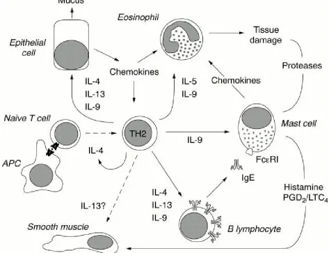
Figure 2.Inflammatory and immune cells involved in asthma.
III. CELLS INVOLVED IN ASTHMA
The main cells involved in asthma pathogenesis are inflammatory cells such as mast cells, eosinophils, T lymphocytes, neutrophils, macrophages and structural cells such as, epithelial cells, smooth muscle cells and fibroblasts.
A. Mast cells
871
©IJRASET (UGC Approved Journal): All Rights are Reservedsynthesis of vascular endothelial factor (VEGF) which increase vascular permeability and also cause vascular remodeling in chronic asthmatic condition. Persistent exposure of allergen promotes the synthesis of large amounts of mediators and cytokines. The mast cell cytokine is IL 4 which converts naïve cell into Th2 cells at the sensitization stage. The proinflammatory granules present in mast cells cause mucus secretion, airway smooth muscle constriction and mucosal edema. The preformed mediators that are stored as cytoplasmic granules are secreted upon activation of mast cells which include histamines, tryptase, proteoglycans, chymase, carboxypepditase, and heparin. These mediators cause immediate airway hyper reactivity, vasodilatation, mucosal edema, bronchoconstriction and itching. Mast cells also contain preformed stores of cytokines such as TNF and VPF, as well as many other cytokines including IL-2, IL-3,IL-4, IL-13,GM-CSF and various chemokines. Mast cells also release lipid mediators such as prostaglandin D2, leukotrienes (L4)[6].
B. Eosinophils
Eosinophils are produced in the bone marrow from CD 34+pleuripotent progenitor cells. The various growth factors, interleukin 3 (IL 3), granulocyte-macrophage colony –stimulating factor (GM-CSF), and IL 5 are responsible for the differentiation and development of eosinophils[7].The chemotactic agents for eosinophils include 5-oxo-ETE(5-oxo-eicosotetranoic acid), PAF (platelet activating factor) and PGD2 (prostaglandin 2) and Ig G (eg; immobilized IgG).5-oxo-HETE has the similar effect on migration as that of 5-oxo-ETE and 5-OXO-ETE also plays role in the survival of eosinophils[8].
The number of eosinophils is found to increased in n peripheral blood, sputum, BAL fluid and bronchial tissue of patients with asthma and their numbers have been shown to be correlated with severity of the disease[9,10,11,12]Eosinophil migration is stimulated by IL-5, eotaxin and RANTES[13,14]. Once activated, products from the eosinophils contract the airway smooth muscle, increase vascular permeability and induce airway hyper responsiveness[15]. Eosinophil granule products such as major basic protein and eosinophilic cationic protein also cause epithelial denudation[16].
C. Lymphocytes
There are two major types of Lymphocytes: B cells and T cells. B cells, antigen presenting cell, after processing the antigen, they are differentiated to antibody-producing plasma cells. So naïve B cells are able to convert into IgM-producing plasma cells T cells recognize antigens through its T cell receptor and interact with MHC class I and II molecules of antigen presenting cell. There are two major types: T helper cells (CD4+) and T cytotoxic cells (CD8+), based on their interleukin secretion profile and function. T helper cells are further classified into Th0, Th1 and Th2 cells. CD4+ T helper 0 cells secrete IL-2, IL-4, IFN-γ, IL-3, Th1 cells secretes IL-2, IFN-γ, IL-3 and Th2 cells secrete IL-4, IL-5, IL-9, and IL-13. The imbalance between Th1 and Th2 plays a great role in pathogenesis of asthma. Th 2 lymphocytes are having crucial roles in initiation, progression and persistence of allergic asthma as they activatesIgE antibody-producing B cells, mast cells and eosinophils. Th2 cells also stimulate the immunoglobulin switching to IgE in naïve B cells.
D. Neutrophils
Neutrophils, polymorphonuclear leukocytes, act as the first line of defense against bacterial and fungal infections through their phagocytosis and release of enzymes, other cytotoxic agents. Neutrophil is dominant inflammatory cell type in non-atopic and more severe asthmatic conditions[17].
E. Macrophages
Macrophages are having a central role in host defense against infection and in inflammatory processes. Activated macrophages secrete a wide variety of products and cause tissue repair and remodeling. Macrophages are significantly correlated with severity of asthma[18].
F. Basophils
Basophils are rare type of white blood cell that are involved in airway inflammatory reactions. These cells also possess the FcεR similar to mast cells. These cells are responsible for the secretion of late phase mediators which include histamine, leukotrienes and chemokines and are also the main stores of IL-4 and IL-13.
872
©IJRASET (UGC Approved Journal): All Rights are ReservedDendritic cells are a special type of macrophage-like cells located beneath the the airway epithelium. These cells are very effective in antigen presentation and therefore play an important role in allergen induced responses in asthma. These cells are essential in generating allergen specific-effector Th2 response.
H. Airway Epithelial cells
Airway epithelium is the first line of defense against foreign invaders and plays an important role in the pathogenesis of asthma. Normal airway epithelial layer consists of various cell types such as ciliated columnar epithelia, mucus-secreting cell, surfactant secreting Clara cells and basal cell and this epithelial layer forms a impermeable barrier by forming tight junctions. These cells secrete mucus to drain the foreign invaders. The airway epithelia are also a targets of the inflammatory attack by T-cells [19]. Activated epithelial cells release multiple mediators and express adhesion molecules that can direct and activate the various inflammatory and immunological cells that lead to the progression of airway remodeling and mucus metaplasia. These cells can also interact with their neighboring fibroblasts and extracellular matrix. Epithelial repair is a normal process following inflammatory damage, and this involve several steps such as de-differentiation, flattening and migration followed by the proliferation of new epithelial cells[20].
I. Airway Smooth Muscle cells
In contrast to earlier belief, airway smooth muscle cell has recently been implicated as inflammatory cell in asthma as they also showed synthetic and secretory potential other than their contractile and mitogenic responses[21]. Smooth muscle also can secrete matrix proteins and orchestrate key events in the process of chronic airway remodeling. These cells undergo hypertrophy and hyperplasia leading to the development of airway obstruction and increased nonspecific airway hyper responsiveness. These cells secrete cytokines, chemokines and growth mediators in addition to extracellular proteins. Airway smooth muscle cells also secretes matrix proteins such as fibronectin, elastin and collagens I, III, IV and V that contribute to sub-epithelial fibrosis.
J. Fibroblasts
Fibroblasts are typically spindle-shaped cells with an oval flat nucleus that are present in interstitial spaces of organs. In the lung they reside in highly complex multicellular environments. The greater numbers are found in the sub-epithelial of the conducting airways and interstitium of the lung parenchyma. These cells maintain the form, solidity and integrity of the lungs by the secretion of various extracellular components such as collagen, lamenin, fibronectin, hyaluronic acid and proteoglycans. Airway remodeling in asthma is due to the altered fibroblast. Fibroblasts secrete cytokines, chemokines and surface molecules which are able to activate and attract to stimulate inflammatory stimulation. The sub-epithelial collagen deposition is due to the significant increase in the number of myofibroblasts in asthma patients[22].
K. Extracellular Matrix
Extracellular matrix is a meshwork-like substance in the extra cellular space connected with the basement membrane of the cell surface. There is an intimate relationship between airway smooth muscle and ECM of the airways. Matrix metalloproteinase (MMPs) and their inhibitors, tissue inhibitors of metalloproteinase’s (TIMPs) play a role in the pathogenesis of asthma by influencing the function and migration of inflammatory cells. They also play a role in matrix deposition and matrix degradation. MMPs and TIMPs bind to one another in 1:1 ratio and the increase in TIMP/MMP cause fibrosis[23].
IV. CYTOKINES AND GROWTH FACTORS INVOLVED IN ASTHMA
873
©IJRASET (UGC Approved Journal): All Rights are ReservedIL-4 is expressed by both CD4+ and CD8+ cells, mast cells and eosinophils in both atopic and non atopic asthma. IL-4 plays a critical role in the isotype switching of B lymphocytes from IgG to IgE. The mast cells are activated by the binding of IgE produced in the asthmatic airways to FcɛR1 present on mast cells. IL-4 also increases the expression of an inducible form of low affinity receptor for IgE on B lymphocytes and macrophages, which in turn results in the increased expression of CD23 on alveolar macrophages from asthmatic person, which accounts for the increased release of cytokines from these macrophages. It is also important in driving the differentiation of CD4+ TH precursors into TH2-like cells. It also increases the expression of vascular cell adhesion molecule-1 (VCAM-1) on endothelial and airway epithelial cells. IL-4 induces fibroblast chemotaxis and activation. It also promotes growth of eosinophils and basophils.
IL- 5 along with IL-13 and GM-CSF helps in the differentiation, migration and pathobiological effects of eosinophils. It prolongs the survival of eosinophils and helps in the development of eosinophilia. Exogenous administration of IL-5 produces eosinophilia. IL-13 is synthesized by activated CD4+ and CD8+ cells. It is produced in response to specific antigen or polyclonal stimuli. It is a potent modulator of human monocyte and B cell function. It has profound effects on human monocyte morphological features, surface antigen presentation, antibody dependent cellular cytotoxity and cytokine synthesis. IL-13 expression increase in both atopic and non atopic asthma. There is strong correlation between eosinophil counts and IL-13.
In contrast to the above discussed cytokines, IL-10has anti-inflammatory effects that inhibit the characteristic features of asthma. The production of pro-inflammatory cytokines and chemokines by macrophages, neutrophils, and eosinophils can also be inhibited by IL-10. It also inhibits eosinophil production in bone marrow that ultimately results in decrease eosinophil accumulation.
[image:7.612.70.543.345.708.2]Eotaxin is a chemokine that acts as an eosinophil chemoattractant in asthma. Along with IL-5, eotaxin prolongs the viability of eosinophils.
874
©IJRASET (UGC Approved Journal): All Rights are ReservedUpon recognition of the antigen and activation by antigen presenting cells (APC), naive T cells differentiate into TH2 cells, a process that is promoted by interleukin 4 (IL-4). Activated TH2 cells stimulate B cells to produce IgE antibodies in response to IL-4, and to a lower extend to IL-13 or IL-9. IgE binds the high affinity IgE receptor at the surface of mast cells, the proliferation and differentiation of which is promoted by IL-9, in synergy with other factors such as fibroblast derived mast cell growth factor. At contact with antigen, mast cells release the contents of their granules, including histamine, which will induce a bronchospasm, together with newly synthesized prostaglandins and leukotrienes (PGD2 and LTC4). Mast cells also release chemo tactic factors that contribute to the recruitment of inflammatory cells, particularly eosinophils, whose proliferation and differentiation from bone marrow progenitors is promoted by IL-5 and IL-9. Finally, epithelial cells up regulate their production of mucus and chemokines in responses to TH2 cytokines such as Il-4, IL-13, and IL-9. The presence of the IL-13 receptor at the surface of smooth muscle cells suggests that this factor can also directly affect smooth muscle contractility, but this remains to be demonstrated.
V. PROINFLAMMATORY MEDIATORS OF ASTHMA
Various pro-inflammatory mediators are involved in asthma pathogenesis. These inflammatory mediators are released by inflammatory cells as well as activated structural cells that perform several functions contribute to airway inflammation.
Histamine is synthesized and released by mast cells in the airway wall that leads to the recruitment of inflammatory cells such as eosinophils, neutrophils etc into the airways. Bronchoconstriction was one of the first recognized effects of histamine[25].
Leukotrienes (LTs) are potent lipid mediators that are produced by arachidonic acid metabolism. Cysteinyl leukotrienes (Cys-LTs) stimulate specific receptors which results in bronchoconstriction and increase vascular permeability in airways. It also increases the mucus secretions. Elevated levels of Cys-LTs have been observed in plasma, BAL and sputum of asthmatics[26].
Chemokine’s are the chemotactic cytokines that are involved in attracting leukocytes into the site of inflammation. Mainly CC chemokine’s are involved in chemoattractans of eosinophils, monocytes and T-lymphocytes into the airways in case of asthma.
Lung fibroblast may serve as a source of TGF-β which may be the main cause of airway remodeling in asthma[27].
VI. SMALL MOLECULES INVOLVED IN ASTHMA
A. Reactive Oxygen Species
The reactive oxygen species produced in cells include hydrogen peroxide (H2O2), hypochlorous acid (HOCl), and free radicals such as the hydroxyl radical (·OH) and the superoxide anion (O2−). Various inflammatory cells such as eosinophils, macrophages, mast cells and epithelial cells are known to produce ROS. These contribute to inflammation and may damage airway epithelium that results in epithelial denudation and increased bronchoconstriction[28].
B. Nitric Oxide
Nitric oxide is involved in the pathogenesis of various inflammatory diseases. Nitric oxide (NO) is produced by NOS enzyme which is of two types: constitutive (neural NOS, endothelial NOS [eNOS]) and inducible NOS. In normal airways, constitutive NOS consume L-ARG to maintain airway smooth muscle tone through activation of the cyclic guanosine monophosphate (cGMP) pathway. NO production is limited by the presence of sufficient argentine. NO plays a key role in physiological regulation of airway functions and is implicated in airway diseases, including asthma. In human allergic airway inflammation, there has been an increased exhaled nitric oxide (ENO)[29].
C. Oxidative stress
Oxidative stress is a predominant factor associated with the pathogenesis of asthma. Oxidative stress results due to an imbalance between ROS and antioxidants. It mainly occurs when the production of ROS exceeds the body's natural antioxidant defence mechanisms. It leads to the damage of biomolecules such as lipids, proteins and DNA. During the oxidative phosphorylation, ROS are synthesized in mitochondria. ROS can also participate in immune response as they modulate immune system. Exposure to allergen induces immune system to release Proinflammatory mediators such as histamine, PGs, and leukotrienes, which lead to recruitment and activation of various inflammatory cells into the airways, such as mast cells, eosinophils, neutrophils, lymphocytes, macrophages, and platelets.
875
©IJRASET (UGC Approved Journal): All Rights are Reserved [image:9.612.170.447.126.299.2]effects on airway function, including induction of airway hyper responsiveness, airway smooth muscle contraction, mucus hyper secretion, denudation of epithelium and vascular exudation[30].
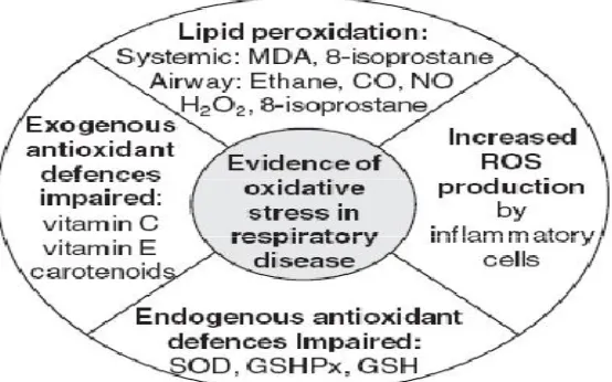
Figure 4: Evidence of oxidative stress in respiratory disease.
(Lisa G. Wood, Peter A.B. Wark, and Manohar L. Garg. Antioxidant and Anti-Inflammatory Effects of Resveratrol in Airway Disease. Vol; 13. 2010.)
D. Lipoxygenases
Lipoxygenases are a class of non heme iron containing deoxygenises that are involved in the hydroperoxidation of fatty acids of lipids. Lipids containing a series of cis double bonds are suitable substrates for these enzymes. These are found in plants, mammals and fungi. There are seven genes in mouse that code for these proteins and there are five homologous in humans. These enzymes cause the per oxidation of linoleic acid rich phospholipid such as cardiolipin which is present in the inner mitochondrial membrane. The increased 15-LOX expression was found in human tracheal epithelial cells as compared with other cell types[31]. 15-LOX also expressed in other cells also that are involved in lung inflammation such as airway epithelial cells, mast cells, dendritic cells, eosinophils etc. IL-4 and IL-13 is known to induce the expression of 15-LOX through STAT-6 phosphorylation that results in epithelial cell apoptosis[32].15-LOX has been predominantly shown to be expressed in bronchial epithelial cells.
In humans, 15-LOX has two different is forms: 15-LOX-1and 15-LOX-2[33]. 15-LOX-1 also referred to as reticulocyte 15-LOX or ALOX15 and 15-LOX-2 also known as epidermis-type 15-LOX. 15-LOX-1 results in degeneration of mitochondria present in reticulocyte to convert them into mature red blood corpuscle by catalyzing the oxygenation of polyenoic fatty acids of phospholipids like cardiolipin[34].
15-LOX-1, a human counterpart of murine 12/15-lipoxygenase (12/15-LOX), preferentially metabolizes linoleic acid to form
HODEs such as 9 and 13-(S)-HODE and minimally metabolizes arachidonic acid to form hydroxyeicosatetraenoic acids (HETEs)
such as 12 and 15-(S)-HETEs. In contrast, 15-LOX-2 preferentially metabolizes arachidonic acid into 15-(S)-HETE. Among the two forms of 15-LOX, 15-LOX-1 is mainly up-regulated in airway inflammation as compared with 15-LOX-2. 15-LOX-1 is also known to induce MUC5AC, which is the marker of goblet cell metaphase[35]. 15-LOX-1 expression in mouse macrophages resulted in 25 fold in 15-LOX-1 specific product 13-HODE.
VII. LIPID PEROXIDATION
876
©IJRASET (UGC Approved Journal): All Rights are Reservedpermeability-transition pore (PTP), and also it alters equilibration of solutes across the membranes to produce matrix swelling and rupture of the outer membrane[38]. Lipid peroxidation is the oxidative degradation of the lipids. It is the process in which ROS extracts the electrons from the lipids. It results in the formation of lipid hydroperoxides which are non-radical intermediates. Lipid peroxidation is used as an indicator of oxidative stress in cells and tissues.
A. Consequences of Lipid Peroxidation
Cardiolipin have a tight association with COXETC and this association is needed for maximum mitochondrial activity. Lipid peroxidation causes the oxidation of cardiolipin. Further, this leads to the reduction in the activity of COXETC and the release of cytochrome c in the cytosol. Peroxidation of lipid membranes also leads to the production of various lipid mediators such as prostaglandin, leukotrienes, lipoxins and lipid hydroperoxides etc.
B. Biomarkers of Lipid Peroxidation
Direct measurement of oxidants is difficult because these are highly reactive. Therefore, it is easy to measure damage caused by oxygen radicals upon various biomolecules, such as lipids, proteins, or DNA. Various other markers of lipid per oxidation include 13(S)-HODE, 9(S)-HODE, 12(S)-HETE, 15(S)-HETE etc.
C. 13-S-HODE (13-S-hydroxyoctadecaenoic acid)
13-S-HODE is a linoleic acid metabolite. Its levels are known to increase both in asthmatic mice and humans. It is also associated with mitochondrial degradation[39]. 13-S-HODE is an endogenous legend for PPARᵧ .The activation of PPARᵧ by 13-S-HODE inhibits cell growth and induces apoptosis[40]. Vitamin E reduces expression of 15-LOX and decrease synthesis of 13-S-HODE in allergic mice model[41].13-S-HODE is known to activate TRPV1 and produce pain in rodents[42].
VIII. APOPTOSIS
[image:10.612.138.473.542.714.2]Apoptosis is a programmed cell death. Apoptosis occurs normally during development and aging and as a homeostatic mechanism to maintain cell populations in tissues. Apoptosis also occurs as a defense mechanism such as in immune reactions or when cells are damaged by disease or noxious agents. The features of apoptosis include DNA fragmentation, protein cleavage at specific sites, increased mitochondrial membrane permeability and the appearance of phosphitidylserine on the membrane of cell surface. There are two major pathways of apoptosis: (a) extrinsic pathway, which relays apoptotic messages via receptors and (b) intrinsic pathway (mitochondrial) which transmits death signals received intracellular. Mitochondria play an important role in apoptosis. It contains Bcl-2 proapoptotic proteins Bad, Bid, Bax and Bim which reside in the cytosol. These proapoptotic proteins translocation into mitochondrial membrane following death signaling, where they promote the release of cytochrome c. These mitochondrial permeability transition pore resulting in mitochondrial swelling and rupture of the outer membrane, or by channels formed by oligomerization of Bax (Bcl-2-associated X protein). Once cytochrome c is released it binds with Apoptotic protease activating factor - 1 (Apaf-1) and deoxyATP, to create a protein complex known as an apoptosome. The apoptosome cleaves the pro-caspase to its active form of caspase-9, which in turn activates the effectors caspase-3.
877
©IJRASET (UGC Approved Journal): All Rights are ReservedMitochondria can participate in apoptosis in two ways: one involves release of cytochrome c and the other release of apoptosis inducing factor (AIP). Cytochrome c binds to Apaf-1 to activate caspase 9 which can cleave and activate caspase. 3 AIP can also activate caspase-3 leading to formation of various death substrates that produce apoptosis. Bcl-2 can interfere with cytochrome c release and can also suppress the actions of Apaf-1 and, in turn, protect from apoptosis.
A. Release of Cytochrome c
Cytochrome c is a water-soluble, basic, heme-containing protein that binds to the phospholipids cardiolipin. Under normal physiological conditions, cardiolipin anchors cytochrome c protein to the mitochondrial membrane where it participates in ETC between complex III and IV of the respiratory chain[43].
Oxidation of the cardiolipin leads to the release of the cytochrome c. Cytochrome c is released from mitochondria via a two-step mechanism. The pore formation by Bax is required, but not sufficient for release of the cytochrome c from the mitochondria. Before cytochrome c can be released into the cytoplasm via pore formation it must first be detached from cardiolipin[44].
In conclusion, the two-step mechanism of cytochrome c release connects the dissociation of cytochrome c with its subsequent release through pores in the outer membrane.
B. Epithelial Mesenchymal Trophic Unit And Airway Remodelling
Airway remodelling involves the structural changes that occurs in asthma include wall thickening, sub-epithelial fibrosis, goblet and squamous cell metaplasia, sub-mucosal, epithelial, goblet and airway smooth muscle (ASM) cell hypertrophy, epithelial cells, goblet cell and mucous gland hyperplasia and angiogenesis[45]. Damaged epithelium results in increased release of growth factors that contribute to airway remodeling. EMTU are the factors that are required for epithelial-mesenchymal interactions. In chronic asthma, activation of EMTU (involve the communication between damaged epithelium and underlying fibroblast sheath) that leads to mesenchymal proliferation and ultimately to remodeling. Airway mesenchymal cells including smooth muscle cells are able to secrete various Proinflammatory cytokines[46].
878
©IJRASET (UGC Approved Journal): All Rights are ReservedGoblet cells are glandular simple columnar epithelial cells that secrete mucin. In asthmatic conditions, there is an increase in mucus secretion due to goblet cell metaphase. It is the condition in which one differentiated cells converted into another mature differentiated cell type. In asthmatics, goblet cells converted into more mucus secreting cells that leads to increase in mucus production.
C. Concluding Remarks
In conclusion, the need of the hour is to discover non-steroidal drugs that could target lipoxygenases. The progression of the disease needs to be studied as that could also help in finding novel pathways involved in the pathophysiology of asthma and that will in turn lead to the discovery of new promising targets for curing the disease in a better way.
IX. AKNOWLEDGEMENT
We acknowledge funding by grants, NWP0033 and MLP5502, from the Council of Scientific and Industrial Research (CSIR), Government of India. We thank Dr. Balaram Ghosh and Dr. Anurag Agrawal for their conceptual supports and help.
REFERENCES
[1] Braman SS. The Global Burden of Asthma. CHEST (2006) 130: 4S-12S.
[2] Gupta BM and Bala A. Mapping of asthma in India. A scientometric analysis of publications output during 1999-2008 Lung India (2011) 28. [3] O’Byrne PM, and Inman MD. Airway hyperresponsiveness. CHEST (2003) 123: 411S-416S.
[4] Kalesnikoff J and Galli SJ. New developments in mast cell biology. NatureImmunology (2008) 9, 1215 – 1223 [5] Elias JA, Zhu Z, Chupp G and Homer RJ.Airway remodeling in asthma. J ClinInvest.(1999) 104:1001–1006. [6] Venarske D, and deShazo RD. Molecular Mechanisms of Allergic Disease.South Med J (2003) 96, 1049-54. [7] Kay AB. The role of eosinophils in the pathogenesis of asthma. Trends in Molecular Medicine (2005) 11, 148-152. [8] Filipovic M and Cekic S. The role of eosinophils in asthma. Medicine and Biology (2001) 8, 6– 10.
[9] Monneret G, Gravel S, Diamond M, Rokach J, and Powell WS. Prostaglandin D2 is a potent chemoattractant for human eosinophils that acts via a novel DP receptor.BLOOD (2001) 98, 1942-8.
[10] Bradley BL, Azzawi M, Jacobson M, Assoufi B, Collins JV, Irani AM, Schwartz LB, Durham SR, Jeffery PK and Kay AB. Eosinophils, T-lymphocytes, mast cells, neutrophils, and macrophages in bronchial biopsy specimens from atopic subjects with asthma: comparison with biopsy specimens from atopic subjects without asthma and normal control subjects and relationship to bronchial hyperresponsiveness.J Allergy Clin Immunol (1991) 88:661-674.
[11] Chanez P, Vignola AM, Vic P, Guddo F, Bonsignore G, Godard P and Bousquet J Comparison between nasal and bronchial inflammation in asthmatic and control subjects. Am J RespirCrit Care Med (1999)159: 588-595.
[12] Louis R, Lau LC, Bron AO, Roldaan AC, Radermecker M and Djukanovic R. The relationship between airways inflammation and asthma severity. Am J RespirCrit Care Med (2000 )161: 9-16.
[13] Vignola AM, Chanez P, Campbell AM, Souques F, Lebel B, Enander I and Bousquet J. Airway inflammation in mild intermittent and in persistent asthma. Am J RespirCrit Care Med (1998) 157: 403-409.
[14] Hamid QA and Minshall EM. Molecular pathology of allergic disease: I: lower airway disease.J Allergy Clin Immunol (2000)105:20-36. [15] Wardlaw AJ. Molecular basis for selective eosinophil trafficking in asthma: A multistep paradigm.J Allergy Clin Immunol (1999 )104:917-926.
[16] Trautmann A, Schmid-Grendelmeier P, Kruger K, Crameri R, Akdis M, Akkaya A, Brocker EB, Blaser K and Akdis CA.) T cells and eosinophils cooperate in the induction of bronchial epithelial cell apoptosis in asthma. J Allergy Clin Immunol (2002)109:329-337.
[17] Kamath AV, Pavord ID, Ruparelia PR, Chilvers ER. Is the neutrophil the key effector cell in severe asthma? Thorax (2005) 60:529-530. [18] Lane SJ, Sousa AR and Lee TH. The role of the macrophage in asthma. Allergy (1994) 49, 201–209.
[19] Kelly C, Ward C, Stenton CS, Bird G, Hendrick DJ and Walters EH. Number and activity of inflammatory cells in bronchoalveolar lavage fluid in asthma and their relation to airway responsiveness. Thorax (1988)43:684-692.
[20] Trautmann A, Schmid-Grendelmeier P, Kruger K, Crameri R, Akdis M, Akkaya A, Brocker EB, Blaser K and Akdis CA. T cells and eosinophils cooperate in the induction of bronchial epithelial cell apoptosis in asthma.J Allergy Clin Immunol (2002)109:329-337.
[21] Holgate ST, Lackie PM, Davies DE, Roche WR and Walls AF. The bronchial epithelium as a key regulator of airway inflammation and remodelling in asthma.ClinExp Allergy (1999)29:90-95.
[22] John M, Hirst SJ, Jose PJ, Robichaud A, Berkman N, Witt C, Twort CH, Barnes PJ and Chung KF. Human airway smooth muscle cells express and release RANTES in response to T helper 1 cytokines: regulation by T helper 2 cytokines and corticosteroids.J Immunol (1997) 158:1841-1847
[23] Johnson PR, and Burgess JK. Airway Smooth Muscle and Fibroblasts in the Pathogenesis of Asthma. Curr Allergy Asthma Rep (2004) 4:102-8. [24] Jarjour NN and Kelly EA. Role of matrix metalloproteinases in asthma. CurrOpinPulmMed.(2003) 9:28-33.
[25] Barnes PJ, Chung KF and Page CP. Inflammatory Mediators of Asthma. Pharmacological Reviews (1998) 50: 515-596. [26] Barnes PJ. Histamine receptors in airways. Agents Actions Suppl (1991) 33:103–122.
[27] Taylor GW, Taylor I, Black P, Maltby NH, Turner N, Fuller RW and Dollery CT. Urinary leukotriene E4 after antigen challenge and in acute asthma and allergic rhinitis. Lancet (1989) 1:584–588.
[28] Kelley J, Fabisiak JP, Hawes K and Abscher M. Cytokine signalling in lung: Transforming growth factor-b secretion by lung fibroblasts.Am J Physiol (1991)260: L123–L128.
879
©IJRASET (UGC Approved Journal): All Rights are Reserved[30] Leuppi JD, Downs SH, Downie SR, Marks GB, Salome CM. Exhaled nitric oxide levels in atopic children: relation to specific allergic sensitization, AHR, and respiratory symptoms.Thorax( 2002) 57:518–523.
[31] Katsumata U, Miura M, Ichinose M, et al. Oxygen radicals produce airway constriction and hyperresponsiveness in anesthetized cats. Am Rev Respir Dis (1990) 141:1158–1161.
[32] Lindley AR, Pregont MC, YanjunLiu and Kuperman DA. 12/15-lipoxygenase is an interleukin-13 and interferon-γ counterregulated-mediator of allergic airway inflammation. Mediators of Inflammation. (2010) PMID: 20953328.
[33] Shankaranarayan P, and Nigam S. IL-4 induces apoptosis in A549 lung adenocarcinoma cells:evidence for the pivotal role of 15-hydroxytetraenoic acid binding to activated peroxisome proliferator-activated receptor γ transcription factor.Immunol. J (2003) 170:887-894.
[34] Watson A and Doherty FJ. Calcium promotes membrane association of reticulocyte 15-lipoxygenase.Biochem J (1994) 298:377–383.
[35] Zhao J, Maskrey B, Balzar S, Chibana K, Mustovich A, Hu H, Trudeau JB, Donnell V, and Wenzel SE. Interleukin-13 induced MUC5AC is regulated by 15-lipoxygenase 1 pathway in human bronchial epithelial cells. Am. J. Respir. Crit. Care. Med. (2009) 179:782–790.
[36] Hatley ME, Srinivasan S, Reilly KB, BolickDT, Hedrick CC. Increased production of 12/15 lipoxygenase eicosanoids accelerates monocyte/endothelial interactions in diabetic db/db mice. J BiolChem (2003) 278: 25369-25375.
[37] Praticò D, Zhukareva V, Yao Y, Uryu K, Funk CD, Lawson JA, Trojanowski JQ, and Lee VM. 12/15-lipoxygenase is increased in Alzheimer's disease: possible involvement in brain oxidative stress.Am J Pathol (2004) 164: 1655-1662.
[38] Géminard C, de Gassart A, Vidal M. Reticulocyte maturation: mitoptosis and exosome release.Biocell (2002) 26: 205-215.
[39] Mabalirajan U, Dinda AK, Kumar S, Roshan R, Gupta P, Sharma SK, Ghosh B Mitochondrial structural changes and dysfunction are associated with experimental allergic asthma. J. Immunol (2008)181:3540-3548
[40] Yuan H, Li MY, Ma LT, Hsin MK, Mok TS, Underwood MJ, Chen GG.15-Lipoxygenases and its metabolites 15(S)-HETE and 13(S)-HODE in the development of non-small cell lung cancer.Thorax (2010) 65:321-326.
[41] Mabalirajan U, Aich J, Leishangthem GD, Sharma SK, Dinda AK, Ghosh B. Effects of vitamin E on mitochondrial dysfunction and asthma features in an experimental allergic murine model. J Appl Physiol. (2009) 107:1285-92.
[42] Patwardhan AM, Akopian AN, Ruparel NB, Diogenes A, Weintraub ST, Uhlson C, Murphy RC, Hargreaves KM.
Heat generates oxidized linoleic acid metabolites that activate TRPV1 and produce pain in rodents. J Clin Invest. (2010) 120:1617-26.
[43] Iverson SL and Orrenius S. The cardiolipin-cytochrome c interaction and the mitochondrial regulation of apoptosis.Arch. Biochem. Biophys (2004) 423:37-46. [44] Iverson SL, Enoksson, M., Gogvadze, V., Ott, M., and Orrenius, S. Cardiolipin is not required for Bax-mediated cytochrome c release from yeast mitochondria.
J. Biol. Chem. (2004);279:1100–1107.
[45] Elias JA, Zhu Z, Chupp G, and Homer RJ. Airway remodeling in asthma. J. Clin. Invest. 1999;104:8.
[46] Holgate ST, Holloway J, Wilson S, Bucchieri F, Puddicombe F, and Davies DE. Epithelial–Mesenchymal Communication in the Pathogenesis of Chronic Asthma. Proc Am Thorac Soc. (2004) 1: 93–98.