6107
Introduction
In vertebrates, somites are transient embryonic structures that originate from dorsal paraxial mesoderm and yield a variety of tissues, including muscles and bones. They are segmented, being made of a series of quasi-identical, metameric units, along the anteroposterior axis. Somites form from the anterior part of the presomitic mesoderm (PSM), during rostro-caudal elongation of the PSM.
A recent report demonstrated a role for the Wnt/β-catenin signalling pathway in somite segmentation (Aulehla et al., 2003). However, the segmentation process largely relies on the dynamic expression of genes of the Notch signalling pathway (Maroto and Pourquie, 2001; Saga and Takeda, 2001). Notch protein is a cell surface receptor that, when bound by its ligand from the Delta family, undergoes a proteolytic cleavage that releases the intracellular domain (ICD) in the cell. Notch ICD then associates with a CSL (CBF1/RBP-Jκ, Suppressor of Hairless, Lag1) protein. The Notch ICD-CSL complex transactivates a number of genes, including enhancer of split/hairy-related genes. CSL proteins that are not associated with Notch ICD repress the transcription of these target genes (for a review, see Mumm and Kopan, 2000).
Some segmentation genes belonging to the Notch pathway display particular expression patterns in the PSM of chick or mouse. Their corresponding mRNAs are cyclically expressed, progressing as a wave in the PSM toward the anterior part of
the embryo once during the formation of each somite. This wave of expression does not rely on cell migration. Rather, cells in the PSM are able to successively express and repress segmentation mRNAs, and this periodicity is thought to be the cause for periodic somite formation (Maroto and Pourquie, 2001; Saga and Takeda, 2001). The combination of the expression of these genes with the local concentration of FGF8 commits certain cells to form the boundary between two adjacent somites (Dubrulle et al., 2001). FGF8 mRNA is transcribed only at the posterior extremity of the embryo, leading to a protein gradient that decreases toward the anterior extremity (Dubrulle and Pourquié, 2004).
In Xenopus laevis, the only cycling mRNAs described to date are ESR9and the closely related mRNA ESR10(Li et al., 2003). Other segmentation mRNAs show a different expression pattern in the PSM (Jen et al., 1999; Jen et al., 1997; Lamar et al., 2001; Sparrow et al., 1998). Some are expressed in the caudal-most region of the PSM (tailbud domain). They are also expressed very transiently in the anterior or posterior part of one or a few presumptive somites (somitomeres). These segmentation mRNAs may be expressed in a given somitomere and completely absent in the following one, demonstrating a very dynamic expression pattern.
This dynamic expression pattern of segmentation genes requires: (1) that the promoters controlling the expression of these genes can rapidly be switched on and off; (2) that the corresponding mRNAs are unstable, so that their expression EDEN-BP is a XenopusRNA-binding protein that triggers
deadenylation [poly(A) tail shortening], and thereby translational repression and degradation, of a subset of maternal mRNAs soon after fertilization. We show here that this factor is expressed in the presomitic mesoderm of older embryos, the site where somitic segmentation takes place. Inhibiting EDEN-BP function using either antisense morpholino oligonucleotides or neutralizing antibodies leads to severe defects in somitic segmentation, but not myotomal differentiation. This is associated with defects in the expression of segmentation markers belonging to the Notch signalling pathway in the presomitic mesoderm. We show by a combination of approaches that the mRNA
encoding XSu(H), a protein that plays a central role in Notch signalling, is regulated by the EDEN-BP pathway. Accordingly, XSu(H) is overexpressed in EDEN-BP knock-down embryos, and overexpressing XSu(H) causes segmentation defects. We finally give data indicating that, in addition to XSu(H), other segmentation RNAs are a target for BP. These results show that EDEN-BP-dependent post-transcriptional regulation of gene expression is required for the process of somitic segmentation.
Key words: mRNA stability, Poly(A) tail, Metamerization, Notch, Suppressor of Hairless/RBP-Jκ
Summary
EDEN-BP-dependent post-transcriptional regulation of gene
expression in Xenopus somitic segmentation
Carole Gautier-Courteille, Christophe Le Clainche*, Carine Barreau, Yann Audic, Antoine Graindorge, Dominique Maniey, H. Beverley Osborne and Luc Paillard†
CNRS UMR 6061, IFR 97, Faculté de Médecine, Université Rennes 1, 2 avenue Léon Bernard, CS 34317, 35043 Rennes Cedex, France
*Present address: 16 Barker Hall, Department of Molecular and Cell Biology, University of California Berkeley, Berkeley, CA 94720-3202, USA
†Author for correspondence (e-mail: luc.paillard@univ-rennes1.fr)
Accepted 12 October 2004
Development 131, 6107-6117
Published by The Company of Biologists 2004 doi:10.1242/dev.01528
patterns follow the transient activity of the promoters; and (3) that the encoded proteins are unstable for the same reason. The first point was illustrated for the lunatic fringegene. In mice, the transcription of this gene is activated in a periodic fashion, and elements within the promoter that are responsible for cyclic expression were identified (Cole et al., 2002; Morales et al., 2002). Abolishing the cyclic pattern of expression of lunatic fringe strongly affects somitic segmentation (Dale et al., 2003; Serth et al., 2003). The second point was observed for the Xenopus Hairy2agene. In transgenic Xenopusembryos, only the combination of the Hairy2a gene promoter with a mRNA destabilizing element [originating from Hairy2a 3′ untranslated region (3′UTR) or from the 3′UTR of another unstable mRNA] was able to confer to a reporter gene an expression pattern similar to that of endogenous Hairy2a (Davis et al., 2001). It was also noticed in mice that lunatic fringe mRNA was almost as unstable as intron sequences (Cole et al., 2002; Morales et al., 2002). Finally, both lunatic fringe and Hes7 proteins show a dynamic expression pattern, and, in the case of Hes7, this relies on ubiquitin-proteasome-mediated degradation of the protein (Bessho et al., 2003; Dale et al., 2003).
The molecular mechanisms leading to instability of the segmentation mRNA have not been investigated. However, in vertebrates, mRNA degradation is frequently triggered by mRNA deadenylation (shortening of the poly(A) tail) (Beelman and Parker, 1995). Accordingly, a deadenylation mechanism may be required for somitic segmentation. We have described a sequence-specific mRNA deadenylation mechanism that is active in early Xenopusembryos. A family of Xenopus maternal mRNAs (the Eg/c-mos family) is deadenylated after fertilization of Xenopus eggs. This is mediated by a short sequence, EDEN (Embryo Deadenylation ElemeNt) that is present within the 3′UTR of Eg/c-mos mRNAs. The associated RNA-binding protein, EDEN-BP (EDEN-binding protein), was purified and shown by immunodepletion and rescue experiments to be required for EDEN-dependent deadenylation (Paillard et al., 2003; Paillard et al., 1998). In this article, we adressed the requirement of the EDEN-BP-dependent mechanism for somitic segmentation in
Xenopusembryos.
Materials and methods
Primer sequences19, ATACCgCCAATTTTgTAgACATCCT; 20, TgACTgCATTCTgCCACCTAgT; 141, CAgATTggTgCTggATATgC; 142, ACTgCCTTgATgACTCCTAg;
279, gAAgATCTTAATCTTCCCCAgAAACATgAC;
280, gCTCTAgAAATCAAAATgTAAAAAAAAgAgTgATCACC; 283, gAAgATCTTAACCATAAACTTTggACTTTTAC;
284, gCTCTAgAgTATTgCAggCAAggTAgAggC; 289, gAAgATCTTgAAACTTATTgggggTTTTC; 291, gCTCTAgAAAAggTTAAATATACAAAggC; 449, TgCgCTCTAAAgTATgTgTAg;
474, gTAgaCATCAATAgTTTgTATTg; 482, gCCAgAgCCCGgACCTA; and 483, TTggATgCCAggAgACTTgAA.
Plasmid construction and RNA synthesis
ESR5, X-Delta2 and XSu(H) 3′UTRs were PCR-amplified from
Xenopusembryos cDNA using the primers 289 and 291 (ESR5), 279 and 280 (X-Delta-2), and 283 and 284 [XSu(H)]. The PCR products were directly cloned in pGEM-T (Promega) to be used as matrices for cross-linking analysis. The XSu(H) plasmid was digested with BglII and XbaI, and cloned into the same sites of pGbEg2-497 (Bouvet et al., 1994) (between the 5′UTR of Xenopus β-globin and a 65-nucleotide poly(A) track) for deadenylation analysis. The resulting plasmid pGbXSu(H) used for deadenylation analysis was linearized by restriction digest using EcoRV for the synthesis of poly(A)+RNAs. 32P-labelled and capped in vitro transcripts were obtained from these matrices using the Riboprobe transcription kit (Promega) in the presence of cap analog (Biolabs). GFPmut plasmid and pCS2-XSu(H)1 plasmid were linearized with NotI, and the resulting matrices were used to obtain capped mRNA with the Message Machine kit (Ambion). EDEN-BP mRNA for rescue experiments was transcribed from pT7T-EDEN-BP (Paillard et al., 1998).
Morpholinos
Morpholinos sequences were:
EDEN-BP morpholinos, TTGTGCCATTCATTATCTTAGAAAT and ATTGTGCCATTCATTTTCTTGGAAA (Accession number BG513033);
standard control, CCTCTTACCTAGTTACAATTTATA; XSu(H)1, CCAGGTTGCATAGAACAATATGATG; and XSu(H)2, GCCTCTCCCCAAACTTCATTCCGCT.
The CAT bases shown in bold are complementary to the initiating ATG codons. Morpholino oligonucleotides were dissolved in water.
Embryo manipulations, embryo staining and histological methods
Injections were made into the animal hemisphere of Xenopus laevis embryos, into one or two blastomeres at the 2-cell stage, or one blastomere at the 4-cell stage. Depending on the experiment, the injected solution contained one or several of the following: 0.5-1.0 fmol of reporter transcript, 0.5-2.0 fmol of EDEN-BP, GFP or Xsu(H) mRNA, 100 or 200 ng of antibodies, and 1 to 50 ng of morpholinos, in a final volume of 9.2 to 36 nl. Embryos were allowed to develop at 22°C. Following injection with a GFP mRNA-containing solution, embryos were sorted, using a fluorescence microscope, at about stage 22 according to injection site (left/right, dorsal/ventral).
Whole-mount in situ hybridization was performed as described (Harland, 1991). The EDEN-BP probe corresponds to the whole open-reading frame cloned into the pT7TS vector (Paillard et al., 1998). The sense probe was obtained by in vitro transcription from the T7 promoter after linearization with EcoRI, and the antisense probe from the SP6 promoter after linearization with BglII. The ESR5, ESR9 and X-Delta2 probes were made from plasmids that were either constructed by RT-PCR or provided by the IMAGE consortium. The MHC4 antisense probe was obtained from the pGEM-MHC4 plasmid (a kind gift of Anna Philpott, Cambridge, UK). Whole-mount immunohistochemistry was performed as described (Jen et al., 1999), using the 12/101 (Kintner and Brockes, 1985) hybridoma (obtained from the Developmental Studies Hybridoma Bank, University of Iowa) supernatant at a dilution of 1/10. The secondary antibodies used were HRP-conjugated and staining was performed with Diaminobenzidine.
Stage 39 embryos were fixed and embedded in paraffin wax and horizontal sections were stained by Hematoxylin and Eosin. Microscopy analysis was performed through the serial sections made on several different whole embryos.
Analytical methods
Embryos used for western analysis were homogenized in 100 mM Tris-HCl (pH 7.5), 4 mM EDTA, 16 mM KCl, 0.01% Triton X100, 10 µg/ml Pepstatin, 10 µg/ml Leupeptin, 10 µg/ml Chymostatin. Unsoluble particles were removed by centrifugation (10 minutes at 12,000 g) and proteins were extracted by one volume of 1,1,2-Trichloro-trifluoroethane (Fluka). Proteins were resolved by 10 or 12% SDS-PAGE, blotted onto nitrocellulose, probed by a primary antibody and revealed either by alkaline phosphatase-coupled secondary antibodies using ECF for analysis on a STORM 860, or peroxydase-coupled secondary antibodies using ECL. Primary antibodies used were 83 antiserum against EDEN-BP (Paillard et al., 1998) or the cdc2 A17 monoclonal antibody (Abcam).
Real-time PCR was performed with an ABI Prism 7000 apparatus (Applied), using SybrGreen mastermix and the primers 482 and 483 [XSu(H)], or 19 and 20 (EF1α). For each mRNA sample from individual embryos, quantifications were done in triplicate. Relative mRNA quantities were given by the difference of the Ct with a standard Ct (Ct0) according to the formula [relative quantity=2 exp(Ct0-Ct)]. The results are expressed as the ratio of relative quantity of XSu(H) to relative quantity of EF1α.
Immunoprecipitations were made using purified antibodies against EDEN-BP, bound and chemically cross-linked (with dimethyl pimelimidate) to magnetic protein A-Dynabeads according to the manufacturer’s instructions (Dynal). After the incubation of embryo extract (2 hours, 4°C), beads were abundantly rinsed, and bound proteins were eluted by boiling in SDS buffer. RNA was extracted with Tri-reagent (Euromedex), reverse-transcribed and amplified with primers 449 and 474 [XSu(H)], or 141 and 142 (EF1α).
Results
Expression pattern of EDEN-BP in Xenopus laevis embryos
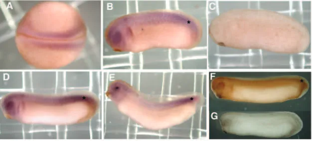
Preliminary western experiments showed that EDEN-BP protein is expressed at approximately constant levels from fertilization to tadpole stages (data not shown; see Fig. S1A in supplementary material). The localization of EDEN-BPmRNA in different embryonic stages was analyzed by whole-mount in situ hybridization and immunohistochemistry (Fig. 1). In blastulae and gastrulae, EDEN-BPmRNA was homogenously present in the whole embryos (data not shown). At the neurula stage, EDEN-BPmRNA was more concentrated in the paraxial mesoderm flanking the neural tube (Fig. 1A). In early tailbuds,
EDEN-BP mRNA was clearly more abundant in the dorsal mesoderm and in the head region (Fig. 1B). This preferential localization in the dorsal mesoderm and head region (including the eye) was more evident in late tailbud or tadpole embryos (Fig. 1D,E). The expression pattern of EDEN-BP protein was
also analyzed by whole-mount immunohistochemistry and found to be very similar to that of EDEN-BPmRNA (Fig. 1F). No signal was detected in control in situ hybridizations using a sense EDEN-BP probe, and in control immunohistochemistry experiments using a pre-immune serum (Fig. 1C,G). The expression of EDEN-BP mRNA and protein in dorsal mesoderm is particularly evident in the posterior PSM (Fig. 1B,D-F). It is very probable that the relocalization of EDEN-BP mRNA and protein to preferred sites at the neurula and tailbud stages relies on zygotic transcription and translation in these sites, rather than on transport in a multicellular embryo. In summary, EDEN-BP shows a preferential localization in paraxial mesoderm, especially in the PSM, and in head regions.
EDEN-BP morpholinos cause somite segmentation defects
The expression pattern of EDEN-BP in the PSM suggested that this protein may be involved in somitic segmentation. This involvement was tested by analyzing the consequences of functionally inhibiting EDEN-BP on segmentation. We used morpholinos (modified antisense oligonucleotides) to inhibit the translation of this protein. EST database searches revealed the presence of a cDNA very similar (>98% identity) to that of the cloned EDEN-BP cDNA. This probably represents the expression product of a second EDEN-BP gene, a frequent situation in Xenopus laevis, whose genome is partly duplicated. A mixture of the two morpholinos directed against the two EDEN-BP mRNAs (E-Mo) was therefore used. To demonstrate the specificity of these morpholinos, co-injections of E-Mo and an mRNA encoding EDEN-BP were also performed. The injected EDEN-BPmRNA (Paillard et al., 1998) possessed the
Xenopusβ-globin 5′untranslated region and was not a target for the morpholinos.
The expression level of EDEN-BP in tailbud (stage 25) embryos after injection of E-Mo into both blastomeres of two-cell embryos was analyzed by western blotting (Fig. 2A, upper panel). Similar loading in all lanes was attested by reprobing the same blot with anti-cdc2 antibodies (lower panel). As compared with control sibling embryos (lane 1), E-Mo strongly decreased the expression level of EDEN-BP (lane 2). A higher expression level was restored by the co-injection of 0.5 or 2
fmol of EDEN-BP mRNA with E-Mo (lanes 3, 4).
[image:3.612.52.365.600.742.2]Accordingly, morpholinos inhibit the expression of EDEN-BP in tailbuds and the microinjection of EDEN-BP mRNA can rescue this inhibition to an almost normal level of expression. The developmental consequences of this inhibition were
Fig. 1.Expression pattern of EDEN-BP during Xenopusearly development. Embryos were collected at different stages: neurula, stage 18 (A); tailbud, stage 25 (B,C,G) and stage 27 (D,F); tadpole, stage 31 (E). (A) Dorsal view, (B-G) lateral views; anterior left. Embryos were processed for whole-mount in situ hybridization using an EDEN-BP antisense (A,B,D,E), or sense (C) probe, or for whole-mount
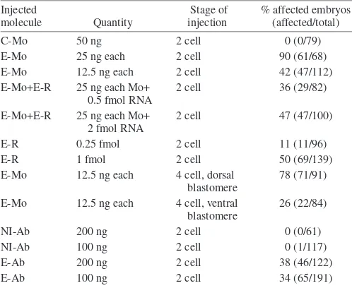
tested by injecting morpholinos into one cell of two-cell embryos and allowing them to develop until tailbud stages. These injected embryos presented an altered somitic segmentation. For better visibility of somitic segmentation, embryos were stained with the myotome-specific 12/101 monoclonal antibody (Kintner and Brockes, 1985) (Fig. 2B-D′). In embryos injected with 50 ng of control morpholinos, segmentation is evidenced by the periodic pattern of somites separated by chevron-shaped borders (Fig. 2B) that is more obvious at a higher magnification (Fig. 2B′). The injection of 25 ng of each E-Mo caused marked defects in somitic segmentation (Fig. 2C,C′). The defects were restricted to the side of the embryos derived from the injected blastomere (data not shown). A large majority (90%) of embryos injected with 25 ng of each E-Mo showed defective segmentation on one side (Table 1). When an mRNA encoding EDEN-BP was co-injected with the E-Mo, the number of embryos with
segmentation defects was significantly decreased (Table 1), and those embryos that no longer showed these defects were indistinguishible from control embryos (Fig. 2D,D′). Interestingly, a significant percentage of the embryos injected with EDEN-BP mRNA alone harbored segmentation defects, which may account for the fact that the percentage of affected embryos observed was higher when the morpholinos were injected with 2 fmol of mRNA than when they were injected with 0.5 fmol mRNA (Table 1).
The absence of externally apparent somitic segmentation in injected embryos may reflect an impairment of somitic differentiation. However, the observation that E-Mo injected embryos were stained with the 12/101 antibody, which is specific for myotomal cells (Kintner and Brockes, 1985), does not support this hypothesis. In addition, myosin heavy chain 4 (MHC4), a marker of terminal muscle differentiation (Vernon and Philpott, 2003), was expressed in E-Mo-injected embryos (Fig. 2F,F′), as in control (Fig. 2E,E′) and rescued embryos (Fig. 2G,G′). Although less easily detectable than with 12/101 staining, embryos shown in Fig. 2E,E′,G,G′, but not 2F,F′, are segmented. This shows that EDEN-BP morpholinos do not impair somitic differentiation, but rather that they impair segmentation.
[image:4.612.86.464.276.644.2]In addition to the rescue experiments, several experiments were performed to test specificity. First, as the effects of the
morpholinos are observed in dorsal regions of the embryo, it is expected that injecting morpholinos into presumptive ventral regions should yield reduced effects. Accordingly, injections of morpholinos were repeated in one blastomere at the four-cell stage, together with a GFP mRNA. A smaller amount of morpholinos (12.5 ng of each) was injected, so as to account for the reduced volume of the injected cells. Embryos were sorted at early tailbud according to ventral or dorsal expression of GFP. Injection of E-Mo in a presumptive dorsal blastomere at the four-cell stage yielded a similar percentage of affected embryos as did injection at the 2-cell stage. By contrast, after injection into a ventral blastomere, the percentage of affected embryos was much lower (Table 1). Second, the percentage of affected embryos was dependent on the quantity of injected morpholinos; injecting 12.5 ng (instead of 25 ng) of each morpholino reduced the percentage of affected embryos from 90% to 42% (Table 1). Finally, anti EDEN-BP antibodies specifically inhibited the rapid deadenylation of EDEN-containing RNAs, when injected into
Xenopus embryos (see Data S1 in supplementary material). We therefore analyzed the developmental consequences of injecting these antibodies into one blastomere of two-cell embryos. Anti EDEN-BP, but not control non-immune antibodies, yielded segmentation defects similar to those provoked by the injection of morpholinos (Fig. 3A,C; Table 1). As for morpholinos, only the injected side of the embryo was affected (Fig. 3B,D). Also, both sides of the antibody-injected embryos that showed defects in somitic segmentation were similarly stained with the 12/101 monoclonal antibody (Fig. 3C,D). If the segmentation defects caused by the morpholinos were just side effects, it would be highly improbable that the antibodies would cause the same effect,
as molecularly different tools are expected to cause different side effects.
To further characterize these observations, horizontal sections were made of stage-39 embryos injected with anti-EDEN-BP antibodies into one blastomere at the two-cell stage. The most evident defect was an absence of somite segmentation on the side originating from the injected blastomere (Fig. 3E, arrow). Higher magnification of the somites on both sides (Fig. 3F,G) indicated that myotomal cells on the injected side, although still present and apparently parallel to the anteroposterior axis, were not organized in stacks as on the non-injected side. Together, these data show that inhibiting EDEN-BP function, by using either morpholinos or neutralizing antibodies, impairs somitic segmentation but not differentiation, and implies that EDEN-BP is required for somitic segmentation.
EDEN-BP morpholinos alter the segmented expression pattern of ESR5 and X-Delta2, but not ESR9, in the presomitic mesoderm
The PSM is not morphologically segmented, but harbors a segmented expression pattern of a number of genes. Several zebrafish mutants display somite segmentation defects that are associated with a complete loss of the segmented expression of genes belonging to the Notch pathway in the PSM. By contrast, the Tbx24 mutant fused somites also shows strong somite segmentation defects, but Notch pathway genes retain some segmented expression pattern in the PSM (Nikaido et al., 2002; van Eeden et al., 1998). We therefore tested whether the segmentation defects caused by the functional inhibition of EDEN-BP are associated with an altered expression pattern of segmentation genes in the PSM.
The expression patterns of three segmentation markers (ESR5, ESR9 and X-Delta2) in the PSM of E-Mo-injected embryos were analyzed by in situ hybridization (Fig. 4). ESR5 and X-Delta2 are a Notch target and a Notch ligand, respectively. ESR9is the only XenopusmRNA known to date to have a cycling expression pattern comparable to what is described in higher vertebrates (see Introduction) (Li et al., 2003). As such, it may be the Xenopusoscillator, or a direct output of the Xenopusoscillator, and is probably upstream of most other gene products in the segmentation process.
The expression pattern of ESR5 on the non-injected side of the embryo (Fig. 4A) was similar to that reported for normal embryos (Jen et al., 1999). ESR5 mRNA was detected in the posterior tailbud domain (TBD), and in the anterior parts of the first two somitomeres (S1 and S2), forming two characteristic chevrons. On the injected side of the embryo, only one chevron could be detected (Fig. 4B). Dorsal examination of this embryo showed that this unique chevron faced the S2 chevron of the non-injected side. In addition, the anterior boundary of the large posterior domain of expression on the injected side faced the S1 chevron of the non-injected side (data not shown). Hence, we interpret the pattern of expression on the injected side as a filling of the gap that normally arises between the TBD and the anterior part of S1.
[image:5.612.48.297.465.669.2]The expression pattern of X-Delta2 on the non-injected side was also similar to previous observations (Jen et al., 1997). X-Delta2 was weakly expressed in the anterior parts of S4, and more strongly in the TBD and the anterior parts of S1 to S3 (Fig. 4C). On the injected side, X-Delta2 expression was Table 1. Percentage of embryos affected by the injection of
morpholinos or antibodies
Injected Stage of % affected embryos
molecule Quantity injection (affected/total)
C-Mo 50 ng 2 cell 0 (0/79)
E-Mo 25 ng each 2 cell 90 (61/68)
E-Mo 12.5 ng each 2 cell 42 (47/112)
E-Mo+E-R 25 ng each Mo+ 2 cell 36 (29/82)
0.5 fmol RNA
E-Mo+E-R 25 ng each Mo+ 2 cell 47 (47/100)
2 fmol RNA
E-R 0.25 fmol 2 cell 11 (11/96)
E-R 1 fmol 2 cell 50 (69/139)
E-Mo 12.5 ng each 4 cell, dorsal 78 (71/91)
blastomere
E-Mo 12.5 ng each 4 cell, ventral 26 (22/84)
blastomere
NI-Ab 200 ng 2 cell 0 (0/61)
NI-Ab 100 ng 2 cell 0 (1/117)
E-Ab 200 ng 2 cell 38 (46/122)
E-Ab 100 ng 2 cell 34 (65/191)
Embryos were injected at the indicated stage with the listed molecules. For morpholinos, the results shown (percentages of affected tailbuds) are given for one representative experiment, and similar results were obtained in at least two other experiments.
weaker in the TBD. In the somitomeric region, the expression level of this mRNA was not reduced, but a segmented pattern of expression was no longer detected (Fig. 4D). Defects in the expression of ESR5 and X-Delta2 similar to those shown in Fig. 4 were detected in 48% (34/71) and 54% (36/67), respectively, of the embryos examined.
As a cycling mRNA, ESR9 shows a dynamic mode of expression that can be subdivided into three phases, named I to III (Li et al., 2003; Pourquie and Tam, 2001). These three phases were found in the PSM of embryos injected with E-Mo (Fig. 4E-G′). The distribution between the phases was not different in control and E-Mo-injected embryos [Phase I, 31% (13/41) in control, 41% (44/107) in E-Mo; Phase II, 34%
(14/41) in control, 33% (35/107) in E-Mo; Phase III, 34% (14/41) in control, 26% (28/107) in E-Mo]. Furthermore, the embryos injected with E-Mo were fairly symmetric regarding the expression of ESR9 (Fig. 4E-G′), indicating that the injected and the uninjected sides of the embryos harbored the same expression pattern for ESR9. We conclude from these data that EDEN-BP morpholinos have no effect on the expression of ESR9. Therefore, they act downstream of ESR9, but on or upstream of ESR5 and X-Delta2, in the segmentation process.
XSu(H)mRNA is regulated by EDEN-BP
The above results show that a downregulation of EDEN-BP disrupts the expression pattern of
[image:6.612.43.398.224.743.2]ESR5 and X-Delta2 mRNA. UV cross-linking experiments were performed to test the ability of the 3′UTRs of these mRNAs to bind to EDEN-BP. GbORF-mosEDEN (Paillard et al., 1998) was used as a positive control for EDEN-BP binding (Fig. 5A, lanes 1-4). This mRNA produced a 53-55 kDa signal in the absence of competitor (lane 1), that was efficiently competed by a 5-fold (lane 2) or 50-fold (lane 3) molar excess of the same unlabelled RNA, but not by a 50-fold molar excess of the same unlabelled RNA with the EDEN deleted (lane 4). That this signal corresponds to EDEN-BP has been previously documented by immunoprecipitation and by experiments cross-linking with recombinant protein (Paillard et al., 1998). No signal was detected using
Fig. 3.Analysis of embryos injected with anti-EDEN-BP antibodies. (A-D) Embryos were injected into one blastomere at the two-cell stage with control (non-immune, C-Ab) or anti-EDEN-BP (E-Ab) antibodies. They were allowed to develop until stage 39 and then were stained with the myotome-specific 12/101 monoclonal antibody. C and D are higher
either ESR5 3′UTR (lanes 5-8) or X-Delta2 3′UTR (lanes 9-12) as cross-linking probes, indicating that these 3′UTRs do not bind EDEN-BP and that they are not direct targets of the EDEN-BP-dependent mechanism.
The altered expression pattern of these two mRNAs in EDEN-BP downregulated embryos (Fig. 4) is therefore indirect and is probably due to the deregulation of other mRNAs. The 3′UTRs of other genes belonging to the Notch signalling pathway were tested for EDEN-BP binding by UV cross-linking. As expected from the above data, ESR9 3′UTR did not bind EDEN-BP; this was also the case of ESR4, Hairy1 and NRARP (Jen et al., 1999; Jen et al., 1997; Lamar et al., 2001) (data not shown). By contrast, XSu(H) 3′UTR produced
a 53-55 kDa UV cross-linking signal (Fig. 5A, lane 13). This signal is competed by an unlabelled EDEN-containing RNA, but not by an RNA with the EDEN deleted (lanes 14-16). It is therefore attributable to EDEN-BP binding. XSu(H) is the
XenopusCSL protein (equivalent to mammalian CBF1/RBP-Jκ, see Introduction) for which two closely related forms were described, XSu(H)1 and XSu(H)2 (Wettstein et al., 1997). The 3′UTRs of these mRNAs are identical, so that XSu(H) 3′UTR refers to both the XSu(H)1 and the XSu(H)2 3′UTRs.
As EDEN-BP binds the XSu(H) 3′UTR in an in vitro (UV cross-linking) assay, we next tested the in vivo interaction
of EDEN-BP with XSu(H) mRNA. EDEN-BP was
immunoprecipitated from embryo extracts, and the associated mRNAs identified by RT-PCR. These analyses showed that XSu(H) mRNA was only precipitated by the anti-EDEN-BP antibodies (Fig. 5B, upper panel, lane 8), and not with protein A alone or with non-immune antibodies (lanes 4, 6). An unrelated mRNA, EF1α(Krieg et al., 1989), was not detected in the EDEN-BP immunoprecipitated mRNAs (lower panel). Accordingly, EDEN-BP interacts with XSu(H) mRNA in vivo. We next tested whether the XSu(H) 3′UTR confers a rapid, EDEN-BP-dependent deadenylation to a reporter mRNA after injection into Xenopusembryos. Reporter RNAs containing the XSu(H) 3′UTR [GbXSu(H)] were injected with either control non-immune or anti-EDEN-BP antibodies. The deadenylation behaviour of this reporter transcript was analyzed by denaturing electrophoresis and autoradiography of RNAs extracted at different times after injection (Fig. 5C). The deadenylation activity was quantified as the relative amount of deadenylated transcript at each time point (Fig. 5D). As for the Gb-Eg2-410 transcript (see Fig. S1B in supplementary material), GbXSu(H) RNA was rapidly deadenylated in the presence of control, non-immune antibodies (lanes 6-10). Importantly, deadenylated RNAs are stable in early Xenopus
embryos until the blastula stage (Audic et al., 1997; Voeltz and Steitz, 1998), which allows the deadenylated transcript to be visualized and quantified. By contrast, the injection of anti-EDEN-BP antibodies strongly reduced the deadenylation of the GbXSu(H) (lanes 1-5). These data show that the XSu(H) 3′UTR contains the sequence information required to direct a reporter RNA to the EDEN deadenylation pathway in Xenopus
embryos.
As mRNA deadenylation leads to degradation after the blastula stage, inhibiting the deadenylation of endogenous XSu(H) mRNA may increase its steady-state level in tailbuds. Steady-state levels of XSu(H), as compared with steady-state levels of EF1α, were assessed in individual embryos by real-time RT-PCR in stage-25 control (non-injected), E-Mo injected, and rescued embryos (Fig. 5E). Some variability occurs between individual embryos, but the average steady-state level of XSu(H) mRNA is clearly higher in E-Mo-injected embryos than in control or rescued embryos. These data show that the functional inhibition of EDEN-BP in tailbuds causes the overexpression of XSu(H), probably via an inhibition of its deadenylation.
Evidences for other targets of EDEN-BP in the segmentation process
[image:7.612.70.271.228.642.2]The functional inhibition of EDEN-BP causes somitic segmentation defects and overexpression of XSu(H). To test whether a causal relationship exists between these events, the Fig. 4.EDEN-BP morpholinos alter presegmentation in the PSM.
effect of overexpressing XSu(H) on segmentation was analyzed. Embryos were injected into one blastomere at the two-cell stage with an in vitro transcribed mRNA containing the XSu(H)1 ORF [but devoid of the XSu(H)1 3′UTR], and were allowed to develop until stage 28. They were then stained with the myotome-specific 12/101 antibody. Sixty-nine percent (130/189) of the embryos injected with XSu(H)1 mRNA displayed segmentation defects (Fig. 6A,B). These defects were similar to those provoked by anti-EDEN-BP morpholinos or antibodies (compare with Figs 2, 3). Embryos injected with
XSu(H)1RNA were also analyzed by in situ hybridization for the expression of ESR5 and X-Delta2. Twenty-seven percent (21/79) and 44% (42/96) of these embryos displayed an expression pattern of ESR5 and X-Delta2 similar to that observed in E-Mo-injected embryos (Fig. 6C-F, compare with Fig. 4A-D).
These data implicate XSu(H) as one of the gene products whose overexpression, following the neutralization of EDEN-BP, causes segmentation defects. If XSu(H) is the only such
gene product, then downregulating XSu(H) in E-Mo-injected embryos should rescue their segmentation defects. A range of concentrations of morpholinos against XSu(H)1 and XSu(H)2 (X-Mo) was therefore injected together with the E-Mo. Fig. 6G shows that none of the concentrations below 5 ng that were tested rescued the segmentation defects caused by the E-Mo. This is probably not due to a poor downregulation of XSu(H) by the X-Mo, as quantities higher than 10 ng were lethal to the embryo during neurulation (to be compared with the lack of effect of 50 ng of a standard control morpholinos, Table 1). These results show that downregulating XSu(H) is not sufficient to restore a correct segmentation in embryos inhibited for EDEN-BP, which suggests the existence of other targets of EDEN-BP in the segmentation process.
Discussion
[image:8.612.56.530.292.739.2]EDEN-BP has been characterized for its role in the post-transcriptional regulation of certain mRNAs in Xenopus
Fig. 5.XSu(H)mRNA is regulated by EDEN-BP. (A) Radiolabelled GbORF-mosEDEN (lanes 1-4), ESR5 3′UTR (lanes 5-8), X-Delta2 3′UTR (lanes 9-12) and XSu(H) 3′UTR (lanes 13-16) transcripts were incubated in Xenopusegg extracts in the absence or the presence of the indicated molar excess of unlabelled RNA with an EDEN (+E, GbORF-mosEDEN) or with the EDEN deleted (–E, GbORF). After UV-cross-linking and RNase treatment, radiolabelled proteins were resolved by denaturing electrophoresis and revealed by autoradiography. The position of the 51 kDa molecular weight marker is indicated on the left. The asterisks indicate the position of the EDEN-BP signal. (B) Embryo extracts were treated for immunoprecipitation with anti-EDEN-BP antibodies (lanes 7-8), or pseudo-immunoprecipitations with beads alone (lanes 3-4) or non-immune serum (lanes 5-6). RNA present in the supernatant (S) or pellet (P) was identified by RT-PCR using primers specific for XSu(H) (upper panel) or EF1α(lower panel). Lanes 1 and 2 show products from PCR performed on the input fraction, either with (lane 2) or without (lane 1) previous reverse transcription. Positions of molecular weight markers are indicated on the left. (C) A capped, radiolabelled,
polyadenylated transcript corresponding to GbXSu(H) was injected into two-cell embryos in the presence of 150 ng of the control (C-Ab, lanes 6-10) or anti-EDEN-BP (E-Ab, lanes 1-5) immunoglobulins, and incubated for the indicated times. RNA was extracted, and analyzed by electrophoresis and
embryos soon after fertilization (Paillard et al., 1998). We showed here that EDEN-BP is also required for the somitic segmentation of older embryos. Inhibition of EDEN-BP dramatically impaired segmentation. This is associated with an altered expression of two markers of presegmented pattern in the PSM, ESR5 and X-Delta2. We finally identified XSu(H)as a segmentation mRNA that is directly regulated by EDEN-BP, and we showed that the overexpression of XSu(H) causes segmentation defects. A model accounting for these observations would be that the overexpression of XSu(H)alone is responsible for the defective segmentation in embryos injected with morpholinos or antibodies against EDEN-BP. However, downregulating XSu(H) using morpholinos did not
restore a normal segmentation in embryos depleted of EDEN-BP (Fig. 6G). In addition, a significant percentage of the embryos injected with E-Mo and EDEN-BP RNA show segmentation defects (Table 1), although the level of XSu(H)
mRNA in embryos injected with E-Mo and EDEN-BP RNA was normal (Fig. 5E). These data imply that, in addition to
XSu(H), there exist unidentified mRNA targets of EDEN-BP that are important for the segmentation process; the coordinated deregulation of these mRNAs following the inactivation of EDEN-BP is probably responsible for the observed segmentation defects. The identification of these unknown targets will represent a major goal in future work
The role of post-transcriptional regulation in somitic segmentation has been described very recently for a gene that is expressed following a precise gradient. A decreasing gradient of FGF8 starting from the posterior extremity of the embryo plays a key role in somitic segmentation and the acquisition of segmental identity (Dubrulle et al., 2001b), and the slope of this gradient is given by the stability of the corresponding mRNA (Dubrulle and Pourquie, 2004). The role of post-transcriptional regulation in segmentation has more often been approached for genes that are cyclically or at least transiently expressed. For instance, Hairy2a mRNA is transiently expressed in two somitomeres of Xenopus
embryos, and an mRNA-destabilizing element localized within the 3′UTR is required to achieve this expression pattern (Davis et al., 2001). Computational modelling showed that mRNA instability is required to achieve sustained oscillations of cyclic genes (Lewis, 2003). The 3′UTRs of several segmentation genes, from Xenopusor other species, were able to replace that of Hairy2a, to confer a transient expression pattern to a reporter gene, showing that they also contain destabilization elements (Davis et al., 2001). This suggests that, in most species, the transient expression pattern of most segmentation mRNAs requires destabilizing elements that are localized within their 3′UTRs. The 3′UTRs of the genes transiently expressed in Xenopus embryos that were tested here (ESR4, ESR9, Hairy1, Hairy2a and NRARP) are not regulated by the EDEN-BP pathway. This implies that the corresponding mRNAs are rapidly degraded by a mechanism different from the EDEN-dependent deadenylation pathway. In Xenopus, at least three deadenylation mechanisms have been described in addition to the EDEN mechanism (Audic et al., 2001; Fox and Wickens, 1990; Varnum and Wormington, 1990; Voeltz and Steitz, 1998) (reviewed by Paillard and Osborne, 2003), and different mRNA destabilization mechanisms may also exist in other species.
[image:9.612.46.322.230.655.2]In contrast to the above examples, XSu(H) mRNA is ubiquitously expressed in Xenopusembryos (Wettstein et al., 1997). How then can XSu(H) overexpression account for the observed segmentation defects? As for CSL proteins in other species (Mumm and Kopan, 2000), XSu(H) protein probably represses the transcription of target genes when not associated with Notch ICD. Stimulating the Notch pathway by overexpressing the Notch ligand X-Delta1 in Xenopus embryos inhibits neuralization (a process termed lateral inhibition), but this effect is reversed by also overexpressing XSu(H) Fig. 6.Evidence for other targets of
EDEN-BP than XSu(H) in the segmentation process.
(A-F) Embryos were injected in one blastomere at the two-cell stage with 2 fmol of XSu(H)1 mRNA. They were allowed to develop to stage 28 (A,B) or stage 26 (C-F), and then stained with the 12/101 monoclonal antibody (A,B), or
(Wettstein et al., 1997), confirming that, in that instance, overexpressing XSu(H) inhibits Notch signalling. As somitic segmentation in Xenopus requires the periodic stimulation/ inhibition of Notch signalling (Jen et al., 1999), constant inhibition of Notch signalling may be the main cause of the defects provoked by overexpressing wild-type XSu(H). However, inhibiting the Notch pathway by expressing a dominant-negative mutant of XSu(H) abolishes ESR5 expression and stimulates X-Delta2 expression in the somitomeric region (Jen et al., 1999; Jen et al., 1997) (also our unpublished data), which is different from what we observe after overexpressing wild-type XSu(H). Consequently, inhibiting the Notch pathway by overexpressing wild-type XSu(H) or expressing a dominant-negative mutant of this protein leads to dissimilar consequences on the expression pattern of these two segmentation markers. Mutant Xsu(H) has lost its capacity to interact with DNA, and supposedly acts by sequestering Notch ICD. By contrast, overexpressed wild-type Xsu(H) still interacts with DNA, and inhibits the transcription of target genes (Wettstein et al., 1997). This difference may be used as a working hypothesis in future experiments designed to explain the different expression patterns of ESR5 and X-Delta2 after expression of wild-type or mutant XSu(H).
We show here, for the first time, the involvement of a molecular mechanism of post-transcriptional regulation in vertebrate somitic segmentation. This work was conducted in
Xenopus, but its conclusions might be generalized to other species. The zebrafish equivalent of EDEN-BP mRNA was reported to be expressed uniformly in embryos up to 24 hours post-fecondation (Suzuki et al., 2000). These results are consistent with the zebrafish EDEN-BP protein being expressed in PSM at the time of segmentation. Similarly, EST database searches reveal that the murine equivalent of EDEN-BP is expressed during embryogenesis, and preliminary experiments show that murine EDEN-BP is strongly expressed in the PSM during segmentation (C.G.-C., unpublished). In addition, in in vitro deadenylation assays, the human and murine equivalents of EDEN-BP can behave as deadenylation factors (Paillard et al., 2003) (data not shown). From these results, it can be postulated that the fish and mammalian equivalents of EDEN-BP could be responsible for the destabilization of segmentation mRNAs in these species. If this were the case, it would reveal a strong conservation of post-transcriptional control in vertebrate somitic segmentation.
We thank Mike Klymkowsky and Chris Kintner for the pCS2-GFPmut and pCS2-XSu(H) vectors. We are grateful to Olivier Pourquié and Nicolas Pollet for discussions and for sharing results before publication. Serge Hardy, Michelle Lesimple, Laurent Richard-Parpaillon and Vincent Legagneux are acknowledged for fruitful discussions. We are also grateful to Professor Jacques-Philippe Moulinoux and the technical staff of his laboratory (Marie-Thérèse Lavault) for access to their histological apparatus. The ESR5 and ESR9 plasmids (5047232 and 4085131) were provided by the IMAGE consortium. This work was supported by grants from the ARC to H.B.O. (number 9529) and L.P. (number 4791). Y.A. was a recipient of an ARC post-doctoral fellowship.
Supplementary material
Supplementary material for this article is available at http://dev.biologists.org/cgi/content/full/131/24/6107/DC1
References
Audic, Y., Omilli, F. and Osborne, H. B. (1997). Postfertilization deadenylation of mRNAs in Xenopuslaevis embryos is sufficient to cause their degradation at the blastula stage. Mol. Cell. Biol.17, 209-218. Audic, Y., Anderson, C., Bhatty, R. and Hartley, R. S. (2001). Zygotic
regulation of maternal cyclin A1 and B2 mRNAs. Mol. Cell. Biol.21, 1662-1671.
Aulehla, A., Wehrle, C., Brand-Saberi, B., Kemler, R., Gossler, A., Kanzler, B. and Herrmann, B. G. (2003). Wnt3a plays a major role in the segmentation clock controlling somitogenesis. Dev. Cell4, 395-406. Beelman, C. A. and Parker, R. (1995). Degradation of mRNA in eukaryotes.
Cell81, 179-183.
Bessho, Y., Hirata, H., Masamizu, Y. and Kageyama, R. (2003). Periodic repression by the bHLH factor Hes7 is an essential mechanism for the somite segmentation clock. Genes Dev.17, 1451-1456.
Bouvet, P., Omilli, F., Arlot-Bonnemains, Y., Legagneux, V., Roghi, C., Bassez, T. and Osborne, H. B. (1994). The deadenylation conferred by the 3′untranslated region of a developmentally controlled mRNA in Xenopus embryos is switched to polyadenylation by deletion of a short sequence element. Mol. Cell. Biol.14, 1893-1900.
Cole, S., Levorse, J., Tilghman, S. and Vogt, T. (2002). Clock regulatory elements control cyclic expression of lunatic fringe during somitogenesis. Dev. Cell3, 75-84.
Dale, J. K., Maroto, M., Dequeant, M. L., Malapert, P., McGrew, M. and Pourquie, O. (2003). Periodic notch inhibition by lunatic fringe underlies the chick segmentation clock. Nature421, 275-278.
Davis, R., Turner, D., Evans, L. and Kirschner, M. (2001). Molecular targets of vertebrate segmentation: two mechanisms control segmental expression of XenopusHairy2 during somite formation. Dev. Cell1, 553-565. Dubrulle, J. and Pourquie, O. (2004). fgf8 mRNA decay establishes a
gradient that couples axial elongation to patterning in the vertebrate embryo. Nature427, 419-422.
Dubrulle, J., McGrew, M. and Pourquié, O. (2001). FGF signaling controls somite boundary position and regulates segmentation clock control of spatiotemporal Hox gene activation. Cell106, 219-232.
Fox, C. A. and Wickens, M. (1990). Poly(A) removal during oocyte maturation: a default reaction selectively prevented by specific sequences in the 3′UTR of certain maternal mRNAs. Genes Dev.4, 2287-2298. Harland, R. M. (1991). In situ hybridization: an improved whole-mount
method for Xenopusembryos. Methods Cell Biol.36, 685-694.
Jen, W. C., Wettstein, D., Turner, D., Chitnis, A. and Kintner, C. (1997). The Notch ligand, X-Delta-2, mediates segmentation of the paraxial mesoderm in Xenopusembryos. Development124, 1169-1178.
Jen, W., Gawantka, V., Pollet, N., Niehrs, C. and Kintner, C. (1999). Periodic repression of Notch pathway genes governs the segmentationof Xenopusembryos. Genes Dev.13, 1486-1499.
Kintner, C. R. and Brockes, J. P. (1985). Monoclonal antibodies to the cells of a regenerating limb. J. Embryol. Exp. Morphol.89, 37-55.
Krieg, P. A., Varnum, S. M., Wormington, W. M. and Melton, D. A. (1989). The mRNA encoding elongation factor 1-a(EF-1a) is a major transcript at the midblastula transition in Xenopus. Dev. Biol.133, 93-100.
Lamar, E., Deblandre, G., Wettstein, D., Gawantka, V., Pollet, N., Niehrs, C. and Kintner, C. (2001). Nrarp is a novel intracellular component of the Notch signaling pathway. Genes Dev.15, 1885-1899.
Lewis, J. (2003). Autoinhibition with transcriptional delay: a simple mechanism for the zebrafish somitogenesis oscillator. Curr. Biol.13, 1398-1408.
Li, Y., Fenger, U., Niehrs, C. and Pollet, N. (2003). Cyclic expression of ESR9 gene in Xenopuspresomitic mesoderm. Differentiation71, 83-89. Maroto, M. and Pourquie, O. (2001). A molecular clock involved in somite
segmentation. Curr. Top. Dev. Biol.51, 221-248.
Morales, A., Yasuda, Y. and Ish-Horowicz, D. (2002). Periodic Lunatic fringe expression is controlled during segmentation by a cyclic transcriptional enhancer responsive to notch signaling. Dev. Cell3, 63-74. Mumm, J. S. and Kopan, R. (2000). Notch signaling: from the outside in.
Dev. Biol.228, 151-165.
Nikaido, M., Kawakami, A., Sawada, A., Furutani-Seiki, M., Takeda, H. and Araki, K. (2002). Tbx24, encoding a T-box protein, is mutated in the zebrafish somite-segmentation mutant fused somites. Nat. Genet.31, 195-199.
Paillard, L. and Osborne, H. B. (2003). East of EDEN was a poly(A) tail. Biol. Cell95, 211-219.
that mediates sequence-specific mRNA deadenylation in Xenopusembryos. EMBO J.17, 278-287.
Paillard, L., Legagneux, V. and Osborne, H. (2003). A functional deadenylation assay identifies CUG-BP as a deadenylation factor. Biol. Cell
95, 107-113.
Pourquie, O. and Tam, P. P. (2001). A nomenclature for prospective somites and phases of cyclic gene expression in the presomitic mesoderm. Dev. Cell
1, 619-620.
Saga, Y. and Takeda, H. (2001). The making of the somite: molecular events in vertebrate segmentation. Nat. Rev. Genet.2, 835-845.
Serth, K., Schuster-Gossler, K., Cordes, R. and Gossler, A. (2003). Transcriptional oscillation of lunatic fringe is essential for somitogenesis. Genes Dev.17, 912-925.
Sparrow, D. B., Jen, W. C., Kotecha, S., Towers, N., Kintner, C. and Mohun, T. J. (1998). Thylacine 1 is expressed segmentally within the paraxial mesoderm of the Xenopus embryo and interacts with the Notch pathway. Development125, 2041-2051.
Suzuki, H., Maegawa, S., Nishibu, T., Sugiyama, T., Yasuda, K. and Inoue,
K. (2000). Vegetal localization of the maternal mRNA encoding an EDEN-BP/Bruno-like protein in zebrafish. Mech. Dev.93, 205-209.
van Eeden, F. J., Holley, S. A., Haffter, P. and Nusslein-Volhard, C. (1998). Zebrafish segmentation and pair-rule patterning. Dev. Genet.23, 65-76.
Varnum, S. M. and Wormington, W. M. (1990). Deadenylation of maternal mRNAs during Xenopusoocyte maturation does not require specific cis-sequences: a default mechanism for translational control. Genes Dev.4, 2278-2286.
Vernon, A. E. and Philpott, A. (2003). A single cdk inhibitor, p27Xic1, functions beyond cell cycle regulation to promote muscle differentiation in Xenopus. Development130, 71-83.
Voeltz, G. K. and Steitz, J. A. (1998). AUUUA sequences direct mRNA deadenylation uncoupled from decay during Xenopusearly development. Mol. Cell. Biol.18, 7537-7545.