4651
Introduction
Polarity pervades the cell much as a magnetic field pervades space, without the help of iron filings that bring it to light
A. D. Hershey
Cells in an epithelium are polarised – both in the basal/apical axis and in the plane of the sheet. This latter or planar polarity (Nübler-Jung et al., 1987) is exemplified by the oriented outgrowth of hairs and the asymmetric action of cilia (Drubin, 2000). Planar polarity is not always so conspicuous – for example, the epithelial cells of the elongating germ band of Drosophila appear unpolarised, yet when they are made to express the Slam protein, they display planar polarity (Lecuit et al., 2002). We think of planar polarity, not as a special characteristic that occurs only when anisotropic structures are being made, but instead as an ever-present property of all or nearly all epithelial sheets, even though it is usually invisible. How is planar polarity generated and organised? Studies of different systems [wing, eye, abdomen in Drosophila, and the stereocilia of the ear (Lewis and Davies, 2002) as well as convergent extension in vertebrates (Wallingford et al., 2002)]
find that many of the same genes or their homologues act in all systems, arguing that there are common elements; however there is no real understanding of the mechanism.
In the fly, polarity has been mainly studied in wing and eye, but we have chosen the abdomen. Unlike the wing (an appendage), the abdomen represents the atavistic body plan, a continuous epithelium that is subdivided into a succession of anterior (A) and posterior (P) compartments. Pattern is organized by signalling across A/P compartment boundaries and depends on the different abilities of A and P cells to send and interpret these signals (Struhl et al., 1997a; Struhl et al., 1997b; Lawrence et al., 2002).
In the anteroposterior axis, both the pattern and the gradient of cell affinity are determined by a morphogen, Hedgehog (Hh) (Struhl et al., 1997a; Struhl et al., 1997b; Lawrence et al., 1999b; Lawrence et al., 1999a). Hh, secreted by P cells, spreads as a gradient into the A compartment and specifies stripes of distinct A cell types. Hh also directs expression of a second morphogen, Wingless (Wg), which then spreads back into P, forming another gradient to specify pattern in the P compartment (Lawrence et al., 2002). Our working model is The integument of the Drosophila adult abdomen bears
oriented hairs and bristles that indicate the planar polarity of the epidermal cells. We study four polarity genes, frizzled ( fz), prickle (pk), Van gogh/strabismus (Vang/stbm) and starry night/flamingo (stan/fmi), and note what happens when these genes are either removed or overexpressed in clones of cells. The edges of the clones are interfaces between cells that carry different amounts of gene products, interfaces that can cause reversals of planar polarity in the clone and wild-type cells outside them. To explain, we present a model that builds on our earlier picture of a gradient of X, the vector of which specifies planar polarity and depends on two cadherin proteins, Dachsous and Fat. We conjecture that the X gradient is read out, cell by cell, as a scalar value of Fz activity, and that Pk acts in this process, possibly to determine the sign of the Fz activity gradient.
We discuss evidence that cells can compare their scalar readout of the level of X with that of their neighbours and can set their own readout towards an average of those. This
averaging, when it occurs near the edges of clones, changes the scalar response of cells inside and outside the clones, leading to new vectors that change polarity. The results argue that Stan must be present in both cells being compared and acts as a conduit between them for the transfer of information. And also that Vang assists in the receipt of this information. The comparison between neighbours is crucial, because it gives the vector that orients hairs – these point towards the neighbour cell that has the lowest level of Fz activity.
Recently, it has been shown that, for a limited period shortly before hair outgrowth in the wing, the four proteins we study, as well as others, become asymmetrically localised in the cell membrane, and this process is thought to be instrumental in the acquisition of cell polarity. However, some results do not fit with this view – we suggest that these localisations may be more a consequence than a cause of planar polarity.
Key words: frizzled, Van gogh, starry night, prickle
Summary
Cell interactions and planar polarity in the abdominal epidermis of
Drosophila
Peter A. Lawrence1,*, José Casal1and Gary Struhl2
1MRC Laboratory of Molecular Biology, Hills Road, Cambridge CB2 2QH, UK
2Howard Hughes Medical Institute, Columbia University College of Physicians and Surgeons, 701 West 168th Street, New York,
NY 10032, USA
*Author for correspondence (e-mail: pal@mrc-lmb.cam.ac.uk)
Accepted 14 July 2004
Development 131, 4651-4664
Published by The Company of Biologists 2004 doi:10.1242/dev.01351
that both these morphogens, via the activation of the transcription factor Optomotor blind (Omb), establish a different gradient, ‘X’ (Lawrence et al., 2002). The nature of the X gradient is unknown, it may depend on a diffusing morphogen or it may not, but in either case, the vector of X at each locale should determine the polarity of each cell (Lawrence, 1966; Stumpf, 1966; Zheng et al., 1995; Struhl et al., 1997b; Wehrli and Tomlinson, 1998). In the present model, the X gradient is reflected across compartment boundaries, giving opposing slopes in A and P (Casal et al., 2002). Evidence suggests that X is neither Hh, Dpp nor any of the seven Wnts in Drosophila, nor any of the fly EGF or FGF homologues, nor does it appear to depend in any way on Notch (Lawrence et al., 2002).
Polarity genes can tentatively be divided into two groups: those required for generating the X gradient, and those required for responding to it. In the first group, consisting of four jointed (fj), dachsous (ds) and fat (ft), polarity changes produced by clones of mutant cells in the A and P compartments are of different sign – that is like the opposing slopes of the presumed gradients of X in each compartment. For example, clones overexpressing fj in the A compartment reverse the polarity of cells in front of the clone, while, in the P they reverse cells behind. Building on results from the eye (Yang et al., 2002) we speculated that these three genes help form the gradient or gradients that constitute X (Casal et al., 2002).
Genes in the second group might be responsible for reading and responding to the X gradient, a process that would require cells to sense the vector of X, and to orient accordingly. Because most cells in both A and P make hairs (Struhl et al., 1997b), all of which point posteriorly, clones of cells mutant for these genes should affect polarity in the same way in both A and P. We selected four polarity genes that strongly affect polarity, frizzled (fz), prickle (pk), Van gogh/strabismus (Vang/stbm) and starry night/flamingo (stan/fmi), and show that all belong to the second group, in which mutant clones cause the same polarity effects in A and P. However, the properties of each gene are distinct, providing insights that allow us to build a model of planar polarity that is fundamentally different from previous ones (e.g. Adler et al., 1997; Tree et al., 2002; Ma et al., 2003). This model is based on the patterns of repolarisations seen following experiments. We conjecture that in normal development, cells align their polarity, using Stan, to detect a difference between the perceived levels of X of their neighbours (X being measured by a scalar readout related to the activity of Fz). We propose that the cells read X but also reset their own scalars towards an average of the scalar levels of their neighbours.
It has been shown in recent years that the proteins encoded by all four of these genes become asymmetrically localised in wing cells just before they make polarised hairs (reviewed by Mlodzik, 2002; Strutt, 2002). We present evidence that this localisation may not be as instrumental as is commonly assumed and may be more a consequence than a cause of planar polarity.
Materials and methods
Mutations, insertions and transgenes
The FlyBase (FlyBase, 1999) entries of the mutations, insertions and transgenes referred in the text are as follows.
Vang–: Vangstbm-6is a 2 bp deletion, resulting in a frameshift. UAS.Vang: VangScer\UAS.cWa.
fz–: fz15 is a single nucleotide substitution; fz21 is an insertion into the first exon; fz37 is an imprecise excision of a P element, resulting in the deletion of more than 50 kb flanking genomic sequences.
UAS.fz: fzScer\UAS.cZaand fzScer\UAS.cSa. fz2–: fz2C1.
stan–: stan3 and stan71 are hypomorphic alleles; stanE59 has a nonsense mutation in the ectodomain.
UAS.stan: stanScer\UAS.cUa.
pk–: pkpk-sple–13is a small aberration affecting both pk and sple open reading frames; pkGO12 is a P-element insertion and pk1 is a spontaneous mutation.
UAS.pk: pkScer\UAS.cGa. UAS.sple: pksple.Scer\UAS. vg.Gal4: Scer\GAL4vg.PM.
abx/ubx>f+>Gal4-lacZ: Scer\Gal4Scer\FRT.Ubx. 1407.Gal4: Scer\GAL41407.
tub.Gal4: Scer\GAL4alphaTub84B.PL. act.Gal4: Scer\GAL4Act5C.PP. ptc.Gal4: Scer\GAL4ptc-559.1. ci.Gal4: a gift from Bob Holmgren. en.Gal4: Scer\GAL4en-e16E. hh.Gal4: Scer\GAL4hh-Gal4.
tub.Gal80: Scer\GAL80alphaTub84B.PL. ptc.lacZ: Ecol\lacZptc-AT96.
hh.lacZ: hhP30.
UAS.FLP: Scer\FLP1Scer\UAS.cCa. hs.FLP: Scer\FLP1hs.PS.
UAS.GFP: Avic\GFPScer\UAS.T:Hsap\MYC,T:SV40\nls2. UAS.lacZ: Ecol\lacZScer\UAS.T:SV40\nls2.
CD2y+: Rnor\CD2hs.PJ. FRT18A: P{neoFRT}18A. FRT42: P{FRT(whs)}42D. FRT2A: P{FRT(whs)}2A. FRT80: P{neoFRT}80B.
Experimental genotypes
(1) tub.Gal4 UAS.fz: w/y w hs.FLP; tub.Gal4/+; UAS.fz/TM2. (2) fz–clones: y w hs.FLP; fz15trcC1ri FRT2A/CD2y+ hs.GFP ri FRT2A.
(3) abx/ubx>Gal4 UAS.fz clones: males w/y hs.FLP; UAS.fz/abx/ubx>f+>Gal4-lacZ.
(4) tub.Gal4 UAS.fz clones: w/y w hs.FLP; FRT42 pwn cn bw/FRT42 tub.Gal80 CD2y+; UAS.fz/tub.Gal4.
(5) act.Gal4 UAS.fz clones: y w hs.FLP; FRT42 pwn cn bw/FRT42 tub.Gal80 CD2y+; fz15ri FRT2A act.Gal4/UAS.fz.
(6) act.Gal4 UAS.fz clones in fz–flies: y w hs.FLP; FRT42 pwn cn bw/FRT42 tub.Gal80 CD2y+; fz15 ri FRT2A Act.Gal4/UAS.fz fz15 fz2C1ri FRT2A.
(7) en.Gal4 UAS.fz: y w hs.FLP; en.GAL4 wgCX4pr/FRT42 pwn; UAS.fz fz15fz2C1FRT2A/+.
(8) en.Gal4 UAS.fz in fz– flies: y w hs.FLP; en.GAL4 wgCX4 pr/FRT42 pwn; UAS.fz fz15fz2C1FRT2A/fz15ri FRT2A hh.lacZ.
(9) hh.Gal4 UAS.fz: y w hs.FLP; FRT42 pkpk-sple–13/CyO; hh.Gal4/UAS.fz.
(10) hh.Gal4 UAS.fz in fz–flies: y w hs.FLP; ptc.lacZ/CyO; fz21 CD2y+ UAS.GFP hh.Gal4/UAS.fz fz21fz2C1ri FRT2A.
(11) pk–: pkGO12; hh.lacZ/+.
(12) tub.Gal4 UAS.sple: w/y w hs.FLP; tub.Gal4/If; UAS.sple/TM2.
(13) en.Gal4 UAS.sple: y w/w; UAS.sple/+; en.Gal4/+.
(14) hh.Gal4 UAS.sple: y w hs.FLP; UAS.sple/Sp; fz21CD2y+ ri FRT2A hh.Gal4/MKRS.
(16) tub.Gal4 UAS.pk: y w/y w hs.FLP; tub.Gal4/+; UAS.pk/TM2. (17) pk–fz–: FRT42 pkpk-sple–13; fz21CD2y+ ri FRT2A.
(18) hh.Gal4 UAS.sple in fz– flies: y w FLP; UAS.sple/Sp; fz21 CD2y+ ri FRT2A hh.Gal4/fz21 CD2y+ ri FRT80.
(19) fz–clones in pk–flies: y w hs.FLP; FRT42 pkpk-sple–13; fz15 trcC1ri FRT2A/CD2y+ hs.GFP ri FRT2A.
(20) hh.Gal4 UAS.fz in pk–flies: y w hs.FLP; FRT42 pkpk-sple–13/ FRT42 pkpk-sple–13 sha; hh.Gal4/UAS.fz.
(21) pk–: y w hs.FLP; FRT42 pkpk-sple–13/FRT42 pkpk-sple–13 sha; hh.Gal4/ TM2.
(22) pk–clones: y hs.FLP/+; FRT42 pk1pwn/FRT42 CD2y+; and (23) y hs.FLP/+; FRT42 pkpk-sple–13/FRT42 CD2y+; and
(24) y hs.FLP; FRT42 pkpk-sple–13sha/FRT42 CD2y+.
(25) tub.Gal4 UAS.sple clones: w/y w hs.FLP; FRT42 pwn cn bw/FRT42 tub.Gal80 CD2y+; UAS.sple/tub.Gal4.
(26) Vang–: y hs.FLP; FRT42 Vang–.
(27) tub.Gal4 UAS.Vang: w/y w hs.FLP122; UAS.Vang/tub.Gal4; +/TM2.
(28) Vang–clones: y/y hs.FLP; FRT42 pwn Vang–/FRT42 CD2y+; and
(29) y w hs.FLP; FRT42 Vang–/FRT42 CD2y+.
(30) pwn clones: y w/+; smo3b FRT42 cn pwn y w; FRT42 sha; hs.FLP/+.
(31) tub.Gal4 UAS.Vang clones: w/y w hs.FLP; FRT42 pwn cn bw/FRT42 tub.Gal80 CD2y+; UAS.Vang/tub.Gal4.
(32) tub.Gal4 UAS.Vang clones in Vang–: y w/y w hs.FLP tub.Gal4 UAS.GFP; UAS.Vang Vang–/FRT42 Vang–; mwh jv CD2y+ FRT2A/tub.Gal80 FRT2A.
(33) fz–clones in Vang–: y w hs.FLP; FRT42 Vang–; fz15trcC1ri FRT2A/CD2y+ hs.GFP ri FRT2A.
(34) tub.Gal4 UAS.fz clones in Vang–: w/y w hs.FLP tub.Gal4 UAS.GFP; FRT42 Vang–/FRT42 pwn Vang–; tub.Gal4 FRT2A/UAS.fz trc FRT2A.
(35) Vang– tub.Gal4 UAS.fz clones: w/y w hs.FLP; FRT42 pwn Vang–/FRT42D tub.Gal80 CD2y+; UAS.fz trc FRT2A/tub.Gal4.
(36) tub.Gal4 UAS.Vang clones in fz–: y w hs.FLP/y w hs.FLP tub.Gal4 UAS.GFP; FRT42 pwn/FRT42 tub.Gal80; UAS.Vang fz21 CD2y+ ri FRT2A/fz21CD2y+ FRT80.
(37) Vang–clones in fz–: y w hs.FLP; FRT42 pwn Vang–/FRT42; fz21CD2y+ ri FRT80/fz21ri FRT2A
(38) stan–: y w hs.FLP; stan3CD2y+/stanE59; +/TM6B; and (39) stan3; and
(40) y w hs.FLP; ptc.Gal4 stanE59/FRT42 pwn stan71; and (41) y w; stanE59/stanE451407.Gal4; UAS.stan/+.
(42) tub.Gal4 UAS.stan: y w/y w hs.FLP; UAS.stan/tub.Gal4/; +/TM2.
(43) stan–clones: y w hs.FLP; FRT42 stanE59/FRT42 hs.GFPy+; vg.Gal4 UAS.FLP/+; and
(44) y w FL122; FRT42 pwn stanE59/FRT42 CD2y+ RpL19; vg.Gal4/UAS.FLP/+; and
(45) y w/y hs.FLP; FRT42 pwn stanE59/FRT42 CD2y+.
(46) stan– tub.Gal4 UAS.fz clones: w/y w hs.FLP; FRT42 pwn stanE59/FRT42 tub.Gal80 CD2y+; UAS.fz/tub.Gal4.
(47) fz– clones in stan–: y w hs.FLP; stan3; fz15 trcC1 ri FRT2A/hs.CD2y+ hs.GFP ri FRT2A; and
(48) y w hs.FLP; stan3; fz15ri FRT2A/CD2y+ hs.GFP ri FRT2A. (49) ptc.Gal4 UAS.fz in stan–: y w hs.FLP; ptc.Gal4 stanE59/stan3; UAS.fz CD2y+ ri FRT2A/+.
(50) tub.Gal4 UAS.stan clones: w/y w hs.FLP; FRT42 pwn cn bw/FRT42 tub.Gal80 CD2y+; UAS.stan/tub.Gal4.
(51) stan–tub.Gal4 UAS.Vang clones: w/y w hs.FLP; FRT42 pwn stanE59/FRT42 tub.Gal80 UAS.Vang/tub.Gal4.
(52) tub.Gal4 UAS.stan clones in fz–: y w hs.FLP tub.Gal4 UAS.GFP/y w hs.FLP; FRT42 pwn/FRT42 tub.Gal80; UAS.stan fz21 CD2y+ ri FRT2A/fzP21 CD2y+ FRT80.
(53) ptc.Gal4 UAS.stan: y w hs.FLP; Sp/ptc.Gal4; UAS.stan fz37/TM6B.
(54) ptc.Gal4 UAS.stan in fz–: y w hs.FLP; Sp/ptc.Gal4; UAS.stan fz37/fz21.
Clones were induced by heat-shocking third instar larvae or pupae for 1 hour at 34, 35 or 37°C. β-galactosidase activity was developed as described by Lawrence et al. (Lawrence et al., 1999a). Abdominal cuticles were dissected, mounted in Hoyer’s and images captured with Auto-Montage (Syncroscopy).
Results and Discussion
Dorsal and ventralThe dorsal surface of the abdomen includes the pigmented tergites as well as regions of flexible cuticle; it is mostly decorated with polarised hairs and bristles (Fig. 1A). From landmarks, we can estimate positions of the cells vis-à-vis the compartments and their boundaries. The ventral surface consists of the sternites and the pleura, the latter a flexible cuticle forming a featureless lawn of polarised hairs. Both dorsally and ventrally, hairs point posteriorly, indicating the polarity of the epidermal cells (Struhl et al., 1997b).
Even though the effects of experiments are concordant, the tergites and sternites are less sensitive to mutations in polarity genes than the pleura. For example, the pleurae of flies mutant for fz or stan bear hairs with randomised orientation, while, in the tergites and sternites, there are large areas with near-normal polarity – this implies that there may be some additional polarising mechanism in those parts. Therefore we use the pleura when we can, but we must use the tergites when it is important to know the exact position of the clone relative to the compartments.
We can remove or overexpress a gene or genes in marked clones of cells. These clones generate sharp disparities in gene function at their edges that cause changes in polarity. We can also overexpress the gene under Gal4/UAS control using Gal4 drivers, which act in either the A (ci.Gal4) or in the P compartment (en or hh.Gal4) – these drivers create disparities across the A/P compartment boundaries. We can also make gradients within the A compartment (ptc.Gal4). We summarise the main results in Fig. 1B.
Building a model for how Fz, Pk, Vang and Stan act to polarise cells
frizzled (fz)
Fz protein
fz encodes one of the Fz family of transmembrane proteins (Adler et al., 1990; Park et al., 1994; Wodarz and Nusse, 1998), at least two of which, Fz and Fz2, function as redundant Wg receptors, each capable of bearing the full burden of Wg transduction (Bhanot et al., 1996; Bhanot et al., 1999; Chen and Struhl, 1999). Fz, but not Fz2, is also required for normal cell polarity (Gubb and Garcia-Bellido, 1982; Vinson and Adler, 1987; Chen and Struhl, 1999). In the pupal wing, Fz protein accumulates transiently along the distal edge of each cell, prefiguring the proximodistal outgrowth of a single hair from this position (Strutt, 2001; Strutt, 2002).
fz–and UAS.fz flies
compartment. However, ventrally, the hairs of the pleura lack all anteroposterior bias; in some places they are randomly oriented, in others they point mostly laterally. General overexpression (tub.Gal4) of a UAS.fz transgene in the fly abdomen causes only a little dishevelment and, in the pleura, occasional patches of hairs of disturbed polarity (genotype 1 in Materials and methods).
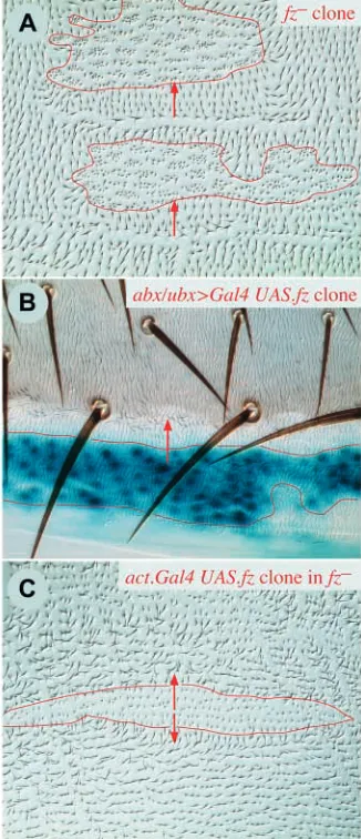
Clones of fz–and UAS.fz cells
Clones of fz–cells have strong effects: hairs at the back of the clone are reversed, with reversal extending beyond and behind the clone for about 2-4 rows of cells (Fig. 2A). The hairs point into the clone, that is, towards the cells with no Fz. This phenotype occurs wherever the clones are made, dorsally or ventrally, regardless of compartment (A or P), or of the
[image:4.612.354.517.171.549.2]presence of compartment boundaries. For example, fz–clones at the back of the A or at the back of the P compartment will reverse the polarity of the cells behind, which will be the most anterior P and the most anterior A cells, respectively. fz–clones in the pleura have randomised hairs within the clone, except for the most anterior row of cells within the clone (normal polarity) and the most posterior row of cells within the clone (reversed polarity).
Fig. 1. Summary model and results. (A) Pattern formation in the
abdomen. Two segments are shown, each with an A and a P (blue) compartment stratified into different types of cuticle (shown at bottom of the panel). The Hh and Wg gradients pattern the cuticle (Struhl et al., 1997b; Lawrence et al., 2002). The gradient of ‘X’ may have opposing slopes in A and P; its vector determines the
orientation of the hairs in A (up the gradient) and P (down the gradient) as shown by the arrows. Anterior is to the left. (B) A field guide to the main results; the genotypes of clones are shown inside the ellipses. Genotype symbols outside the clone, say, fz–, indicate the genetic background in which the clone was induced (fz–). Red arrows mark where polarity is reversed or normalised. Anterior is up.
[image:4.612.55.279.252.605.2]UAS.fz clones cause a phenotype that is as strong, but opposite to fz–clones. Up to several rows of cells in the anterior half of the clone, as well as beyond and in front, show reversal of polarity (Fig. 2B); hairs lateral to the clone also point outwards. Again the phenotype is the same, no matter where the clones are located; for example, clones at the anterior limit of the P compartment will reverse hairs of the A cells in front of them (Fig. 2B).
UAS.fz clones in fz–flies
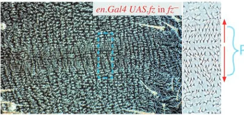
These clones behave like UAS.fz clones in a wild-type background, except in their capacity to repolarise surrounding cells. Inside the front part of the clone, several rows of hairs are reversed, but, outside the clone, repolarisation of the mutant cells is limited to only one cell (Fig. 2C). We also compared the effect of en.Gal4 or hh.Gal4 driving UAS.fz in the P compartment of wild-type and fz–flies (genotypes 7-10). In fz+ flies we see reversal straddling the anterior border of the P compartment and affecting two to three cells on either side. In fz–flies there is reversal anterior to the compartment border, but it is more limited and more difficult to define (the reversal is noisy and the hairs are somewhat dishevelled). In the pleura, imposed on the rather chaotic hairs of the fz– territory, there are two differently sized zones of oriented hairs, the smaller pointing anteriorly near the front of the P compartment, and the larger one pointing posteriorly behind it (Fig. 3).
These results in fz–flies show that a group of fz-expressing cells affects the cells both anterior and posterior to the clone, and directs hairs to point away. This makes clear that, in most other experiments, polarity changes are only seen on one side of the clone because any effects on the other side are invisible, being concordant with normal polarity. Also, they show that the fz-expressing cells can repolarise cells that lack Fz protein, but only if they are in direct contact.
Fz: discussion
Fz, a receptor for X?
Fz is a transmembrane receptor for Wnt genes and it has therefore been reasonably argued (Adler et al., 1997; Tomlinson et al., 1997) that, in planar polarity, it also might be a receptor for X, leading to the hypothesis that X is a Wnt. In support of this, dishevelled (dsh), which functions in the Wnt pathway, also affects planar polarity (Klingensmith et al., 1994; Theisen et al., 1994). However, tests in Drosophila (Lawrence et al., 2002) indicate that X is not a Wnt. Nevertheless, in addition to its function in the Wnt pathway, Fz could also be a receptor for X and, if so, fz–cells should show randomised polarity – and this is true of the pleura and the eye (Zheng et al., 1995), as well as parts of the tergite.
Fz, polarity and the borders of compartments
All our results suggest that cells compare their relative levels of Fz activity and form hairs pointing away from neighbours that have a higher activity. fz–cells can be repolarised as long as they are in direct contact with UAS.fz or fz+cells: a cell with no Fz can still be compared with a cell that has some. Moreover, as observed, repolarisation should not spread further than one cell into fz–territory because, within that territory, all cells being compared have no Fz.
In the normal fly, all cells make hairs and bristles that point posteriorly, suggesting that there is a continuous gradient of Fz
activity from high to low, from anterior to posterior. Given that the pattern repeats every metamere, this would give a gradient repeating once per unit, with a precipice at either the A/P or the P/A border. Hence, if all cells secrete hairs that point towards that neighbour with the lowest Fz activity, we would expect cells flanking the A/P or P/A boundaries to point anteriorly, but this is not the case. One could postulate barriers or other special properties for the A/P and P/A boundaries that would insulate cells in different compartments. Indeed, there is some evidence that the A/P boundary may function as a barrier: UAS.fj clones can repolarise adjacent cells across the A/P boundary, but only in one direction (A to P, but not P to A) (Casal et al., 2002). However, fz– and UAS.fz clones repolarise cells across compartment boundaries just as effectively as they do within compartments. Perhaps we could argue that such clones cause an abnormally large disparity in Fz activity, and as a consequence can override any special conditions at the boundaries. But this is not a satisfying argument.
Non-autonomy
Why do changes in polarity spread more than one cell into fz+ territory, and what limits this non-autonomy to only a few cells? These are important questions and we discuss them later.
prickle (pk)
Pk and Sple proteins
[image:5.612.317.562.73.189.2]These are homologous proteins that derive from two transcripts constituting the pk locus, they are cytosolic and contain a LIM domain (Gubb et al., 1999). The pk–mutation we use removes both transcripts. In wing cells, shortly before hair outgrowth, Pk accumulates at the proximal cell boundary, which is opposite to the site of Fz accumulation (Tree et al., 2002). In pk–wing cells, Fz protein is still localised to the membrane of cells, but does not accumulate asymmetrically (Strutt, 2001; Tree et al., 2002). Similarly, Stan and Vang proteins fail to accumulate asymmetrically in pk–cells (Shimada et al., 2001; Bastock et al., 2003).
pk–, UAS.sple and UAS.pk flies
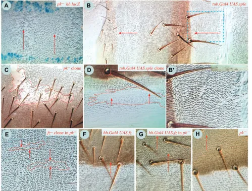
Flies that lack pk are viable and have reorganised polarity in the abdomen: dorsally there is a large zone of reversed polarity near the back of the A compartment, while polarity in the rest of the segment is normal. Ventrally, the same zones are observed, but they are more regular and better demarcated (Fig. 4A). Uniform overexpression of sple transcript produces a near-reciprocal phenotype: both dorsally and ventrally, the polarity of the entire P compartment is reversed, while, apart from the front, the A compartment is largely unaffected (Fig. 4B). Overexpression of sple in the P compartment alone gives the reversed phenotype there (genotypes 13, 14), while overexpression in the A compartment (ci.Gal4) has relatively little effect, causing only some localised disturbances at the fronts of both A and P compartments in the tergite, but no effect
in the pleura (genotype 15). General overexpression of the pk transcript causes reversal of polarity in part of the A compartment, but the P compartment is unaffected (genotype 16).
pk–flies that also lack fz do not show areas of consistently reversed polarity and, both dorsally and ventrally, have a pattern similar to fz–flies, indicating that the pk–phenotype is dependent on Fz (genotype 17). Similarly, if the sple transcript is overexpressed in the P compartment of fz– flies, the fz– phenotype is largely or entirely unaffected, indicating that the gain-of-function phenotype is also dependent on Fz (genotype 18).
fz–clones and UAS.fz in pk–flies
[image:6.612.56.555.251.630.2]fz clones cause hairs around the clone to point inwards, towards
Fig. 4. Clones involving prickle (pk). (A) Part of the pleura of a fly lacking pk and stained for hh.lacZ to demarcate the P compartments
the clones, irrespective of whether they are located in regions of normal or reversed polarity (Fig. 4E). The range appears to be longer, as also noted by Adler et al. (Adler et al., 2000) in hypomorphic pk mutants. Also, we used hh.Gal4 to drive UAS.fz in pk– flies; this causes a zone of reversed polarity straddling the A/P compartment boundary (compare Fig. 4G,H). The range of repolarisation is somewhat longer than that induced in wild-type flies (Fig. 4F). Thus, the loss of both Pk and Sple does not block the ability of sharp disparities in Fz activity to repolarise several rows of cells.
pk–, UAS.pk and UAS.sple clones
pk1and pk– clones have disturbed polarity and usually there are whorls associated with the clones. However, the effects are almost entirely autonomous to the clone (genotypes 22, 23 and Fig. 4C).
UAS.pk clones have a strong phenotype in the wing, the hairs point towards the clone, giving some reversal of polarity of hairs distal to it (Tree et al., 2002). However, in the P compartment of the abdomen, UAS.sple clones are almost entirely reversed, apart from some of their most anterior cells. Immediately behind these clones, some wild-type cells are usually reversed (Fig. 4D). UAS.sple clones in the A compartment are mostly normal but those at the back of the A compartment reverse a zone of P cells behind them (resembling, piecemeal, the phenotype caused by uniform expression of UAS.sple in the A compartment).
Pk: discussion
General loss of Pk and Sple leads to a reversal of polarity in a large portion of the A compartment, the rest of the segment being unaffected. But, overexpression of Sple leads to reversal of polarity in the P compartment, without affecting the A compartment much. These phenotypes are unrelated to the scalar pattern of cell types in the abdomen, which remain normal, and, at least for the pk–flies, are not associated with changes in either fj or ds expression (unpublished results). A clue to understanding these phenotypes may come from studying disparities in Fz activity in pk– flies; these appear to cause polarity changes similar to those they cause in wild-type flies. Therefore, the ability to read the levels of Fz activity and to respond to it could not depend on Pk and Sple. Perhaps, in pk–animals, many A compartment cells react to an unchanged X gradient with opposite sign? The reversal of P cells that overexpress Sple provides some support, because if such cells were to read the X gradient with opposite sign they should show reversed polarity. This raises a related but unsolved conundrum: as discussed above, the uniform polarity of hairs in the wild type argues that the gradient of Fz activity is itself monotonic. However we previously suggested that gradients of X might have opposing slopes in the A and the P compartment, and that they might be read with different sign – to ensure that hairs in both compartments would point the same way (Casal et al., 2002). Is there no simple relationship between what we earlier called ‘X’ and what we now refer to as ‘Fz activity’? Perhaps Pk and Sple are normally involved in rectification. And so, for example, without Sple, cells in the A compartment interpret X with the opposite sign. Likewise cells in the P compartment misinterpret X when Sple is over expressed. Clearly, we need a better understanding of how X, which we imagine is built by Ds, Ft and Fj, is linked to Fz activity.
Van Gogh (Vang)
Vang protein
This gene, also known as strabismus, encodes a probable transmembrane protein of type IIIa with a PDZ binding motif (Taylor et al., 1998; Wolff and Rubin, 1998; Bastock et al., 2003). Late in development, it becomes localised, like Pk, to the proximal edge of each cell in the wing, which is opposite to the site of accumulation of Fz (Bastock et al., 2003). In Vang– wing cells, some localisation of Fz to the membrane is preserved, but it is no longer concentrated on one edge of the cell (Strutt, 2001). In such cells there is also an increase in the amount of Pk protein (Bastock et al., 2003).
Vang– and UAS.Vang flies
Vang– flies are viable. Dorsally, the tergites resemble those of fz–flies; they are dishevelled, especially in the anterior parts of A, but have largely normal polarity elsewhere. Similarly, in the pleura, polarity is generally disordered, as in fz–, except that there is a weak tendency for the hairs to be organized into zones of alternating polarity along the anteroposterior axis. The hairs in the middle of the A compartment tend to be reversed, with the remaining hairs being more normally polarised (genotype 26). If Vang is universally overexpressed, the flies are viable and polarity is little affected (genotype 27).
Vang–and UAS.Vang clones
Vang–clones give consistent results both dorsally and ventrally. Within Vang–clones marked with pawn, the rows of hairs are jumbled and poorly oriented; some hairs point straight upwards, especially those in clones in anterior regions of the A compartment (to see the effect of pawn, compare Fig. 5A and B). Thus, there is an autonomous loss of polarity. By contrast, when clones are marked with yellow, there is some dishevelment, but the hairs in the clone are largely oriented correctly, resembling pieces of Vang–flies (genotype 29). This difference between pawn and yellow clones suggests there is some interaction between the pawn and Vang mutations. Nevertheless, irrespective of whether we use the pawn or yellow marker, Vang– cells appear largely refractory to neighbouring wild-type cells.
Outside and anterior to both pawn Vang–and yellow Vang– clones there are polarity disturbances: these extend a few cells and are variable, usually including patches with some hairs that are reversed (Fig. 5A). This reversal anterior to the clone is more consistent and extensive in the pleura (Fig. 5C). The amount of reversal appears not to depend on the location of the clone along the A/P axis. Indeed, a clone located at the anterior extreme of the P compartment can reverse the polarity of cells across the border at the back of the A compartment.
UAS.Vang clones show two effects: (i) hairs behind the clone are usually reversed, but (ii) hairs inside and mainly at the back of the clone are dishevelled, with some disorientation within those clones that were marked with pawn. Hairs at the front of the clone are unaffected (Fig. 5D). Clones at the back of the A compartment can cause reversal behind, in P cells.
UAS.Vang clones in Vang–flies
We have studied UAS.Vang clones marked with pawn in Vang– flies. These clones appear to have no effect on the surrounding Vang–cells, indicating that Vang–cells cannot be repolarised by adjacent Vang-expressing cells (Fig. 5E).
fz– and UAS.fz clones in Vang–flies
Can Vang– cells respond to discontinuities in Fz activity between neighbouring cells? Both fz–and UAS.fz clones fail to affect the polarity of Vang–adjacent cells (genotypes 33, 34), suggesting that they cannot (similar results for fz–clones were described in the wing) (Taylor et al., 1998). To check that the failure is specifically due to the responding cells, we gave those cells Vang, and took it away from the cells in the clone: thus we made UAS.fz clones that are Vang– in otherwise wild-type animals. These clones do repolarise the cells in front (genotype 35). Hence, it appears that Vang– cells can communicate disparities in Fz activity to their neighbours, provided that those neighbours have wild-type Vang activity.
Vang–and UAS.Vang clones in fz–flies
The repolarising effects of Vang– and UAS.Vang clones are reciprocal to those of fz–and UAS.fz clones; perhaps Fz activity might be abnormally elevated in Vang–cells and suppressed in
UAS.Vang cells? Consistent with this possibility, we have examined UAS.Vang clones in fz– flies, and found that these clones do not repolarise hairs immediately behind the clones (Fig. 5F). Likewise, Vang–clones fail to reverse hair polarity in front when they were made in fz–flies (genotype 37).
Vang: discussion
Abrupt disparities in the amount of Vang activity, like those in Fz, are sufficient to trigger repolarisation, which can progress a few cell diameters into wild-type territory. As UAS.Vang clones resemble a loss of function of fz, and Vang– clones partially mimic UAS.fz, it could be that Vang acts by suppressing Fz activity. However, the properties of these two proteins differ: we have found no situation in which the polarity of Vang– cells can be reversed, whereas fz–cells can be repolarised. It seems that Vang– cells can send information about their level of Fz activity to neighbouring cells, but cannot receive, or respond to, this information in return. Vang thus appears to be required for a subset of the functions performed by Stan, as described below.
starry night (stan)
Stan protein
[image:8.612.45.374.75.454.2]stan, also known as flamingo, encodes a protein with a large
cadherin-like extracellular region and seven receptor-like transmembrane domains (Chae et al., 1999; Usui et al., 1999). Stan becomes localised transiently along both the distal and proximal edges of each wing cell shortly before hair formation (Usui et al., 1999). In stan– cells, Fz, Pk, Vang and Dsh all fail to accumulate in the membrane, remaining largely cytoplasmic (Shimada et al., 2001; Strutt, 2001; Tree et al., 2002; Bastock et al., 2003). Usui and colleagues made the important observation that only if Stan is present in both adjacent cells will it accumulate along the apical membranes where these two cells abut, suggesting Stan makes homodimers that form a bridge between adjacent cell membranes (Usui et al., 1999).
stan mutant flies
Homozygous stan– zygotes die as embryos. However, hypomorphic stan embryos survive to adults, as do stan– zygotes that are rescued by neural expression of the UAS.stan transgene (Lu et al., 1999; Usui et al., 1999). For both the dorsal and ventral abdomen, the polarity phenotypes of hypomorphic flies (genotypes 38-40) and rescued stan– flies (genotype 41) are the same as fz– flies. When UAS.stan is generally and strongly overexpressed (tub.Gal4), the flies do not emerge as adults (genotype 42).
Clones of stan–cells
It was reported that, in the wing (Usui et al., 1999), stan–cells (marked with pawn) make polarised hairs with randomised
orientation, but we do not find this to be usually so. Although some clones are dishevelled, others have near-normal polarity. We found that large stan–clones marked with yellow (genotype 43) are even more normal, and similar to the equivalent parts of wings that are mutant for stan. This suggests that the pawn marker itself might be contributing to the disarray and indeed, in the wing, pawn clones that are otherwise wild type are sometimes dishevelled. We made wings that were largely pawn– stan–, with only small patches of stan+ cells. These wild-type patches most often have normal polarity, indicating that small clusters of stan+ cells can respond autonomously, and correctly, to the X gradient even when isolated in a sea of mutant tissue (genotype 44).
[image:9.612.46.565.384.653.2]In the abdomen, pawn itself does not noticeably alter polarity (Fig. 5B) and stan– clones marked with yellow resemble pieces of the pattern of stan mutant flies (normal in some areas, abnormal in others). However, stan–clones marked with pawn are dishevelled, both in orientation and in the orderliness of the hair rows (Fig. 6A). Within the clone, they thus resemble patches of pawn Vang– cells. However, unlike pawn Vang– clones, pawn stan– clones have little effect on surrounding wild-type cells, except for rare, local disturbances just outside and mostly in front of the clones. In the pleura of stan mutant flies polarity is lateralised/randomised, and stan– clones, both marked with pawn and unmarked, appear to show this phenotype autonomously (the frequency, size and shape of the unmarked patches indicate that they are clones). These
clones in the pleura do show occasional local non-autonomy, disturbing the polarity of hairs here and there in front of the clone. Thus, stan– cells appear to be unable to receive polarising information from their wild-type neighbours, and have little, or no ability to send such information. In this respect they differ from Vang–cells, which typically repolarise wild-type neighbours.
stan– clones that express UAS.fz or UAS.Vang
Clones of stan– cells that express UAS.fz show randomised polarity inside (owing to the lack of Stan in combination with pawn) but fail to repolarise in front of the clone (Fig. 6B), in contrast to simple UAS.fz clones (Fig. 6C), and to UAS.fz Vang– clones (genotype 35), which normally do. Likewise, if fz– clones are made in flies carrying viable mutations of stan (genotypes 47, 48), repolarisation both inside and outside the clone is blocked [as in the wing (Chae et al., 1999)]. Also, if fz is driven by ptc.Gal4 in stan mutant flies, the repolarisation that occurs in a wild-type background is suppressed (genotype 49). Finally, as observed for UAS.fz stan–clones, UAS.Vang stan– clones show randomized polarity within the clone (marked with pawn), but fail to repolarise wild-type cells outside (Fig. 6E). These results indicate that stan– cells are unable to communicate their level of Fz activity to neighbouring cells, and are unable to respond to such information communicated to them.
UAS.stan clones
Cells within UAS.stan clones show some dishevelment, possibly because of the presence of pawn. The clones induce extensive reversal behind the clone, both in the A and P compartments, and also within the clone, at the back (Fig. 6D). This suggests that an abrupt disparity in the amount of Stan protein is sufficient to polarise cells, with effects spreading outwards from the interface, providing that at least some Stan is expressed in cells on both sides of that interface.
Both UAS.stan and UAS.Vang clones cause a similar phenotype to fz– clones, raising the possibility that overexpression of either of these proteins autonomously suppresses Fz activity. Consistent with this, UAS.stan clones fail to cause repolarisations in fz–flies, even in cells along the clone border (Fig. 6F). We have also expressed UAS.stan at the back of the A compartment using ptc.Gal4, which normally results in a zone of reversed polarity at the back and behind the A compartment (genotype 53). This is expected from the clones (at the back of the A compartment there will be a sharp interface in the amount of Stan), but, again, if stan is expressed in the same pattern in fz–flies, that zone is not seen (genotype 54). These findings show that Fz is required for UAS.stan clones to cause repolarisations.
Stan: discussion
Animals that are entirely mutant for stan or fz have similar phenotypes in the abdomen, notably in the pleura where hair polarity is randomized. Stan and Fz are seven-pass transmembrane proteins with similar structures, making them both valid candidates for receptors for X. However, unlike Fz, we find that Stan appears necessary on both sides of a clone interface (say, a clone that is overexpressing fz) to induce and propagate a change in polarity both into and out of the clone. It is probably important to this function that Stan may form
homodimers that act as a link between cells (Usui et al., 1999). But note that wild-type cells that are adjacent to stan–clones, or patches of stan+ cells in a sea of stan–cells are, usually, normally polarised, suggesting that polarity in one cell does not depend on it having such links with every neighbour.
General Discussion
Outline of a working model for cell polarity in the abdomen
There are a number of simple systems in which isolated cells orient to a polarising signal. These include the localized outgrowth, or ‘schmooing’ of yeast in response to mating pheromone and directed migration of Dictyostelium cells up a gradient of cyclic AMP (Arkowitz, 1999). Small differences (as little as 1-5%) in receptor activation across single cells are sufficient to polarise them (Zigmond, 1974; Parent and Devreotes, 1999; Drubin, 2000), a response that, in yeast and elsewhere probably depends on localised exocytosis (Bretscher, 1984; Valdez-Taubas and Pelham, 2003). It is not known whether the polarisation of single, isolated cells is a model for planar polarity of cells in an epithelium, but it is likely that they share at least some of the mechanisms.
We have proposed that, in the abdomen of Drosophila, morphogen gradients (Hh in the A compartment and Wg in the P compartment) organise a secondary gradient (‘X’); the vector of X specifying the polarity of each cell (Struhl et al., 1997a; Struhl et al., 1997b; Lawrence et al., 2002). Although the composition of X is unknown, at least three proteins, Fj, Ds and Ft, are implicated (Casal et al., 2002; Lawrence et al., 2002; Yang et al., 2002). All three may be expressed, or be active, in bell-shaped distributions that peak near the A/P (Ds) or P/A (Fj, Ft) boundaries (Casal et al., 2002). Ds and Ft are transmembrane proteins in the cadherin superfamily; Fj probably acts in the Golgi (Strutt et al., 2004). Ds and Ft are integrated into the membrane, suggesting that the X gradient itself may not be diffusible but instead might depend on information transfer from cell to cell.
How does Hh set up the X gradient? Although changing the real or perceived level of Hh does affect polarity, many clones (for example clones that lack Smo, an essential component of Hh reception) show there is no simple correlation between Hh concentration and polarity. For instance, large smo–clones in the centre of the A compartment are polarised normally, even though they are blind to Hh. Also, while smo–clones in some regions of the A compartment do affect polarity, both mutant and wild-type cells are repolarised (Struhl et al., 1997a). Both these observations argue for some transfer of information about polarity between cells, a process that would be at least partly Hh independent. This paper explores this process and is concerned with four genes (stan, fz, Vang and pk) that probably act downstream of ds, ft and fj.
the levels of X (recorded in the activity of Fz) in neighbouring cells. Then, in a way analogous to how a Dictyostelium amoeba reads the vector of a cAMP gradient, a cell would determine its polarity from the vector of Fz activity. The results suggest that Vang also acts in this step, helping cells to sense the level of Fz activity in neighbouring cells.
The working model, details and conundrums We now discuss some of the results in terms of the model.
Non-autonomy
Clones that lack, or overexpress Fz cause local and consistent repolarisations of cells that extend from within the clone and affect normal wild-type cells outside it (Gubb and Garcia-Bellido, 1982; Vinson and Adler, 1987). Because simply removing the fz gene from all cells randomizes polarity in the ventral pleura, it is self-evident that these organised polarity reversals must result from an interaction between the clone and the surrounding cells. We have argued that Stan and Fz act in this process, but how? Note that stan and fz are the only mutants that have randomised hairs in the pleura, and our results indicate that neither Stan nor Fz can function properly without the other. Averaging might depend on the capacity of Stan to form homophilic dimers as bridges between neighbouring cells (Usui et al., 1999), with such Stan:Stan dimers serving as a conduit for information about the relative level of Fz activity in each cell. However, with respect to non-autonomy, the results with the two genes differ:
(i) stan– cells cannot be repolarised by, and cannot repolarise, neighbouring cells. This shows that Stan is essential in both neighbouring cells for the transfer of information between them. Without Stan, the cells cannot compare and cannot therefore determine any vector. However, a stan+cell, even if it is adjacent to a stan–cell, can be polarised normally; having Stan it should be able to read the levels of all neighbouring cells except the stan– one and, having Fz, it should be able to set its own level.
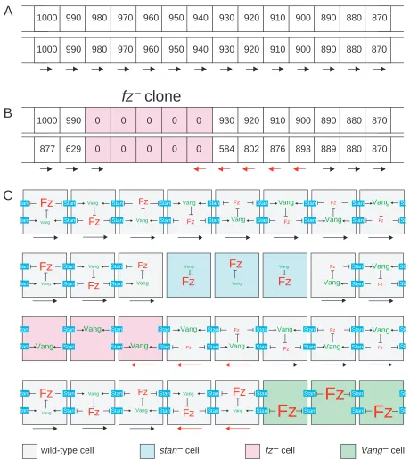
(ii) But, fz– cells, unlike stan– cells, can repolarise neighbouring wild-type cells. Also, a fz–cell, again unlike a stan–cell, can itself be repolarised. The results also show that to be repolarised, or to polarise a neighbour, a fz–cell must be adjacent to a stan+fz+cell. Such a fz–cell will accumulate Stan in that membrane which abuts the stan+neighbour (Usui et al., 1999) so it should be able to read the level of the neighbouring wild-type cell and be polarised accordingly. Consider a fz–cell at the outer edge of a fz– clone (Fig. 7): lacking Fz, its activity level would be zero, but this level would be communicated by Stan to the neighbouring cells. Wild-type cells outside would obviously have a higher level (than zero). The result would be that the two cells abutting the interface, the fz– cell inside, and a fz+ cell outside, would both make hairs that point into the centre of the clone. The nextmost interior cell would not be polarised, as all its neighbours would be cells with level zero. In contrast, the next most exterior cell would be repolarised, as its scalar level would be brought down by the averaging process. How far does the non-autonomy spread into wild-type cells? We have simulated this process (Fig. 7, see also supplementary material). According to the model this range would depend on the value of a single adjustable parameter, a (see supplementary material) that relates to how much a cell’s scalar is read from X. At one extreme for this parameter
(a=0), when the scalar of a cell depends only on X, a wild-type cell just posterior to a clone of fz–cells would reset its scalar as it was before; there could be no averaging and only that cell and its fz– neighbour will be repolarised. Thus the non-autonomy would be limited to one cell. At the other extreme (a=1), any local disturbance produced by a clone would decay rapidly because of averaging, and the repolarisation will tend to be lost altogether. In between these extremes, the non-autonomy spreads more than one cell, but over diverse values for this parameter, the range is near the amount we usually observe (2-4 cell diameters; see supplementary material).
It has been observed that fz–clones have effects over longer range in backgrounds such as ds–(Adler et al., 1998; Ma et al., 2003) where the X gradient might be flatter than normal. Similarly, cells are normally polarised in large smo–clones in the middle of the A compartment, where, because there can be no input from Hh, the X gradient could also be flat. Both these results are consistent with the model, because the range affected by averaging will increase (see supplementary material).
The localisation of proteins in cells of pupal wings Many of the proteins required for normal cell polarity, including Fz, Dsh, Dgo, Pk, Vang and Stan are found to be asymmetrically localised in the proximodistal axis of wing cells (Strutt, 2002). This localisation is restricted to a brief period of just a few hours shortly before the wing hairs grow out, but, nevertheless it is assumed to be mechanistically important to planar polarity (Strutt, 2002). For example, non-autonomy could be explained if localised proteins were components of one or more molecular complexes that propagate polarity from cell to cell (Usui et al., 1999; Axelrod, 2001; Feiguin et al., 2001; Strutt, 2001; Adler, 2002; Strutt, 2002; Tree et al., 2002; Bastock et al., 2003; Ma et al., 2003). In support of this, note that loss of any of these proteins, including the removal of both Pk and Sple, prevents the asymmetric localisation of the others (Strutt, 2002; Bastock et al., 2003).
1000 990 980 970 960 950 940 930 920 910
1000 990 980 970 960 950 940 930 920 910
1000 990 0 0 0 0 0 930 920 910
877 629 0 0 0 0 0 584 802 876
900 900 900 893 890 890 890 889 880 880 880 880 870 870 870 870
fz
–
clone
Fz
Vang Vang Vang VangFz
Fz
Fz
Vang Stan Stan Stan Stan Stan Stan Stan Stan Stan Stan Stan tan tan St St Stan Stan StanFz
Fz
Fz
FzFz
Fz
Vang Vang Fz Vang Fz Vang Fz Vang Fz Vang Fz Vang Fz Vang Stan Stan Stan Stan Stan Stan Stan Stan Stan Stan Stan Stan Stan Stan St St tan tan Vang Vang VangFz
Vang VangFz
VangFz
Vang Stan Stan Stan Stan Sta Sta tan tan Stan StanFz
Fz
Fz Fz Fz Vang Vang Vang Vang Vang VangStan Stan Stan Stan Stan
Stan Stan Stan St St tan tan Stan Stan Stan Stan Stan Stan Fz Fz
Fz Fz Fz
wild-type cell stan – cell fz – cell Vang – cell
[image:12.612.71.531.69.584.2]Vang Vang Vang
A
B
C
Fig. 7. The averaging model. (A) Imagine a gradient of Fz activity from 1000 downwards (see top diagram, upper row). A cell takes into
Are wing cells polarised only briefly just prior to the hair outgrowth? The reason for raising this possibility is that the proteins are apparently only asymmetrically localised at that time. If this localisation were not causal, as we now suggest, it could be that the cells are polarised for all or most of development – again arguing that the ephemeral localisation of the proteins is more a consequence than a cause of polarisation.
We have had much help from the Drosophila community, we mention particularly David Gubb, Bob Holmgren, Xiao-Jing Qiu, David Strutt and Tanya Wolff, as well as the Bloomington Stock Center who have been generous with stocks and advice. Graeme Mitchison has given us a nihil obstat for the maths. We ask that author order here, as well as our four previous publications on the Drosophila abdomen, not be invested with significance: all three of us have contributed differently and we share responsibility. P.A.L. and J.C. are supported by the MRC; G.S. is an HHMI Investigator.
Supplementary material
Supplementary material for this article is available at http://dev.biologists.org/cgi/content/full/131/19/4651/DC1
References
Adler, P. N. (2002). Planar signaling and morphogenesis in Drosophila. Dev.
Cell 2, 525-535.
Adler, P. N., Charlton, J. and Liu, J. (1998). Mutations in the cadherin
superfamily member gene dachsous cause a tissue polarity phenotype by altering frizzled signaling. Development 125, 959-968.
Adler, P. N., Krasnow, R. E. and Liu, J. (1997). Tissue polarity points from
cells that have higher Frizzled levels towards cells that have lower Frizzled levels. Curr. Biol. 7, 940-949.
Adler, P. N., Taylor, J. and Charlton, J. (2000). The domineering
non-autonomy of frizzled and van Gogh clones in the Drosophila wing is a consequence of a disruption in local signaling. Mech. Dev. 96, 197-207.
Adler, P. N., Vinson, C., Park, W. J., Conover, S. and Klein, L. (1990).
Molecular structure of frizzled, a Drosophila tissue polarity gene. Genetics
126, 401-416.
Arkowitz, R. A. (1999). Responding to attraction: chemotaxis and
chemotropism in Dictyostelium and yeast. Trends Cell Biol 9, 20-27.
Axelrod, J. D. (2001). Unipolar membrane association of Dishevelled
mediates Frizzled planar cell polarity signaling. Genes Dev. 15, 1182-1187.
Bastock, R., Strutt, H. and Strutt, D. (2003). Strabismus is asymmetrically
localised and binds to Prickle and Dishevelled during Drosophila planar polarity patterning. Development 130, 3007-3014.
Bhanot, P., Brink, M., Samos, C. H., Hsieh, J. C., Wang, Y., Macke, J. P., Andrew, D., Nathans, J. and Nusse, R. (1996). A new member of the
Frizzled family from Drosophila functions as a Wingless receptor. Nature
382, 225-230.
Bhanot, P., Fish, M., Jemison, J. A., Nusse, R., Nathans, J. and Cadigan, K. M. (1999). Frizzled and Dfrizzled-2 function as redundant receptors for
Wingless during Drosophila embryonic development. Development 126, 4175-4186.
Bretscher, M. S. (1984). Endocytosis: relation to capping and cell locomotion.
Science 224, 681-686.
Casal, J., Struhl, G. and Lawrence, P. A. (2002). Developmental
compartments and planar polarity in Drosophila. Curr. Biol. 12, 1189-1198.
Chae, J., Kim, M. J., Goo, J. H., Collier, S., Gubb, D., Charlton, J., Adler, P. N. and Park, W. J. (1999). The Drosophila tissue polarity gene starry
night encodes a member of the protocadherin family. Development 126,
5421-5429.
Chen, C. M. and Struhl, G. (1999). Wingless transduction by the Frizzled
and Frizzled 2 proteins of Drosophila. Development 126, 5441-5452.
Drubin, D. (2000). Cell Polarity. Oxford: Oxford University Press. Feiguin, F., Hannus, M., Mlodzik, M. and Eaton, S. (2001). The ankyrin
repeat protein Diego mediates Frizzled-dependent planar polarization. Dev.
Cell 1, 93-101.
FlyBase (1999). The FlyBase database of the Drosophila genome projects and
community literature. http://flybase.bio.indiana.edu. Nucleic Acids Res. 27, 85-88.
Gubb, D. and Garcia-Bellido, A. (1982). A genetic analysis of the
determination of cuticular polarity during development in Drosophila melanogaster. J. Embryol. Exp. Morphol. 68, 37-57.
Gubb, D., Green, C., Huen, D., Coulson, D., Johnson, G., Tree, D., Collier, S. and Roote, J. (1999). The balance between isoforms of the prickle LIM
domain protein is critical for planar polarity in Drosophila imaginal discs.
Genes Dev. 13, 2315-2327.
Klingensmith, J., Nusse, R. and Perrimon, N. (1994). The Drosophila
segment polarity gene dishevelled encodes a novel protein required for response to the wingless signal. Genes Dev. 8, 118-130.
Lawrence, P. A. (1966). Gradients in the insect segment: The orientation
of hairs in the milkweed bug Oncopeltus fasciatus. J. Exp. Biol. 44, 607-620.
Lawrence, P. A., Casal, J. and Struhl, G. (1999a). hedgehog and engrailed:
pattern formation and polarity in the Drosophila abdomen. Development
126, 2431-2439.
Lawrence, P. A., Casal, J. and Struhl, G. (1999b). The Hedgehog morphogen
and gradients of cell affinity in the abdomen of Drosophila. Development
126, 2441-2449.
Lawrence, P. A., Casal, J. and Struhl, G. (2002). Towards a model of the
organisation of planar polarity and pattern in the Drosophila abdomen.
Development 129, 2749-2760.
Lecuit, T., Samanta, R. and Wieschaus, E. (2002). slam encodes a
developmental regulator of polarized membrane growth during cleavage of the Drosophila embryo. Dev. Cell 2, 425-436.
Lewis, J. and Davies, A. (2002). Planar cell polarity in the inner ear: how do
hair cells acquire their oriented structure? J. Neurobiol. 53, 190-201.
Lu, B., Usui, T., Uemura, T., Jan, L. and Jan, Y. N. (1999). Flamingo
controls the planar polarity of sensory bristles and asymmetric division of sensory organ precursors in Drosophila. Curr. Biol. 9, 1247-1250.
Ma, D., Yang, C. H., McNeill, H., Simon, M. A. and Axelrod, J. D. (2003).
Fidelity in planar cell polarity signalling. Nature 421, 543-547.
Mlodzik, M. (2002). Planar cell polarization: do the same mechanisms
regulate Drosophila tissue polarity and vertebrate gastrulation? Trends
Genet. 18, 564-571.
Nübler-Jung, K., Bonitz, R. and Sonnenschein, M. (1987). Cell polarity
during wound healing in an insect epidermis. Development 100, 163-170.
Parent, C. A. and Devreotes, P. N. (1999). A cell’s sense of direction. Science 284, 765-770.
Park, W. J., Liu, J. and Adler, P. N. (1994). The frizzled gene of Drosophila
encodes a membrane protein with an odd number of transmembrane domains. Mech. Dev. 45, 127-137.
Shimada, Y., Usui, T., Yanagawa, S., Takeichi, M. and Uemura, T. (2001).
Asymmetric colocalization of Flamingo, a seven-pass transmembrane cadherin, and Dishevelled in planar cell polarization. Curr. Biol. 11, 859-863.
Struhl, G., Barbash, D. A. and Lawrence, P. A. (1997a). Hedgehog acts by
distinct gradient and signal relay mechanisms to organise cell type and cell polarity in the Drosophila abdomen. Development 124, 2155-2165.
Struhl, G., Barbash, D. A. and Lawrence, P. A. (1997b). Hedgehog
organises the pattern and polarity of epidermal cells in the Drosophila abdomen. Development 124, 2143-2154.
Strutt, D. I. (2001). Asymmetric localization of Frizzled and the establishment
of cell polarity in the Drosophila wing. Mol. Cell 7, 367-375.
Strutt, D. I. (2002). The asymmetric subcellular localisation of components
of the planar polarity pathway. Semin. Cell Dev. Biol. 13, 225-231.
Strutt, H., Mundy, J., Hofstra, K. and Strutt, D. (2004). Cleavage and
secretion is not required for Four-jointed function in Drosophila patterning.
Development 131, 881-890.
Strutt, H. and Strutt, D. (2002). Nonautonomous planar polarity patterning
in Drosophila: dishevelled-independent functions of frizzled. Dev. Cell 3, 851-863.
Stumpf, H. F. (1966). Mechanism by which cells estimate their location within
the body. Nature 212, 430-431.
Taylor, J., Abramova, N., Charlton, J. and Adler, P. N. (1998). Van Gogh:
a new Drosophila tissue polarity gene. Genetics 150, 199-210.
Theisen, H., Purcell, J., Bennett, M., Kansagara, D., Syed, A. and Marsh, J. L. (1994). dishevelled is required during wingless signaling to establish
both cell polarity and cell identity. Development 120, 347-360.
Tomlinson, A., Strapps, W. R. and Heemskerk, J. (1997). Linking Frizzled
and Wnt signaling in Drosophila development. Development 124, 4515-4521.
Tree, D. R., Shulman, J. M., Rousset, R., Scott, M. P., Gubb, D. and Axelrod, J. D. (2002). Prickle mediates feedback amplification to generate
Usui, T., Shima, Y., Shimada, Y., Hirano, S., Burgess, R. W., Schwarz, T. L., Takeichi, M. and Uemura, T. (1999). Flamingo, a seven-pass
transmembrane cadherin, regulates planar cell polarity under the control of Frizzled. Cell 98, 585-595.
Valdez-Taubas, J. and Pelham, H. R. (2003). Slow diffusion of proteins in
the yeast plasma membrane allows polarity to be maintained by endocytic cycling. Curr. Biol. 13, 1636-1640.
Vinson, C. R. and Adler, P. N. (1987). Directional non-cell autonomy and the
transmission of polarity information by the frizzled gene of Drosophila.
Nature 329, 549-551.
Wallingford, J. B., Fraser, S. E. and Harland, R. M. (2002). Convergent
extension: the molecular control of polarized cell movement during embryonic development. Dev. Cell 2, 695-706.
Wehrli, M. and Tomlinson, A. (1998). Independent regulation of
anterior/posterior and equatorial/polar polarity in the Drosophila eye;
evidence for the involvement of Wnt signaling in the equatorial/polar axis.
Development 125, 1421-1432.
Wodarz, A. and Nusse, R. (1998). Mechanisms of Wnt signaling in
development. Annu. Rev. Cell Dev. Biol. 14, 59-88.
Wolff, T. and Rubin, G. M. (1998). Strabismus, a novel gene that regulates
tissue polarity and cell fate decisions in Drosophila. Development 125, 1149-59.
Yang, C., Axelrod, J. D. and Simon, M. A. (2002). Regulation of Frizzled
by Fat-like cadherins during planar polarity signaling in the Drosophila compound eye. Cell 108, 675-688.
Zheng, L., Zhang, J. and Carthew, R. W. (1995). frizzled regulates
mirror-symmetric pattern formation in the Drosophila eye. Development 121, 3045-3055.
Zigmond, S. H. (1974). Mechanisms of sensing chemical gradients by